ITL-46983f4cd258
Juan Li, MD · Updated November 18, 2026
Key points
- The signature. An echo-bright, thickened wall with a hypoechoic space is typical, and the space is anechoic during active bleeding and hypoechoic once thrombosed.[1], [2]
- Use criteria. Echo diagnosis requires at least three listed signs, including a thin, mobile endomyocardial border around the cavity.[3]
- Not the cavity. No Doppler flow between the echo-free space and the LV cavity argues against pseudoaneurysm.[4]
- Not a pseudoaneurysm. The distinction holds only if the surrounding myocardial wall is proven continuous; a true pseudoaneurysm breaks wall continuity.[5]
- Easy to miss. In one series only 55% of septal hematomas were found intraoperatively, and they can enlarge rapidly after surgery.[1]
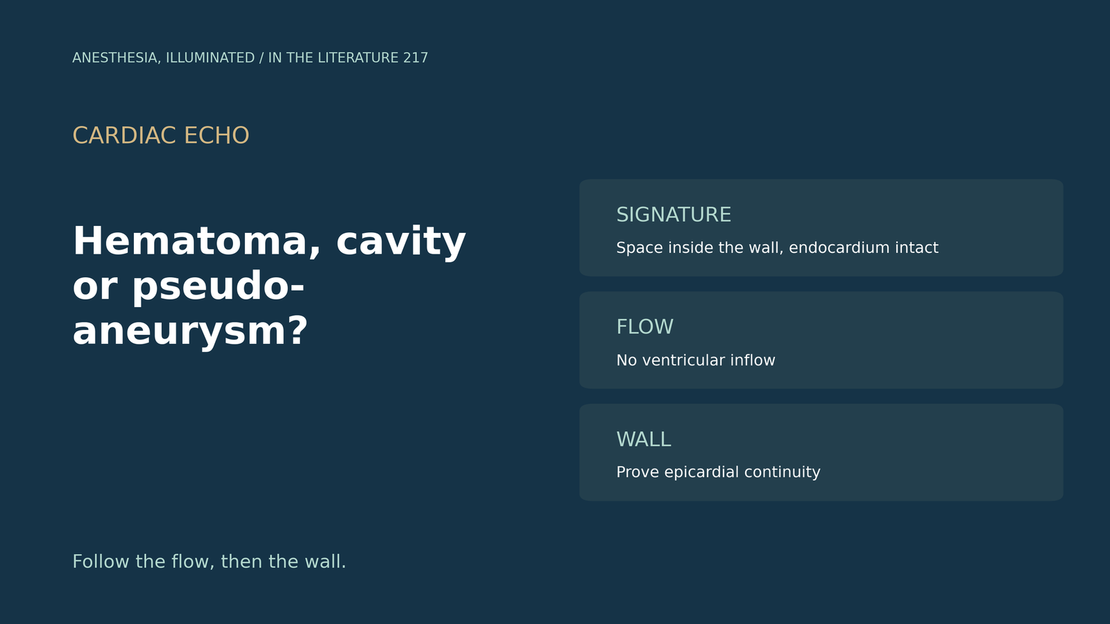
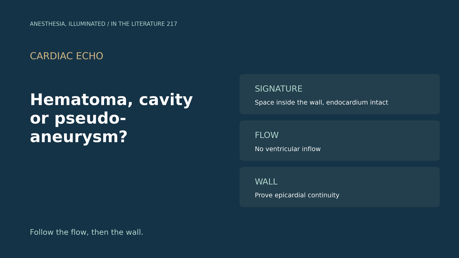
After a septal procedure, a new echo-free space appears inside the wall on a postoperative echo. Is it a hematoma, part of the cavity, or a pseudoaneurysm?[1], [4]
What a hematoma looks like
Akam-Venkata et al. (CASE 2021) describe an echo-bright, thickened septum with a hypoechoic space; in one series only 55% were discovered intraoperatively, and the hematoma can enlarge rapidly after surgery, so close follow-up is warranted.[1] Huang et al. (CASE 2024) note that it appears anechoic during active bleeding and hypoechoic once thrombosed and, unlike thrombus, has an overlying endocardial layer.[2] Tanabe et al. (CASE 2021) require at least three listed signs for echo diagnosis; a thin, mobile endomyocardial border surrounds the cavity, and the myocardium over the space may move outward in systole and inward in diastole.[3]
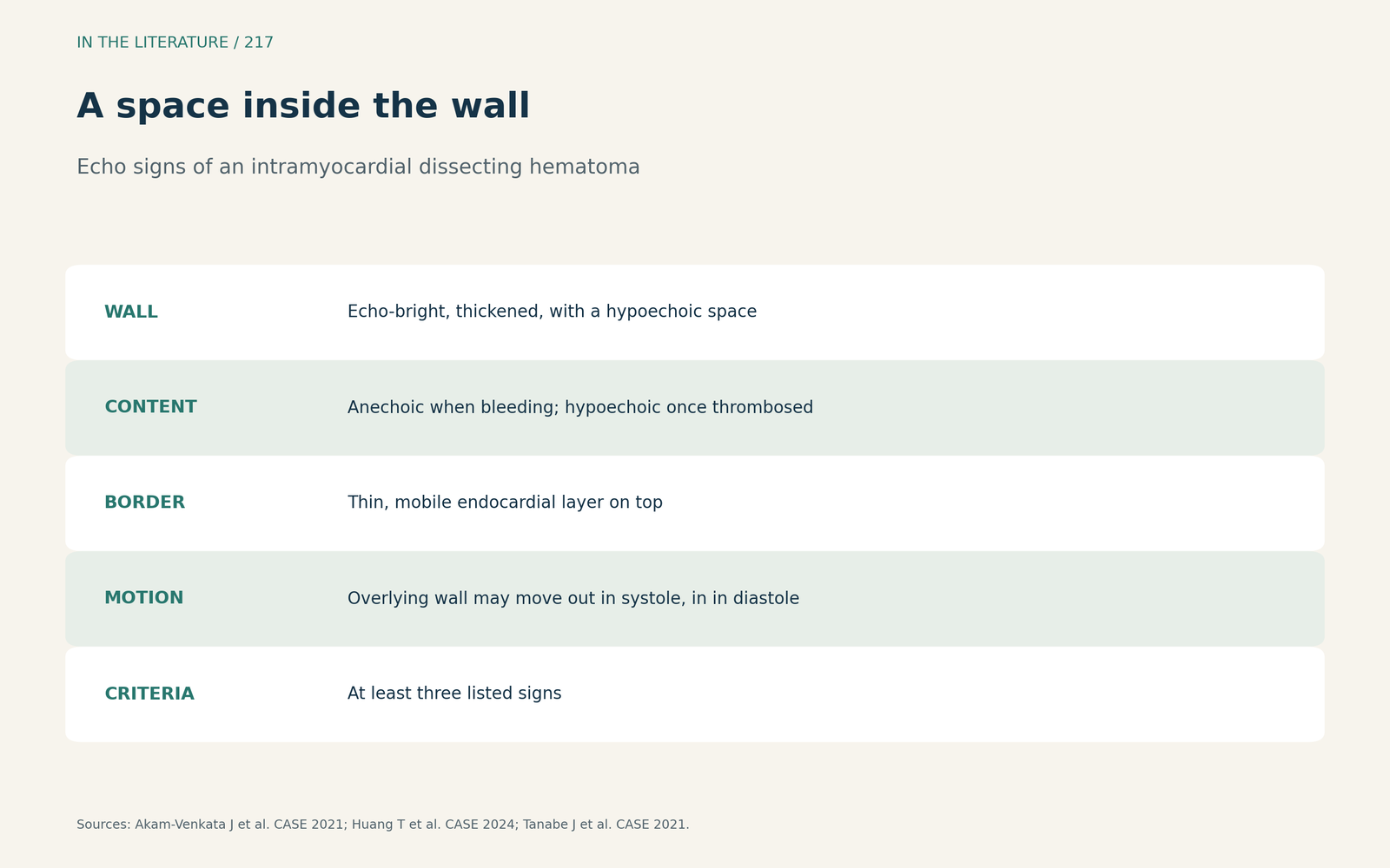
Figure 1. A space inside the wall. Synthesized from Akam-Venkata et al., Huang et al. and Tanabe et al.[1], [2], [3] View larger.

Cavity, thrombus or pseudoaneurysm?
Yuen et al. (CASE 2022) list LV thrombus, pseudoaneurysm, prominent trabeculation and dissecting hematoma as the differential: no Doppler flow between the echo-free space and the LV cavity argues against pseudoaneurysm, and a new finding absent on an earlier echo makes trabeculation unlikely.[4] On contrast echo, filling of the hematoma right after the chamber indicates communication, whereas sparse microbubbles appearing only after several cycles suggest no continued bleeding.[2] Flow is not decisive alone, because the diagnostic criteria also list continuity with a ventricular cavity.[3] Carrión-Sánchez et al. (Eur Heart J Case Rep 2024) define a closed dissection by absence of communication with the LV; it is distinguished from pseudoaneurysm only if the surrounding wall is proven continuous, whereas a true pseudoaneurysm breaks wall continuity with a neck-to-depth ratio below 50%. Cardiac MRI is the gold standard.[5] Yu et al. (Medicine 2025) stress careful examination of the endocardial and epicardial layers; if epicardial integrity is hard to confirm, the hematoma may be missed.[6]
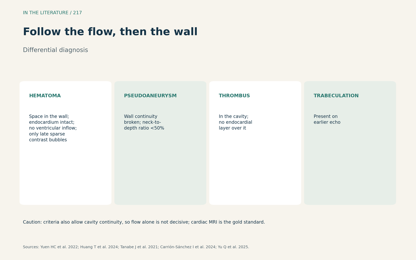
Figure 2. Follow the flow, then the wall. Synthesized from Yuen et al., Huang et al., Tanabe et al., Carrión-Sánchez et al. and Yu et al.[4], [2], [3], [5], [6] View larger.

Course and management
Hemodynamic stability is the decisive factor in managing a septal hematoma.[2] Apical dissecting hematomas have a high probability of spontaneous reabsorption.[3] A pooled analysis of 77 patients from case reports and series (Zavar et al., ARYA Atheroscler 2024) found that pericardial effusion significantly increased the risk of death,[7] and an expanding hematoma with pericardial effusion warrants consideration of urgent surgical repair.[6] The evidence is case reports, small series and reviews of them.
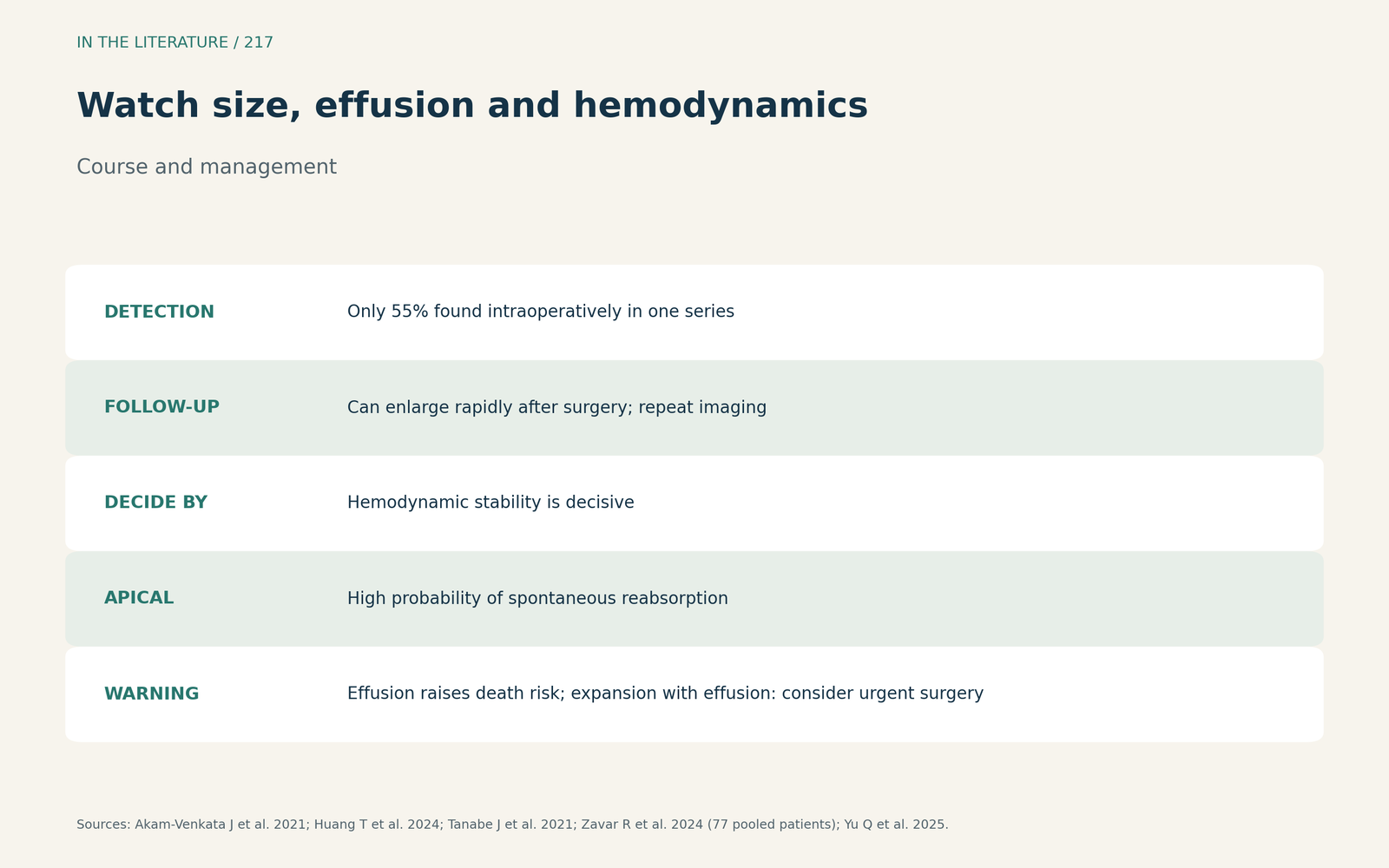
Figure 3. Watch size, effusion and hemodynamics. Synthesized from Huang et al., Tanabe et al., Zavar et al. and Yu et al.[2], [3], [7], [6] View larger.

Three takeaways
- A hypo- or anechoic space inside a thickened wall under a thin endocardial layer suggests a hematoma.[1], [2], [3]
- Check for ventricular inflow, then prove wall and epicardial continuity before excluding pseudoaneurysm.[4], [5], [6]
- Repeat imaging after surgery, and treat new pericardial effusion or expansion as a warning.[1], [7], [6]
References
- Akam-Venkata J, Lemler M, Pirolli T, et al. Evolution of interventricular septal hematoma: echocardiographic diagnosis. CASE (Phila). 2021;5(1):39–42. doi:10.1016/j.case.2020.10.003
- Huang T, Yang L, Wen S, et al. Interventricular septal hematoma following correction of Ebstein anomaly. CASE (Phila). 2024;8(3):170–173. doi:10.1016/j.case.2023.12.005
- Tanabe J, Okazaki K, Endo A, et al. Left ventricular intramyocardial dissecting hematoma. CASE (Phila). 2021;5(6):349–353. doi:10.1016/j.case.2021.07.016
- Yuen HC, Shek J, Wong CK, et al. An unusual case of intramyocardial dissecting hematoma. CASE (Phila). 2022;6(1):16–20. doi:10.1016/j.case.2021.07.015
- Carrión-Sánchez I, Tejada-González C, Gómez-Delgado J, et al. Role of multimodality cardiac imaging for evaluation of intramyocardial dissection, from dissecting haematoma to false-pseudoaneurysm: a case series. Eur Heart J Case Rep. 2024;8(5):ytae219. doi:10.1093/ehjcr/ytae219
- Yu Q, Zhou C, Liu R, et al. Clinical significance of intramyocardial dissecting hematoma after MI: a review. Medicine (Baltimore). 2025;104(44):e45650. doi:10.1097/MD.0000000000045650
- Zavar R, Soleimani A, Tajmirriahi M, et al. Intramyocardial dissecting hematoma: a systematic review and pooled analysis of available literature. ARYA Atheroscler. 2024;20(1):62–71. doi:10.48305/arya.2023.42244.2927