ITL-a07e26859d1c
Juan Li, MD · Updated November 17, 2026
Watch the video
Key points
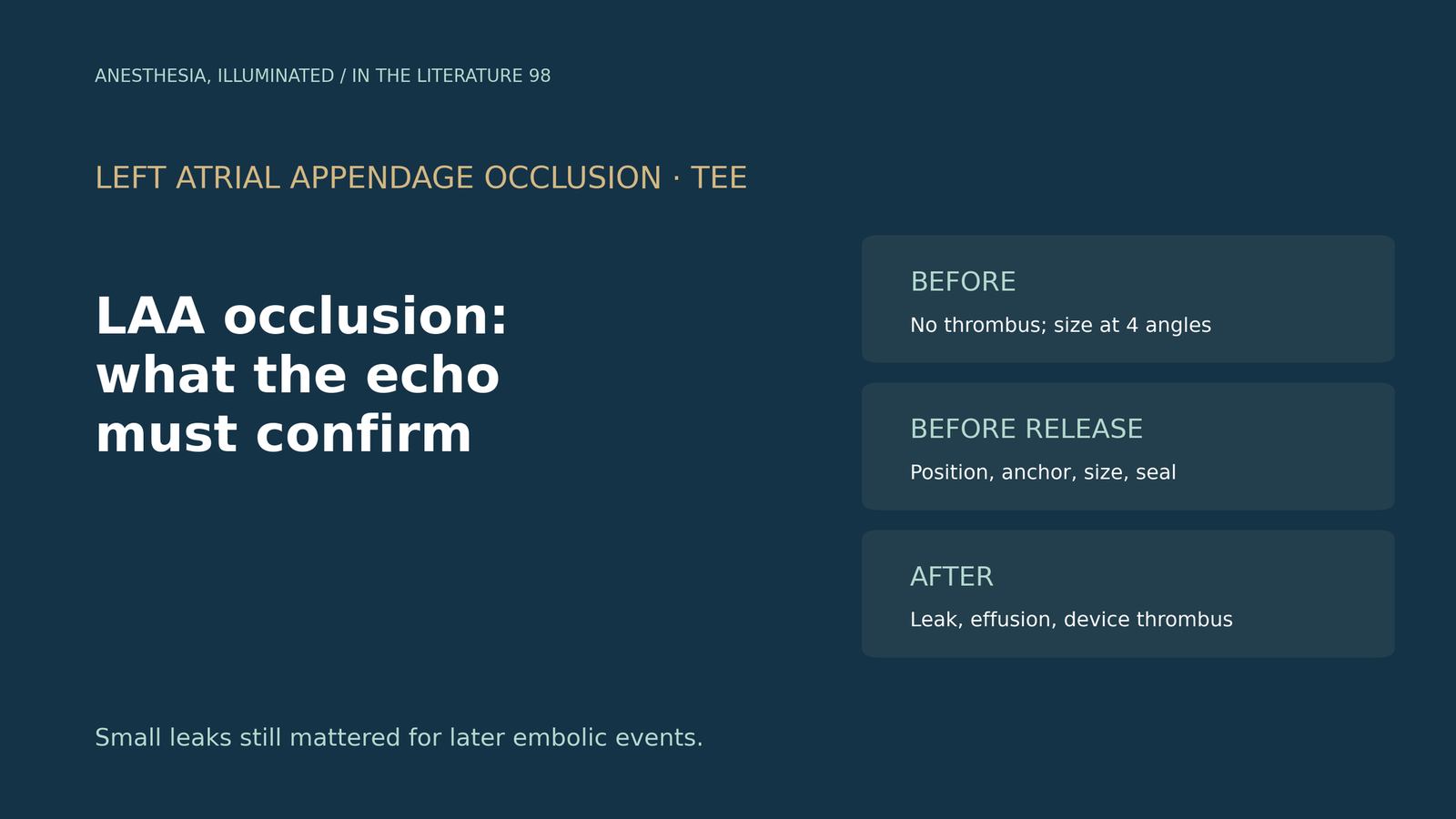
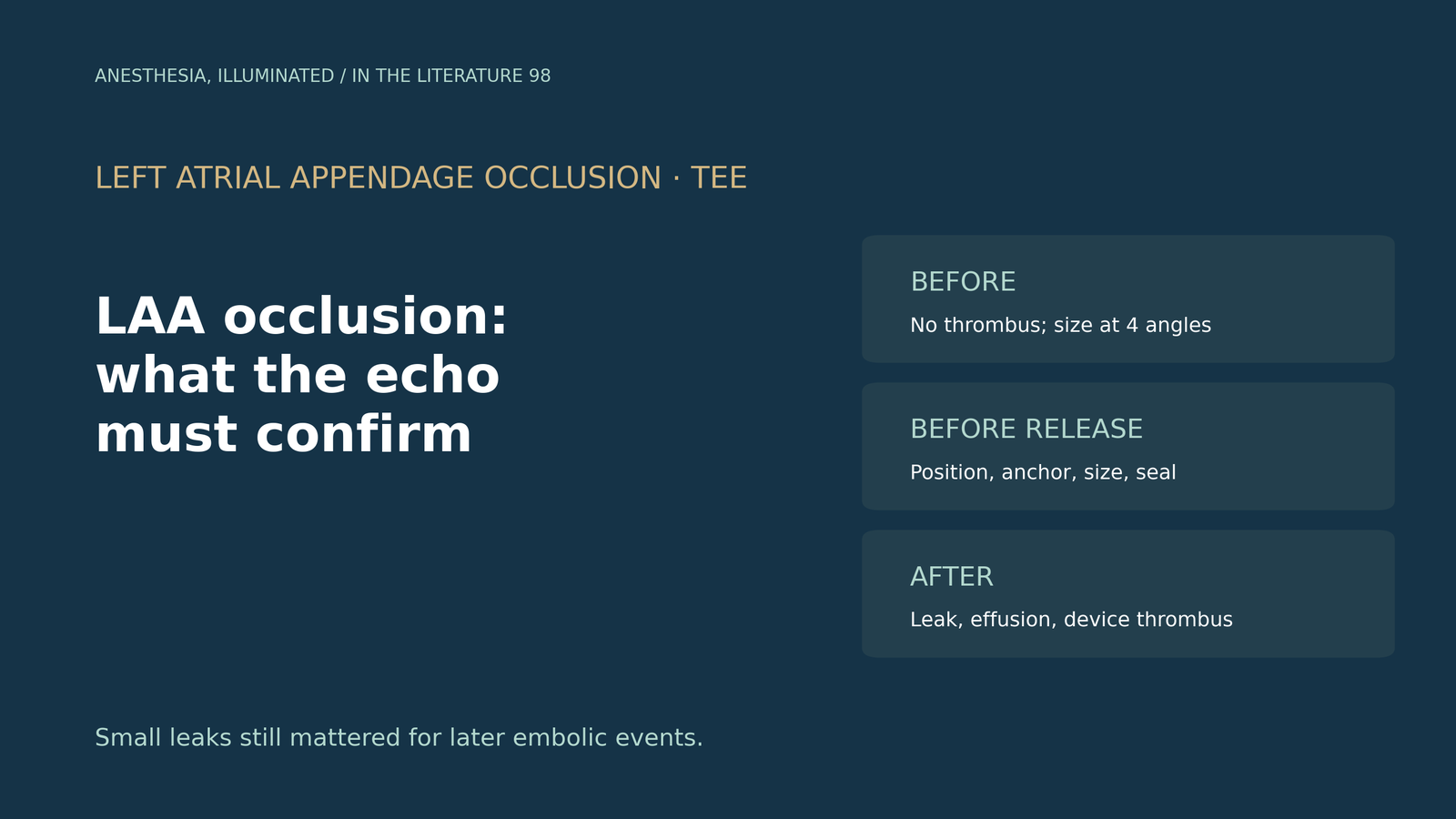
- Before the implant, transesophageal echocardiography (TEE) excludes thrombus and measures the appendage ostium and depth at 0°, 45°, 90° and 135°. Filling conditions change these measurements by up to a few millimetres.[1], [2]
- During deployment, check position, anchor, size and seal before release. A Watchman device should be compressed about 8% to 20%, and a leak greater than 5 mm calls for recapture.[2], [3]
- Coaxial alignment between the delivery catheter and the appendage matters, and pericardial effusion should be watched for throughout the case.[2], [3], [4]
- Small peri-device leaks are not harmless. In a Japanese registry, even leaks of 3 mm or less were associated with more thromboembolic events.[5]
- After the procedure, TEE or CT at about 45 days to 1 year looks for leak and device-related thrombus. Intracardiac echocardiography (ICE) gave similar procedural success to TEE but more effusions needing intervention.[6], [7]
A patient with atrial fibrillation who cannot take long-term anticoagulation comes for left atrial appendage occlusion under general anesthesia. You are holding the TEE probe. What must you show before the device is placed, what must you confirm before it is released, and what should you look for at the end of the case and at follow-up?[1], [2]
Before the device: exclude thrombus and size the appendage
The American Society of Echocardiography screening standards, as summarized by Gartman and colleagues, examine the appendage at the mid-esophagus at 0°, 45°, 90° and 135°, often adding biplane imaging. Measurements depend on loading: volume loading can increase landing-zone diameter and depth by as much as 2.5 mm and 3.6 mm, but is inappropriate with elevated left atrial pressure, heart failure or pulmonary edema.[1] Mitrev and colleagues advise confirming a mean left atrial pressure of approximately 15 mmHg before measuring.[2] In the Canadian Watchman registry, all procedures were done under general anesthesia with TEE guidance.[3]
During deployment: transseptal site, alignment and release criteria
The transseptal puncture determines whether the delivery catheter can line up with the appendage. Pruitt and colleagues describe the standard target as inferior in the bicaval view and mid in the short-axis view, and showed that device buckling relates to the angle of the delivery catheter relative to the appendage axis; a proximal tug with a more coaxial catheter corrected it.[4]
Before release, Mitrev and colleagues list four criteria: position, anchor, size and seal.[2] In the Canadian registry, size meant the device shoulder compressed 8% to 20% of its original size on TEE, and the seal required any leak to be less than 5 mm before release.[3] Gartman and colleagues note that newer devices have different targets, for example 10% to 27% compression for the Watchman FLX.[1] If gaps or leaks greater than 5 mm exist, the device should be recaptured.[2]
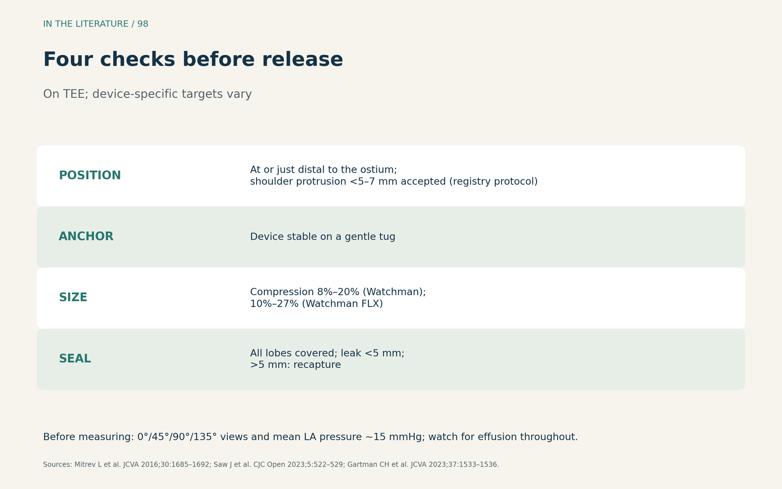
Figure 1. What to confirm before release. Synthesized from Mitrev et al., the Canadian Watchman registry protocol and Gartman et al.; device-specific targets vary.[1], [2], [3] View larger.

Watching for complications
Throughout the procedure, TEE allows rapid recognition of complications such as pericardial effusion, and the echocardiographer should also monitor for device migration or dislodgement.[2] In the Canadian registry, 5 pericardial effusions required drainage (1.8%).[3]
Small leaks may matter
The 5 mm threshold for release does not mean smaller leaks are benign. In the OCEAN-LAAC registry, Saito and colleagues found a peri-device leak at implant in 6.0% of patients, 86.9% of them 3 mm or less. Any leak was associated with transient ischemic attack, stroke or systemic embolism (adjusted subdistribution hazard ratio 4.25; 95% CI 1.91–9.44), including leaks of 3 mm or less (3.53) and leaks of more than 3 to 5 mm (8.48).[5] This was an observational Watchman registry, and the absolute number of events was small.
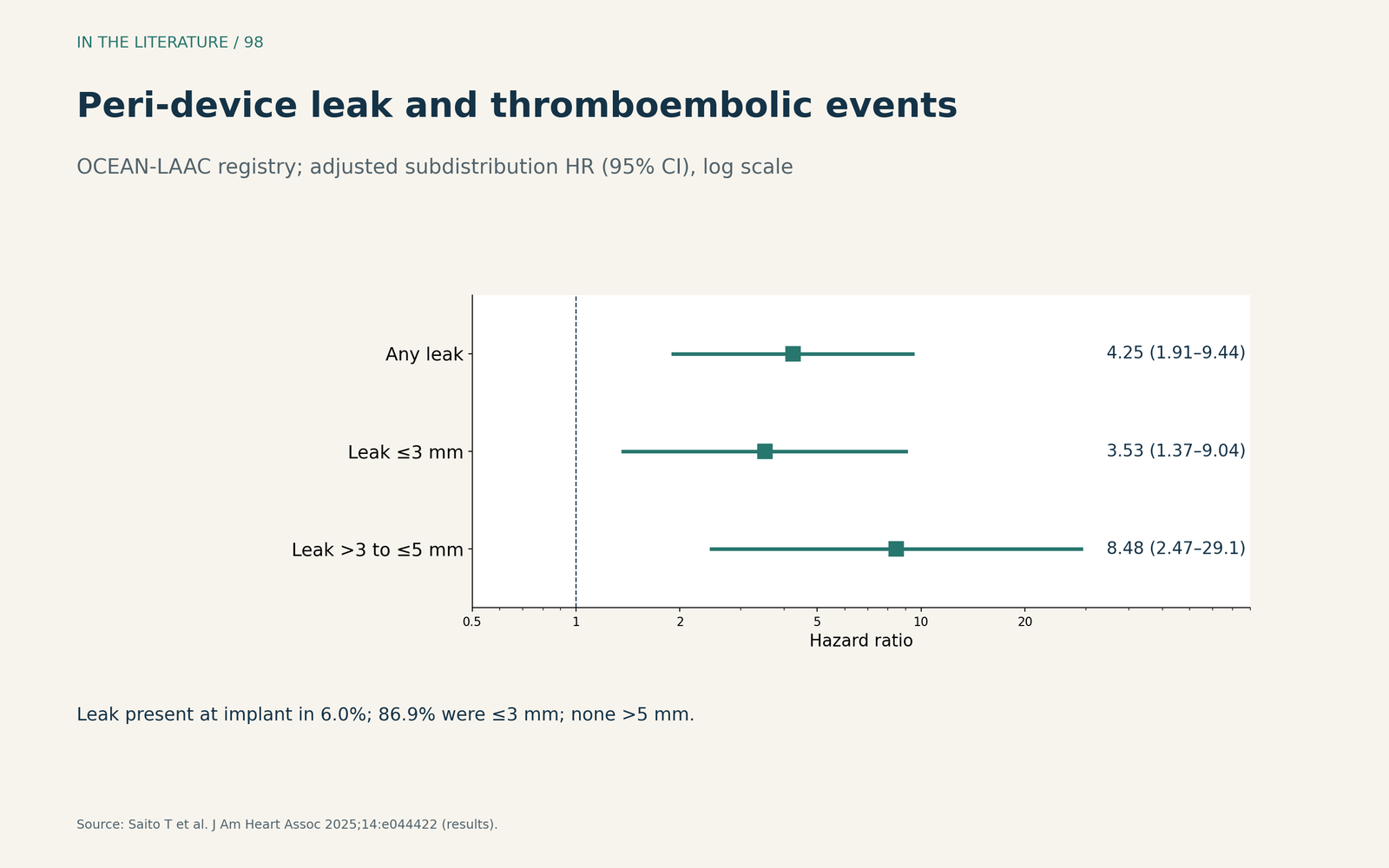
Figure 2. Even small leaks were associated with events. Redrawn from the results of Saito et al. (adjusted subdistribution hazard ratios with 95% confidence intervals, log scale).[5] View larger.

After the procedure: follow-up imaging and TEE versus ICE
In the randomized trials summarized in the 2025 SCAI/HRS technical review, post-procedure imaging was performed at 45 days and again at 1 year. Device-related thrombus, usually defined as an echogenic density in contact with the device, was reported in 4.1% of Watchman patients at a median of 54 days, and the review recommends anticoagulation until resolution is documented on follow-up imaging.[6] The Canadian registry recommended TEE or CT 1 to 6 months after closure.[3]
ICE can replace TEE in experienced centers, with trade-offs. In a meta-analysis by Yaser and colleagues, procedural success was comparable (unadjusted odds ratio 1.16; 95% CI 0.92–1.45), with no significant difference in mortality, stroke or leaks greater than 5 mm, but ICE was associated with more pericardial effusions requiring intervention (odds ratio 1.52) and more residual atrial septal defects. All studies were observational.[7]
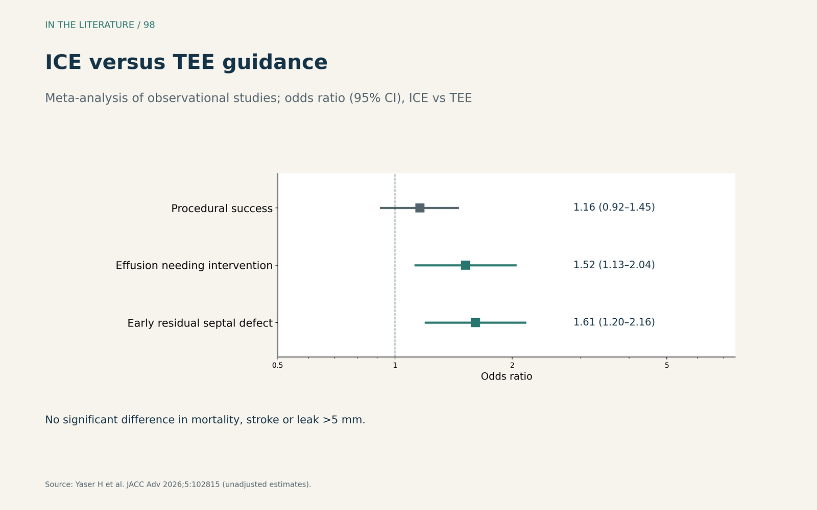
Figure 3. TEE versus ICE. Redrawn from the results of the meta-analysis by Yaser et al. (odds ratios with 95% confidence intervals, observational studies).[7] View larger.

Three takeaways
- Measure under the right filling conditions. Use four TEE angles and a reasonable left atrial pressure; loading changes the size.[1], [2]
- Do not release until position, anchor, size and seal are confirmed. Check coaxial alignment, compression and leak, and look for effusion throughout.[2], [3], [4]
- Treat small leaks and follow-up seriously. Even leaks of 3 mm or less were associated with events; image again for leak and thrombus.[5], [6]
References
- Gartman CH, Pospishil L, Neuburger PJ. Recommendations for transesophageal echocardiographic screening in transcatheter left atrial appendage exclusion, interatrial septum, and interventricular septum interventions: insights for the cardiothoracic anesthesiologist. J Cardiothorac Vasc Anesth. 2023;37(9):1533–1536. doi:10.1053/j.jvca.2023.05.015
- Mitrev L, Trautman N, Vadlamudi R, et al. Anesthesia and transesophageal echocardiography for WATCHMAN device implantation. J Cardiothorac Vasc Anesth. 2016;30(6):1685–1692. doi:10.1053/j.jvca.2016.06.012
- Saw J, Inohara T, Gilhofer T, et al. The Canadian WATCHMAN registry for percutaneous left atrial appendage closure. CJC Open. 2023;5(7):522–529. doi:10.1016/j.cjco.2023.03.016
- Pruitt Z, Bhave P, Daniel K, et al. Left atrial appendage occlusion device buckling: an avoidable deployment complication identified and corrected using intraprocedural transesophageal echocardiography. CASE (Phila). 2023;7(1):27–34. doi:10.1016/j.case.2022.10.004
- Saito T, Tsuruta H, Kajino A, et al. Impact of peridevice leak on clinical outcomes after left atrial appendage closure: the OCEAN-LAAC registry. J Am Heart Assoc. 2025;14(16):e044422. doi:10.1161/JAHA.125.044422
- Cronin EM, Filby S, Field ME, et al. SCAI/HRS technical review on transcatheter left atrial appendage occlusion. J Soc Cardiovasc Angiogr Interv. 2025;4(9):103784. doi:10.1016/j.jscai.2025.103784
- Yaser H, Yaser S, Mohammed HE, et al. Intracardiac versus transesophageal echocardiography for left atrial appendage occlusion: a meta-analysis. JACC Adv. 2026;5(6):102815. doi:10.1016/j.jacadv.2026.102815