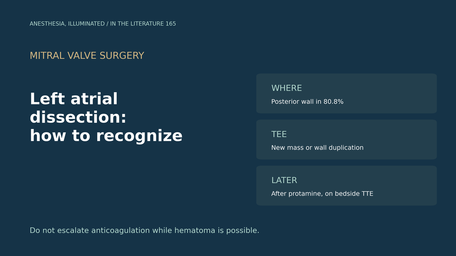
ITL-1a574909395d
Juan Li, MD · Updated November 17, 2026
Watch the video
Key points
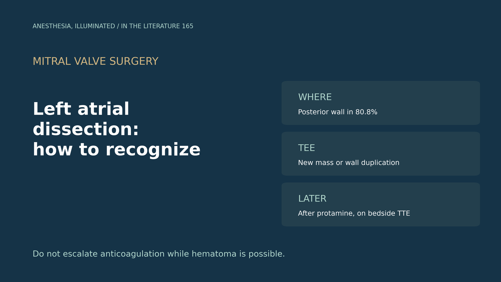
- It is iatrogenic and serious. Left atrial dissection is now mostly a surgical complication, with mortality reported as high as 11.2% to 13.8%, and TEE is the key intraoperative tool.[1]
- Know where and why. It arises in the posterior wall in 80.8% of cases, and annular debridement or an oversized ring can trigger it.[2]
- Look for a second wall. On TEE it may appear as duplication of the atrial wall, and the anterior wall behind the aortic root may be invisible to the surgeon.[3]
- Look again later. It may appear only after protamine, or as a new isoechoic mass on bedside TTE after surgery.[4], [5]
- Think of it in a crisis. A false lumen can obstruct mitral inflow and cause instability or arrest after mitral surgery.[6]
After mitral valve replacement with extensive annular debridement, the post-protamine TEE shows a new echolucent mass in the posterior left atrial wall. Is this a dissection, and how should it be confirmed?[1], [2]
Why it happens and why TEE matters
Kawabori and Ortoleva describe left atrial dissection as now mostly an iatrogenic complication, with mortality reported as high as 11.2% to 13.8%. TEE is the most useful intraoperative modality, a thorough post-bypass examination is critical, and anticoagulation could enlarge the hematoma.[1] Parsons and colleagues report that dissection arises in the posterior wall in 80.8% of cases and that annular debridement and an oversized annuloplasty ring are triggers; in their case, a new left atrial mass appeared that had not been seen earlier after bypass. In stable patients reversing anticoagulation alone may suffice, whereas surgical management carries high mortality.[2]
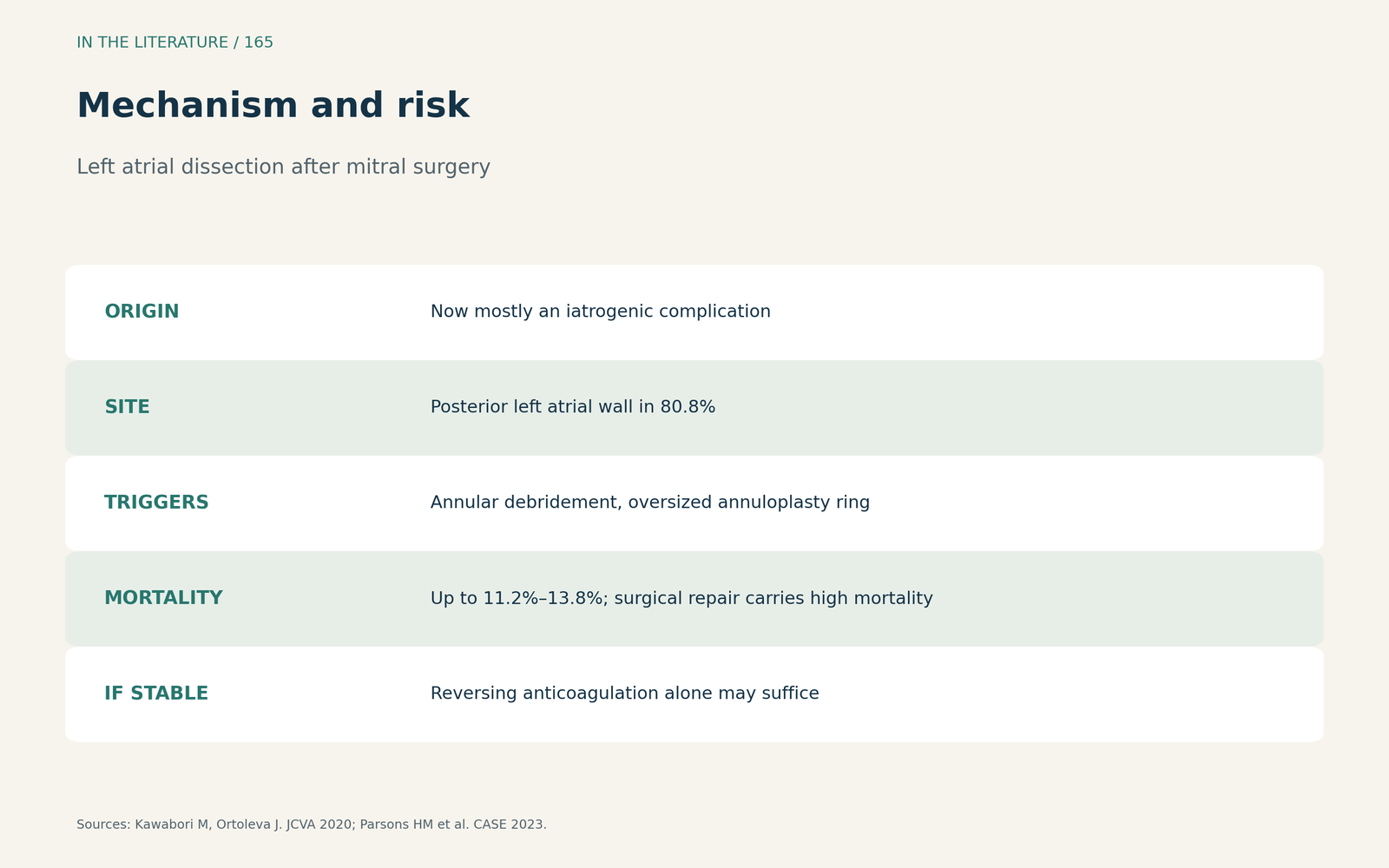
Figure 1. Mechanism and risk. Synthesized from Kawabori and Ortoleva and Parsons et al.[1], [2] View larger.

What to look for on echo
Niyogi and colleagues note that dissection may appear as duplication of an atrial wall, usually posterior; the anterior wall behind the aortic root may not be surgically visible, and the size of the dissected lumen should be assessed.[3] Sarridou and colleagues describe dissection detected by TEE after protamine, when profound hypotension followed within minutes; extensive debridement of a calcified annulus probably triggered it, although most reported cases lack hemodynamic compromise.[4] Wang and colleagues describe bedside TTE showing a new well-circumscribed isoechoic left atrial mass after surgery; imaging alone may not distinguish hematoma from thrombus, escalating anticoagulation or thrombolysis should be withheld if hematoma is possible, and daily point-of-care ultrasound may merit inclusion in postoperative care.[5]
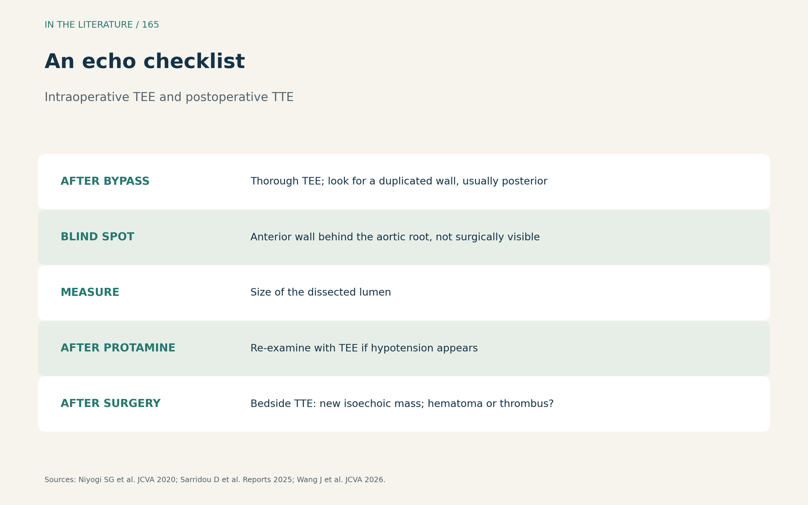
Figure 2. An echo checklist. Synthesized from Niyogi et al., Sarridou et al. and Wang et al.[3], [4], [5] View larger.

When it presents as a crisis
Sahar and colleagues describe a patient whose TEE after arrest showed a mobile membrane and a false lumen into which blood from the left ventricle entered, obstructing the mitral valve. They advise considering dissection in patients with circulatory instability or arrest after mitral surgery, and note that abnormal prosthetic valve motion on TTE should raise suspicion.[6]
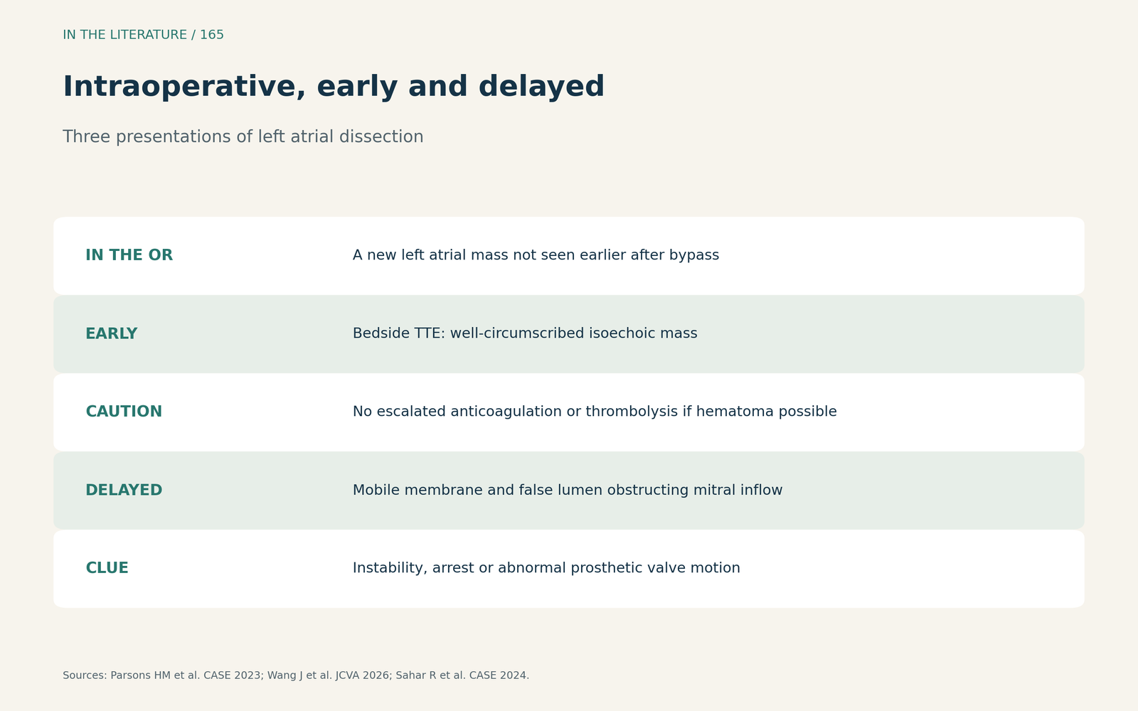
Figure 3. Intraoperative, early and delayed. Synthesized from Parsons et al., Wang et al. and Sahar et al.[2], [5], [6] View larger.

Three takeaways
- Sweep the whole left atrium after bypass. Look for a new mass or a duplicated posterior wall, including behind the aortic root.[1], [3]
- Look again after protamine and after surgery. Dissection can appear late.[4], [5]
- Do not escalate anticoagulation while hematoma is possible. A hematoma can look like a thrombus.[5]
References
- Kawabori M, Ortoleva J. Word of caution: left atrial dissection, an infrequent but critical complication. J Cardiothorac Vasc Anesth. 2020;34(10):2830–2831. doi:10.1053/j.jvca.2020.06.071
- Parsons HM, Aktuerk D, Kyle B. Left atrial dissection during minimally invasive mitral valve surgery. CASE (Phila). 2023;7(8):303–308. doi:10.1016/j.case.2023.02.007
- Niyogi SG, Kumar B, Singh H, Biswas I. Left atrial dissection and rupture following excision of left atrial myxoma: role of transesophageal echocardiography. J Cardiothorac Vasc Anesth. 2020;34(10):2823–2826. doi:10.1053/j.jvca.2020.06.064
- Sarridou D, Mouratoglou SA, Ioannidis R, et al. Left atrial dissection after mitral and aortic valve replacement: the importance of early diagnosis of a rare entity. Reports (MDPI). 2025;8(4):205. doi:10.3390/reports8040205
- Wang J, Lin Z, Qiang H. A rapidly enlarging intramural left atrial hematoma after cardiac surgery. J Cardiothorac Vasc Anesth. 2026;40(8):2489–2492. doi:10.1053/j.jvca.2026.02.027
- Sahar R, Wedin JO, Grinnemo KH. Left atrial dissection resulting in asystole: a rare complication after mitral valve surgery. CASE (Phila). 2024;8(6):369–372. doi:10.1016/j.case.2024.03.005