ITL-2ed2b3bc5fa5
Juan Li, MD · Updated November 18, 2026
Key points
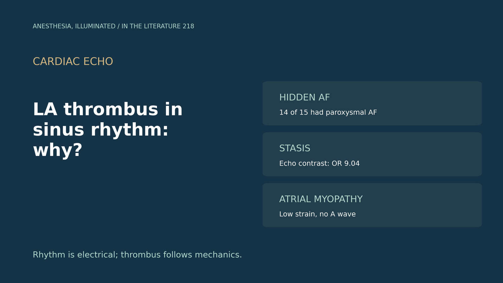
- Sinus rhythm may not mean no AF. Among stroke patients in sinus rhythm, LAA thrombus was found in 6.7%, and paroxysmal AF was detected in 14 of the 15 on 24-hour monitoring.[1]
- Stasis is the marker. Spontaneous echo contrast was the only independent predictor of thrombus (OR 9.04).[1]
- The atrium itself can be the problem. LAA thrombus can form despite maintained sinus rhythm when LA reservoir strain is severely reduced.[2]
- Amyloid is a classic example. In cardiac amyloidosis, atrial paralysis favoring thrombosis can precede AF.[3], [4]
- Image properly. TEE is needed because TTE may not be sensitive enough, and contrast helps exclude thrombus mimics.[5], [6]
A pre-bypass TEE in a patient whose ECG shows sinus rhythm reveals a mass in the left atrial appendage. How can thrombus form without AF, and is it really thrombus?[1], [2]
Hidden AF and stasis
Nguyen et al. (BMJ Open 2021) found LAA thrombus in 15 stroke patients in sinus rhythm (6.7%); paroxysmal AF was detected in 14 of the 15 on 24-hour monitoring, and a 24-hour ECG may be insufficient to detect it. Spontaneous echo contrast was more common with thrombus (53.3% vs 5.3%) and was the only independent predictor (OR 9.04). The authors note that thrombus can also form in sinus rhythm through LV dysfunction and valvular disease, and that electrophysiological remodelling may cause LAA thrombus independent of AF.[1]
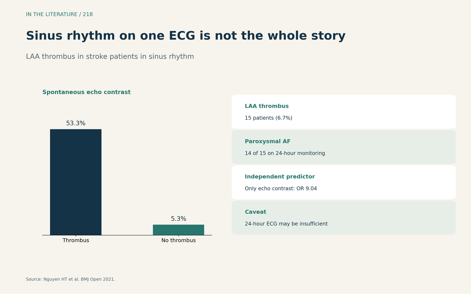
Figure 1. Sinus rhythm on one ECG is not the whole story. Redrawn from Nguyen et al.[1] View larger.

When the atrium is the disease
Ferkh et al. (CASE 2021) describe LAA thrombus that formed despite maintained sinus rhythm, with LA reservoir strain severely reduced (3.2%); the diseased atrium itself can be a nidus for thrombus, atrial myopathy may exist without AF, and LA strain may help identify it.[2] In cardiac amyloidosis, Marchionni et al. (JACC Case Rep 2024) argue that atrial paralysis favoring thrombosis can precede AF, that electromechanical dissociation carries a long-term prognosis similar to AF, and that LA strain should be assessed.[3] Ballantyne et al. (ESC Heart Fail 2020) note that an absent A wave despite sinus rhythm suggests loss of atrial contractility; in one autopsy study 20% of amyloidosis patients without documented AF had thrombi, and at cardioversion atrial thrombi were found in 28% vs 2.5% of controls.[4] Cho et al. (J Am Soc Echocardiogr 2023) add that atrial cardiomyopathy leads to blood stasis and hypercoagulability, that impaired LA functional reserve may be present without LA enlargement, and that AF may be a marker rather than the root cause of stroke.[7]
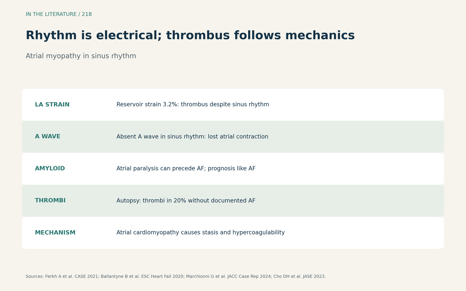
Figure 2. Rhythm is electrical; thrombus follows mechanics. Synthesized from Ferkh et al., Marchionni et al., Ballantyne et al. and Cho et al.[2], [3], [4], [7] View larger.

Imaging the appendage
TTE may not be sensitive enough, so assessment should include TEE.[5] Wehbe and Rigolin (CASE 2020) show that structures next to the left atrium, such as epicardial fat, can be mistaken for LAA thrombus, and that ultrasound enhancing agents add value for excluding thrombus.[6] In amyloidosis with stroke, the threshold for TEE appendage imaging should be low.[4] Mimics are common: only 29.1% of appendages are single-lobed on TEE, and the appendage itself can appear as a mobile mass (Hang et al., J Cardiothorac Vasc Anesth 2022).[8] In uncertain TEE studies, an ultrasound enhancing agent raised reader agreement from 72% to 96% (Catino et al., J Am Soc Echocardiogr 2021),[9] and in persistent AF, stepwise isoproterenol cleared appendage sludge in 95% within about 1 minute (Yamashita et al., J Am Soc Echocardiogr 2023); both studies were in AF populations.[10] The evidence is one cross-sectional study, a small exercise-echo study and case reports.
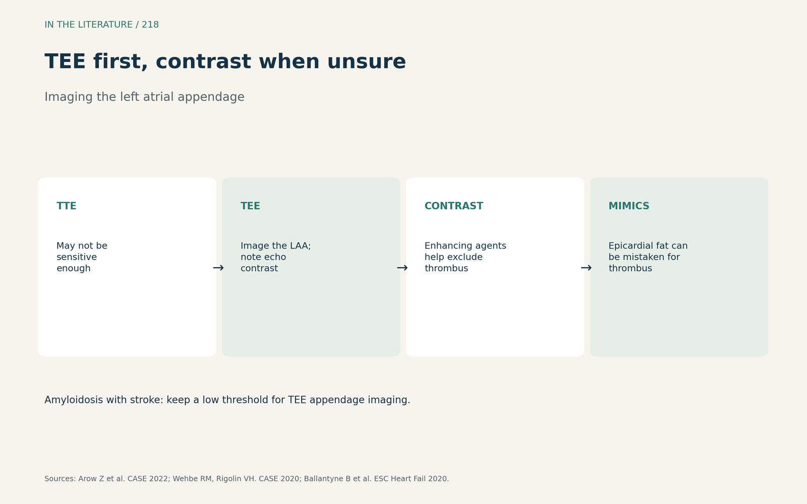
Figure 3. TEE first, contrast when unsure. Synthesized from Arow et al., Wehbe and Rigolin, and Ballantyne et al.[5], [6], [4] View larger.

Three takeaways
- Sinus rhythm on one ECG does not exclude paroxysmal AF.[1]
- Spontaneous echo contrast, low LA strain or an absent A wave point to a thrombogenic atrium.[1], [2], [4]
- Use TEE to look for thrombus, and contrast to separate it from mimics.[5], [6]
References
- Nguyen HT, Nguyen HVB, Nguyen HQ, et al. Prevalence of left atrial appendage thrombus in patients with acute ischaemic stroke and sinus rhythm: a cross-sectional study. BMJ Open. 2021;11(12):e051563. doi:10.1136/bmjopen-2021-051563
- Ferkh A, Whalley D, Warwick N, et al. Left atrial cardiomyopathy with left atrial thrombus despite sinus rhythm in a patient with severe ventricular cardiomyopathy requiring cardiac transplantation. CASE (Phila). 2021;5(4):243–251. doi:10.1016/j.case.2021.03.007
- Marchionni G, Costantino J, Ballatore F, et al. Thromboembolic risk in sinus rhythm. JACC Case Rep. 2024;29(18):102531. doi:10.1016/j.jaccas.2024.102531
- Ballantyne B, Manian U, Sheyin O, et al. Stroke risk and atrial mechanical dysfunction in cardiac amyloidosis. ESC Heart Fail. 2020;7(2):705–707. doi:10.1002/ehf2.12602
- Arow Z, Pereg D, Assali A, et al. Giant left atrial thrombus: a source of systemic emboli. CASE (Phila). 2022;6(5):201–204. doi:10.1016/j.case.2021.12.005
- Wehbe RM, Rigolin VH. A left atrial thrombus mimic: value of ultrasound enhancing agents during transesophageal echocardiography. CASE (Phila). 2020;4(4):263–269. doi:10.1016/j.case.2020.04.005
- Cho DH, Kim YG, Choi J, et al. Atrial cardiomyopathy with impaired functional reserve in patients with paroxysmal atrial fibrillation. J Am Soc Echocardiogr. 2023;36(2):180–188. doi:10.1016/j.echo.2022.09.012
- Hang D, Schmitt MC, Gonzalez LS, Zdanovec A, Pagel PS. Left atrial appendage confusion: a mobile echodensity in a patient with endocarditis. J Cardiothorac Vasc Anesth. 2022;36(8):2829–2832. doi:10.1053/j.jvca.2022.01.008
- Catino AB, Ross A, Suksaranjit P, Wilson BD. Improved thrombus assessment by transesophageal echocardiography: the DOLOP (Detection of Left Atrial Appendage Thrombosis Utilizing Optison) study. J Am Soc Echocardiogr. 2021;34(8):916–917. doi:10.1016/j.echo.2021.03.012
- Yamashita E, Kemi Y, Sasaki T, et al. Stepwise increase of isoproterenol bolus dose method for differentiating left atrial appendage sludge from thrombus. J Am Soc Echocardiogr. 2023;36(5):553–555. doi:10.1016/j.echo.2022.12.009