ITL-8c43123936c2
Juan Li, MD · Updated November 17, 2026
Watch the video
Key points
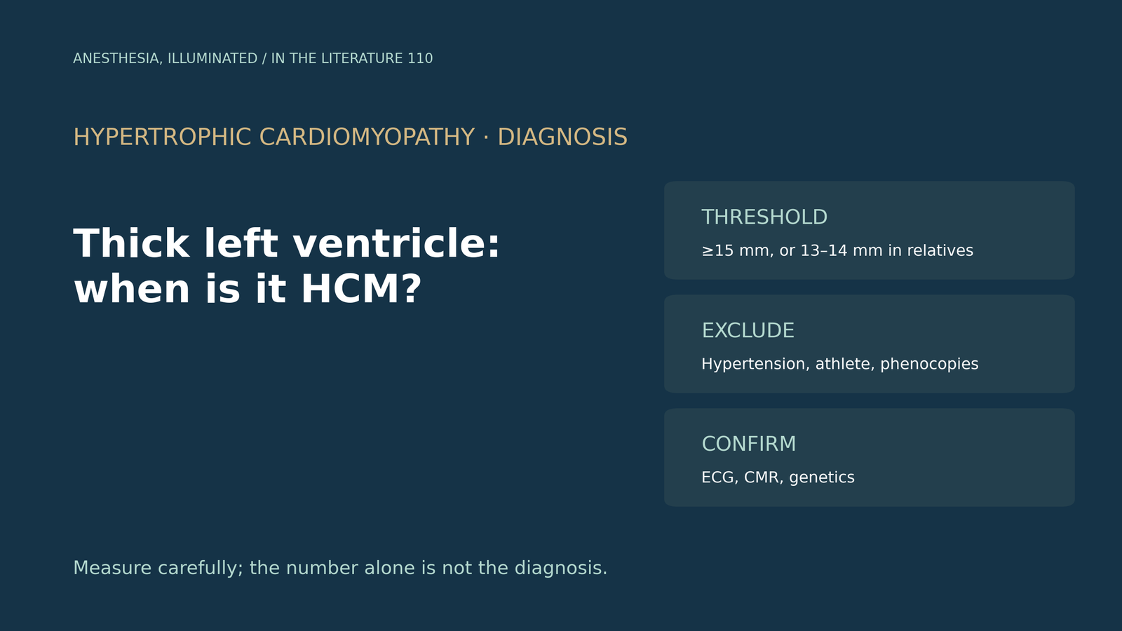
- In adults, hypertrophic cardiomyopathy (HCM) is diagnosed by a maximal wall thickness of 15 mm or more anywhere in the left ventricle, without another cause. In family members or gene carriers, 13–14 mm can be diagnostic. Systolic anterior motion is not required.[1]
- Measure carefully: perpendicular to the septum, including only compacted myocardium, and cross-checking views. Use contrast or cardiac MRI when the apex is poorly seen.[2], [3], [1]
- The pattern helps. Hypertensive hypertrophy is usually symmetric and rarely exceeds 15 mm, whereas a wall above 18 mm with systolic anterior motion and septal contact favors HCM.[2], [4]
- Look for phenocopies: low or normal QRS voltage despite thick walls, diffuse late enhancement and extracellular volume above 30% suggest amyloidosis; basal inferolateral enhancement with low T1 suggests Fabry disease; pre-excitation suggests PRKAG2 or Danon disease. Apical sparing alone is not diagnostic.[4], [5], [6]
- Work up in steps: family history, ECG, echocardiography with provocation, cardiac MRI and genetic testing. For the anesthesiologist, obstruction (a peak outflow gradient of 30 mmHg or more) changes how hypotension is treated.[1]
A 55-year-old with treated hypertension is scheduled for noncardiac surgery, and the preoperative echocardiogram reports a septum of 16 mm with a posterior wall of 11 mm. Is this hypertensive hypertrophy or hypertrophic cardiomyopathy, what else could it be, and what should be done before surgery?[1], [2]
The diagnostic threshold
The 2020 American Heart Association/American College of Cardiology (AHA/ACC) guideline defines HCM in adults by a maximal end-diastolic wall thickness of 15 mm or more anywhere in the left ventricle, in the absence of another cause of hypertrophy. More limited hypertrophy of 13–14 mm can be diagnostic in family members or with a positive genetic test. In children, the threshold is a z-score above 2.5, or above 2 with a family history or positive genetic test. Neither systolic anterior motion nor hyperdynamic function is required, and systemic diseases that cause hypertrophy, such as amyloidosis or Fabry disease, should not be labeled HCM.[1]
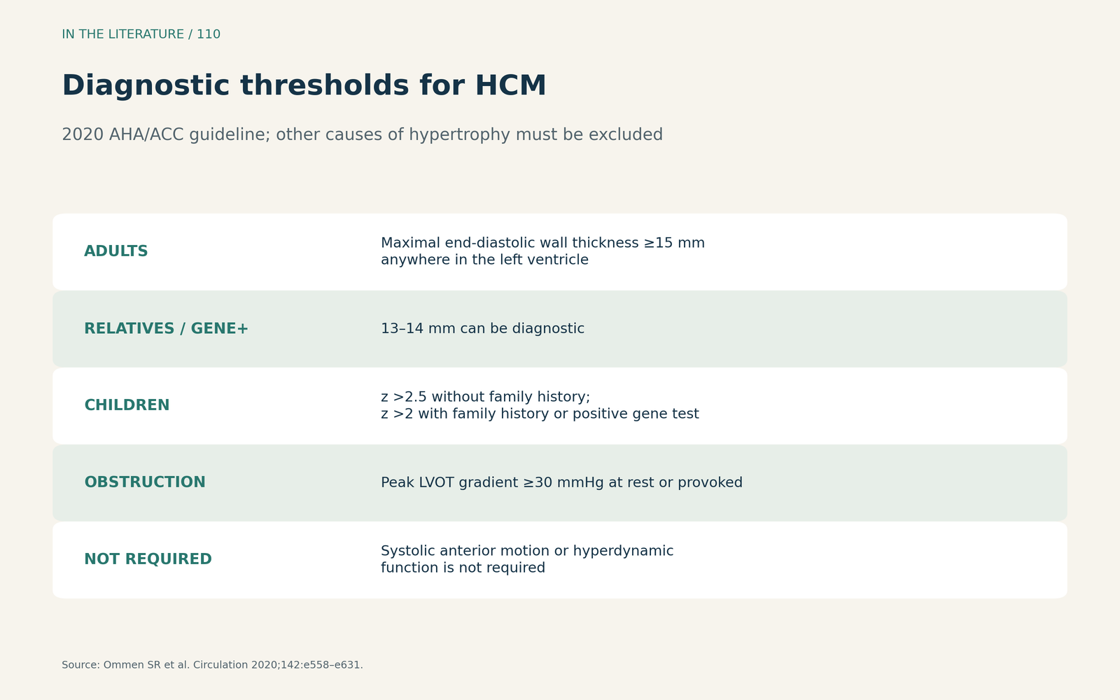
Figure 1. Diagnostic thresholds for HCM. Summarized from the 2020 AHA/ACC guideline.[1] View larger.

Measuring correctly
Maron and colleagues recommend measuring maximal wall thickness perpendicular to the septum at the thickest segment.[2] Mitchell and colleagues add that measurements should include only compacted myocardium, excluding papillary muscles and right ventricular structures, and that values from two planes at a comparable level should match; ultrasound-enhancing agents help define the borders.[3] The guideline notes that apical aneurysms may not be detected on echocardiography and recommends cardiac MRI when an alternative diagnosis is suspected.[1]
Hypertension, athletes and HCM
Hypertension is the common confounder. Banthiya and colleagues note that hypertensive hypertrophy rarely exceeds 15 mm, and describe a septal to posterior wall ratio above 1.3 as characteristic of HCM.[4] Maron and colleagues add that pressure overload usually produces symmetric thickening, whereas a wall above 18 mm with systolic anterior motion and septal contact favors HCM, and regression of hypertrophy with blood pressure control favors hypertensive heart disease.[2] The guideline lists physiological remodeling of the athlete, hypertension, renal disease and infiltrative disease as alternatives to consider.[1]
Phenocopies
Some conditions mimic HCM but are treated differently. In cardiac amyloidosis, low QRS voltage, or normal voltage despite thick walls, is a red flag, and a voltage-to-mass ratio below 1.5 is suggestive.[5], [4] On cardiac MRI, late gadolinium enhancement has a sensitivity and specificity for amyloidosis of up to 86% and 92%, and an extracellular volume above 30% is characteristic.[4] Teresi and colleagues describe basal inferolateral enhancement with low native T1 in Fabry disease, pre-excitation in PRKAG2 disease, and serum and urine immunofixation, serum light chains and bone scintigraphy as the key steps when amyloidosis is suspected.[5]
Relative apical sparing of strain is often used as an amyloid sign. A 2026 meta-analysis by Sonaglioni and colleagues found it is not specific to amyloidosis and should not be used as a stand-alone criterion.[6]
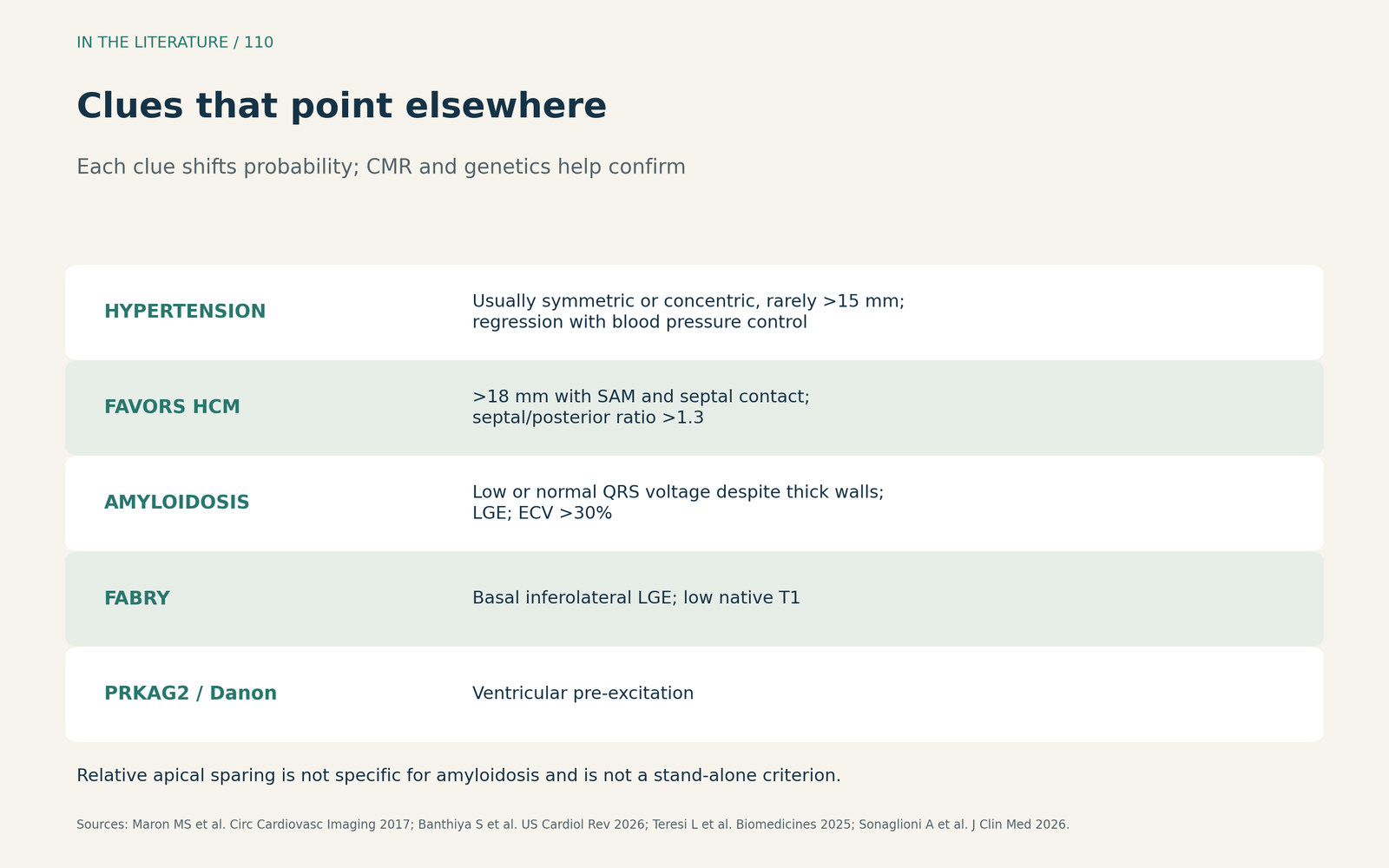
Figure 2. Clues that point elsewhere. Synthesized from Maron et al., Banthiya et al., Teresi et al. and Sonaglioni et al.; each clue shifts probability rather than proving a diagnosis.[2], [4], [5], [6] View larger.

A stepwise workup
The guideline recommends a three-generation family history, a 12-lead ECG (abnormal in 75% to 95% of patients with HCM), and transthoracic echocardiography. Because outflow gradients are dynamic and can be missed at rest, echocardiography with provocation is recommended when the resting gradient is below 50 mmHg. Genetic panels identify a disease-causing variant in about 30% of sporadic and 60% of familial cases.[1] Banthiya and colleagues suggest that genetic testing should be routine in most cases.[4]
For the anesthesiologist, the guideline defines obstruction as a peak outflow gradient of 30 mmHg or more at rest or with provocation. In obstructive HCM with acute hypotension that does not respond to fluids, it recommends intravenous phenylephrine or another vasoconstrictor without inotropic activity.[1]
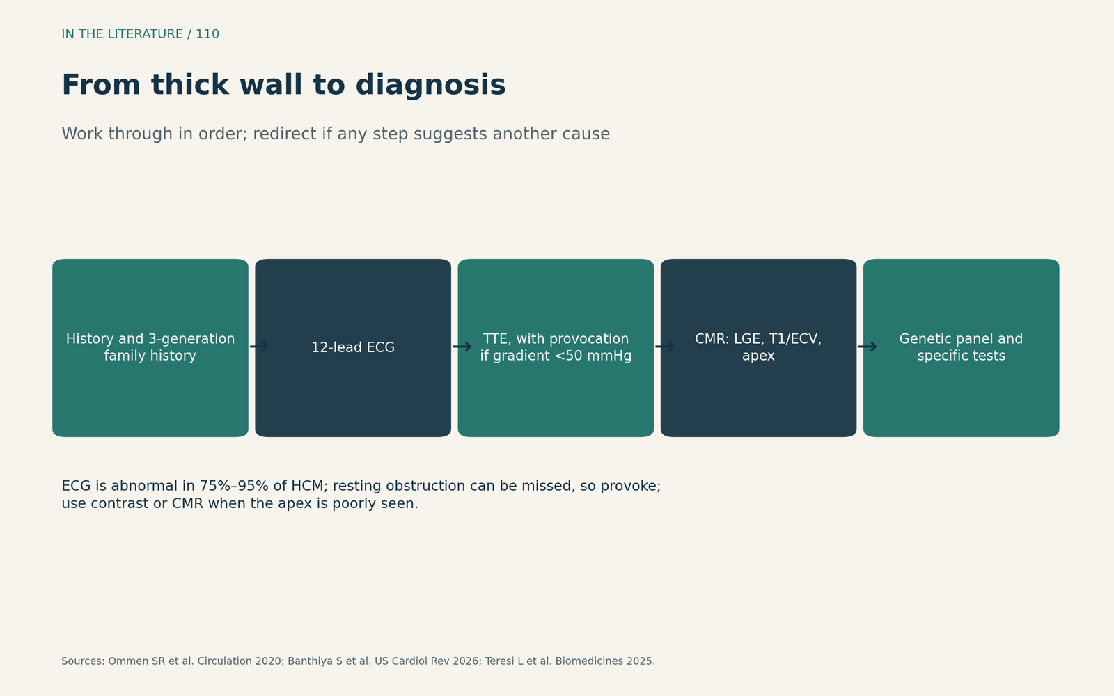
Figure 3. From thick wall to diagnosis. Sequence synthesized from the 2020 AHA/ACC guideline and the reviews by Banthiya et al. and Teresi et al.[1], [4], [5] View larger.

Three takeaways
- Know the threshold and its context. 15 mm or more in adults, 13–14 mm in relatives or gene carriers, and always after excluding other causes.[1]
- Measure well and look at the pattern. Perpendicular, compacted myocardium only; asymmetric hypertrophy above 18 mm with septal contact favors HCM over hypertension.[2], [3], [4]
- Screen for phenocopies and obstruction. ECG voltage, late enhancement and extracellular volume point to amyloidosis; provoke to find obstruction, which changes how hypotension is treated.[1], [4], [5]
References
- Ommen SR, Mital S, Burke MA, et al. 2020 AHA/ACC guideline for the diagnosis and treatment of patients with hypertrophic cardiomyopathy: a report of the American College of Cardiology/American Heart Association Joint Committee on Clinical Practice Guidelines. Circulation. 2020;142(25):e558–e631. doi:10.1161/CIR.0000000000000937
- Maron MS, Rowin EJ, Maron BJ. How to image hypertrophic cardiomyopathy. Circ Cardiovasc Imaging. 2017;10(7):e005372. doi:10.1161/CIRCIMAGING.116.005372
- Mitchell CC, Frye C, Jankowski M, et al. A practical approach to echocardiographic imaging in patients with hypertrophic cardiomyopathy. J Am Soc Echocardiogr. 2023;36(9):913–932. doi:10.1016/j.echo.2023.04.020
- Banthiya S, Agrawal A, Rai D, et al. Diagnostic approach to left ventricular hypertrophy: a review. US Cardiol Rev. 2026;20:e06. doi:10.15420/usc.2024.65
- Teresi L, Trimarchi G, Licordari R, et al. Hypertrophic cardiomyopathy phenocopies: classification, key features, and differential diagnosis. Biomedicines. 2025;13(12):3062. doi:10.3390/biomedicines13123062
- Sonaglioni A, Gramaglia GF, Nicolosi GL, et al. Diagnostic performance of relative apical sparing across cardiac diseases: a multimodality systematic review and meta-analysis. J Clin Med. 2026;15(5):1685. doi:10.3390/jcm15051685