ITL-9c3ac04ceea2
Juan Li, MD · Updated November 17, 2026
Watch the video
Key points
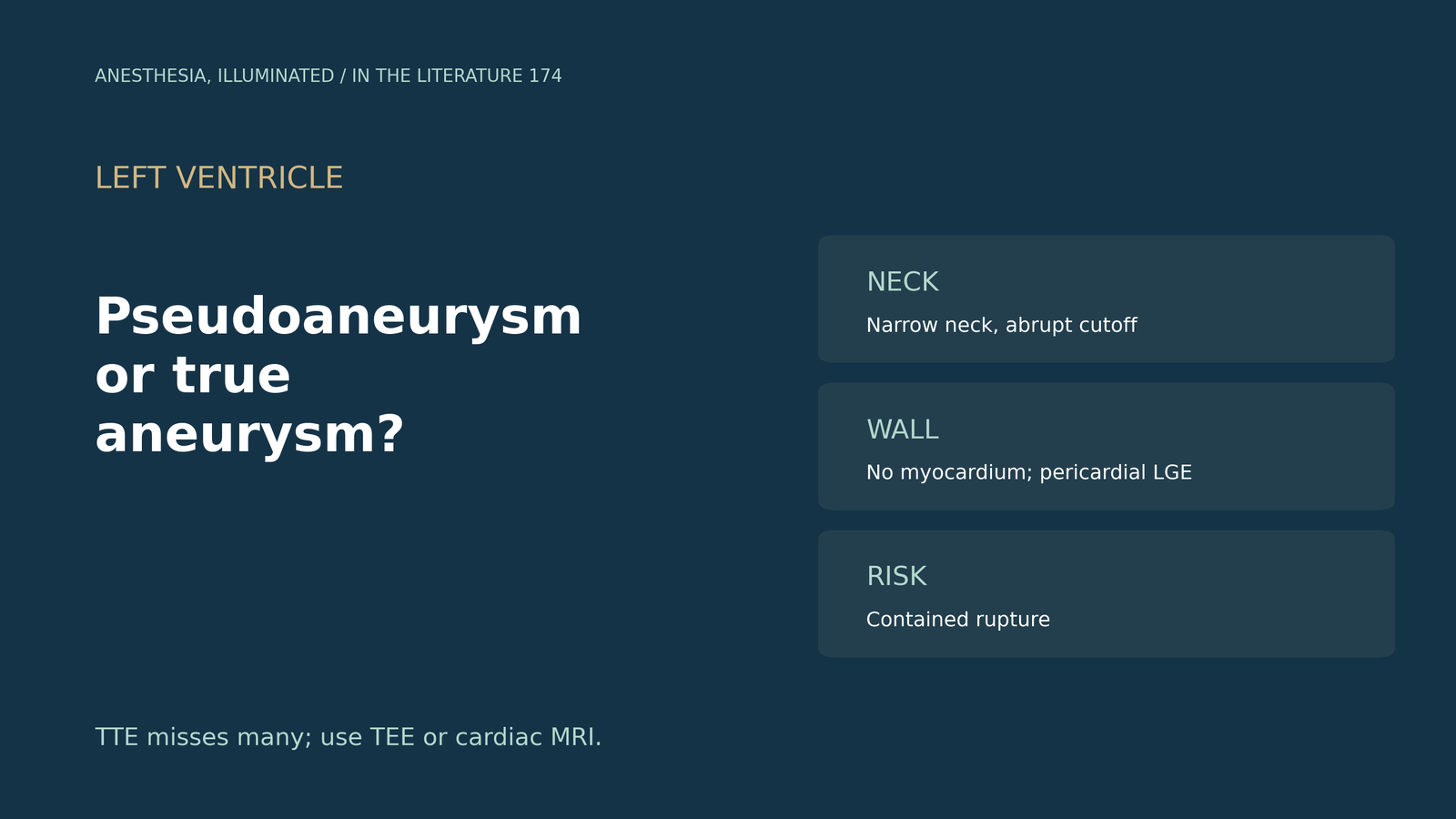
- It is about rupture. The key distinction is whether the heart has ruptured; pseudoaneurysm after infarction occurs in 0.2% to 0.3%, and untreated rupture rates reach 30% to 45%.[1]
- Look at the wall and the neck. A true aneurysm keeps endocardium, epicardium and thinned myocardium and tapers smoothly; a pseudoaneurysm has a narrow neck, with a neck-to-diameter ratio of about 0.5.[2]
- Do not rely on TTE alone. TTE detects pseudoaneurysm in only 18% to 63%, rising to 76% to 100% with TEE, and sharp endocardial discontinuity is a clue.[3]
- CMR helps. Pericardial late enhancement supports pseudoaneurysm, and acute pseudoaneurysms carry a much higher rupture risk.[4]
- The data are thin. In pooled cases echo identified 81.4%, but most studies did not report neck diameters.[5]
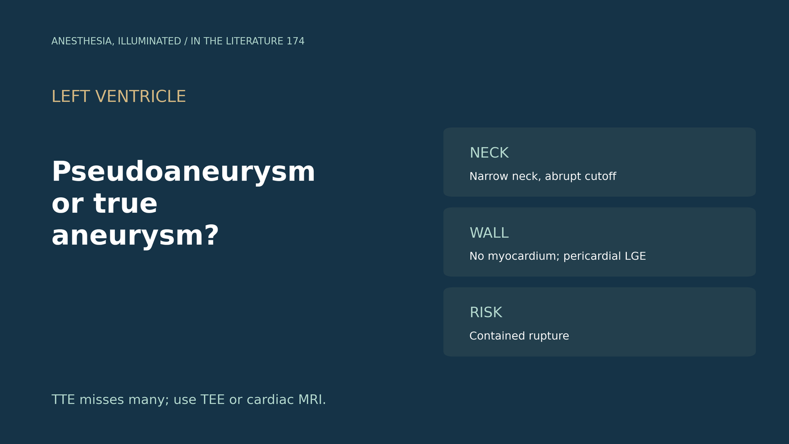
Months after an inferior infarction, echocardiography shows an outpouching of the basal inferolateral wall in a patient scheduled for surgery. Is it a true aneurysm or a pseudoaneurysm, and how can the team tell?[1], [2]
What separates them
Ma and Xu define the distinction as whether the heart has ruptured. Post-infarction pseudoaneurysm occurs in 0.2% to 0.3%; on echo it appears as an echo-free cavity between the ventricular wall and the pericardium, coronary arteries can extend into the wall of a true aneurysm, and untreated pseudoaneurysm rupture rates reach 30% to 45%.[1] Goh and Yew add that a true aneurysm is lined by endocardium, epicardium and thinned scarred myocardium and shows a smoother transition from normal wall, whereas a pseudoaneurysm is a contained rupture covered by adherent pericardium without myocardium; the neck-to-diameter ratio was 0.5 in pseudoaneurysms in a small series, and epicardial fat next to the outpouching on CMR excludes pseudoaneurysm. Noninvasive differentiation is seldom straightforward, and true aneurysms rupture less, with surgery reserved for complications.[2]
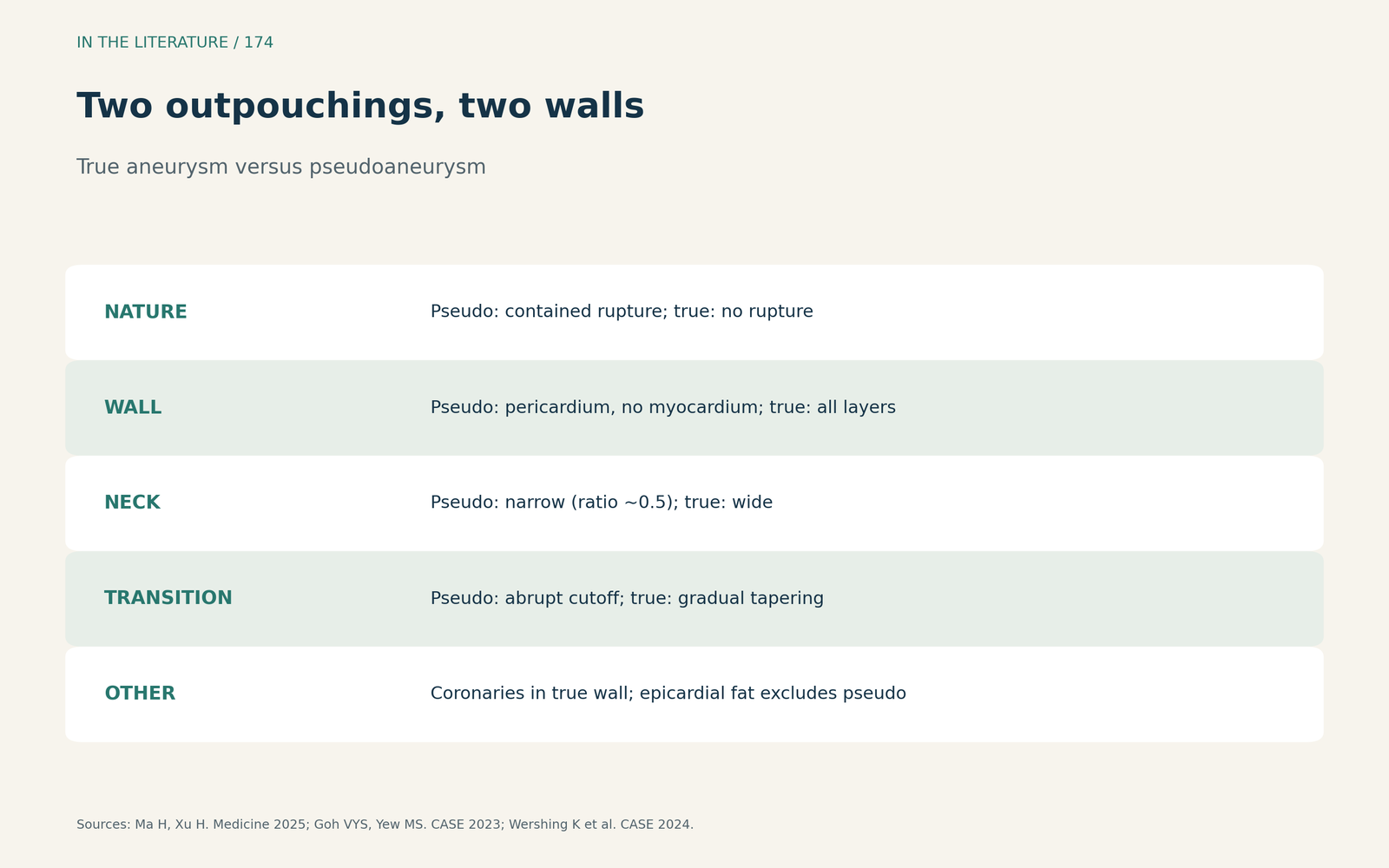
Figure 1. Two outpouchings, two walls. Synthesized from Ma and Xu, Goh and Yew and Wershing et al.[1], [2], [4] View larger.

Which imaging to use
Oluleye and colleagues describe sharp endocardial discontinuity where a pseudoaneurysm communicates with the cavity; TTE detects it in only 18% to 63%, TEE in 76% to 100%, and contrast ventriculography is the gold standard but rarely used. About 55% result from infarction and about 5% from endocarditis, and mortality is about 48% with medical therapy and 23% after surgery.[3] Wershing and colleagues describe a rupture contained by epicardium or pericardial adhesions, diffuse pericardial late enhancement on CMR, and gradual tapering of wall thickness in true aneurysms; an acute pseudoaneurysm carries a much higher rupture risk.[4]
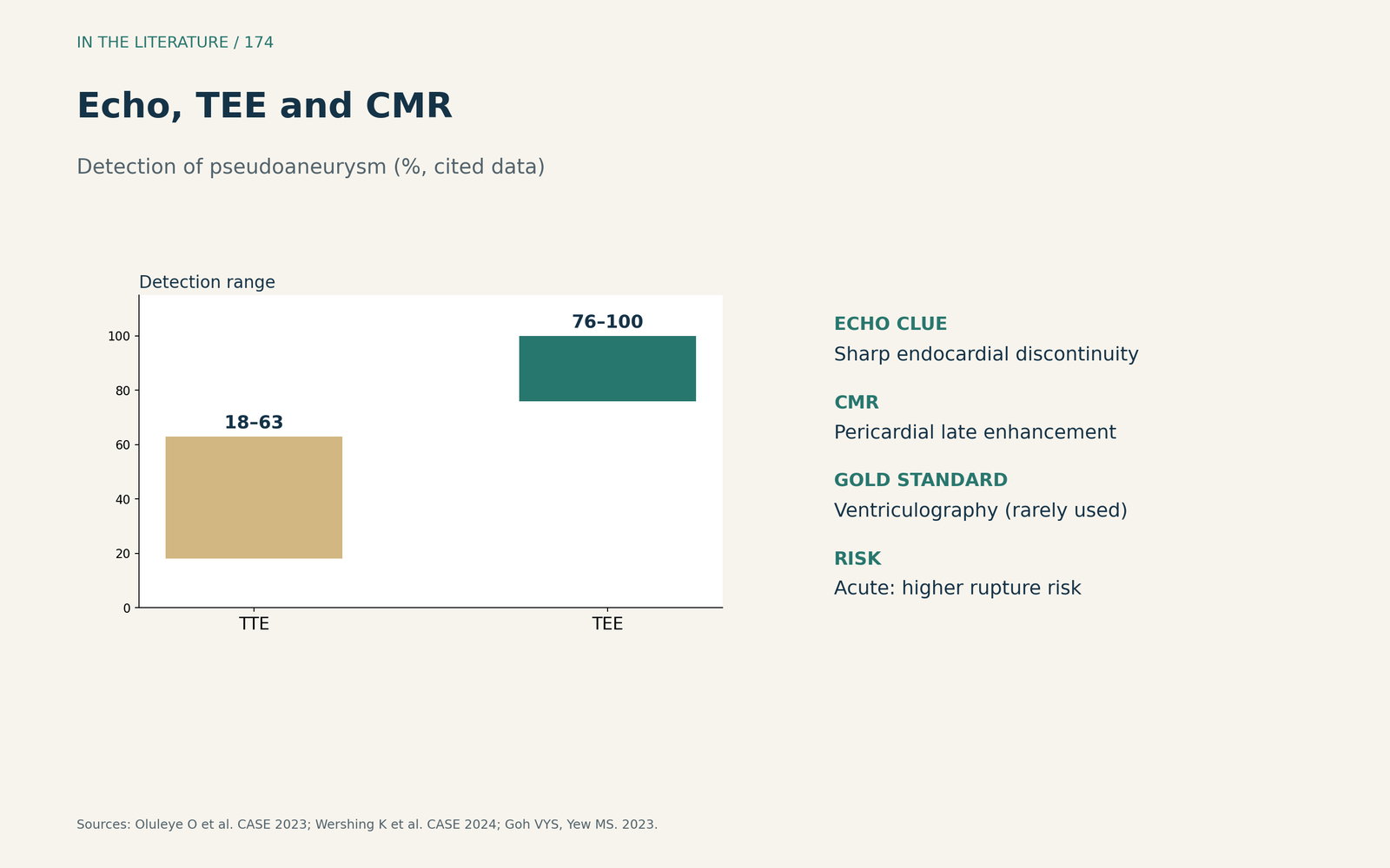
Figure 2. Echo, TEE and CMR. Synthesized from Oluleye et al., Wershing et al. and Goh and Yew.[3], [4], [2] View larger.

What pooled cases add
Afshar and colleagues pooled 504 post-infarction pseudoaneurysms: echo identified 81.4%, less often than true aneurysms; pseudoaneurysms were most often posterior (34.5%) and inferior (21.2%); late enhancement was reported in only 25 of 85 patients with MRI; and most studies lacked neck diameter data.[5]
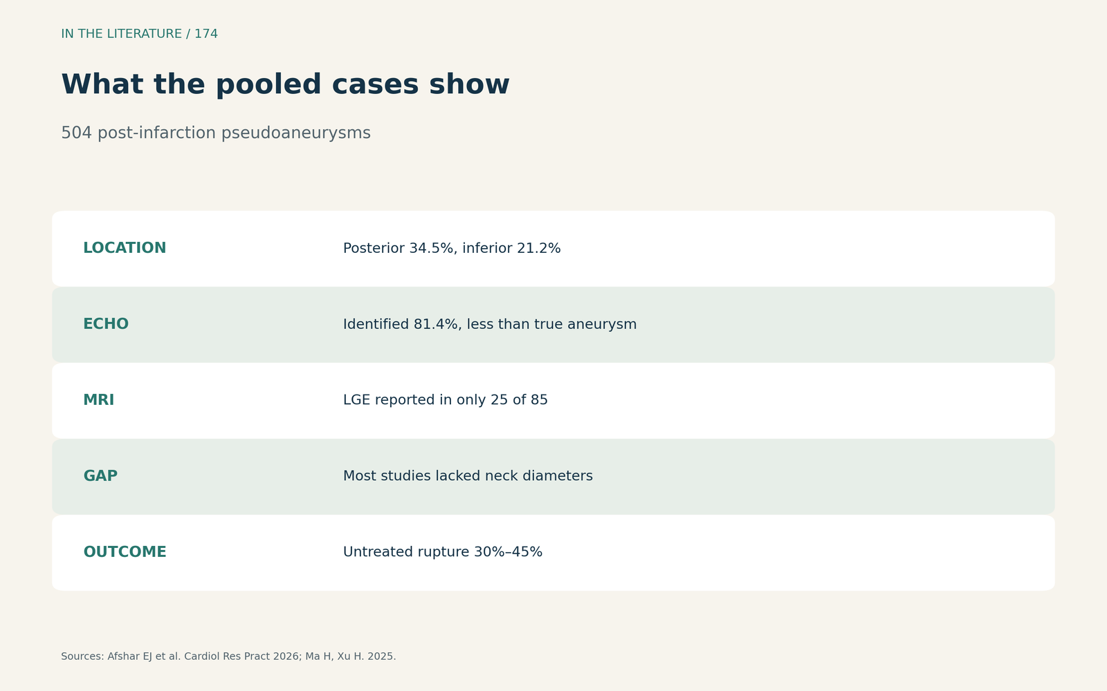
Figure 3. What the pooled cases show. Redrawn from Afshar et al. and Ma and Xu.[5], [1] View larger.

Three takeaways
- Ask whether the wall contains myocardium. A narrow neck and abrupt cutoff suggest pseudoaneurysm.[2], [3]
- Escalate imaging. TTE misses many; use TEE or CMR, looking for pericardial enhancement.[3], [4]
- Treat a pseudoaneurysm as a rupture risk. Especially when it is acute.[1], [4]
References
- Ma H, Xu H. Diagnosis and treatment of post-acute myocardial infarction ventricular aneurysm: a review. Medicine (Baltimore). 2025;104(32):e43696. doi:10.1097/MD.0000000000043696
- Goh VYS, Yew MS. Multimodality imaging of an idiopathic left ventricular aneurysm presenting with frequent premature ventricular beats. CASE (Phila). 2023;7(9):377–382. doi:10.1016/j.case.2023.05.007
- Oluleye O, Danielson D, Lowrie A, et al. Transthoracic echocardiography for diagnosis of infective endocarditis causing late left ventricular pseudoaneurysm of the basal anterolateral wall. CASE (Phila). 2023;7(3):96–100. doi:10.1016/j.case.2022.11.003
- Wershing K, Colon J, Alegria J, et al. Left ventricular pseudoaneurysm in an adult with a repaired partial atrioventricular canal defect. CASE (Phila). 2024;8(3):174–179. doi:10.1016/j.case.2023.12.009
- Afshar EJ, Tayebi A, Samimisedeh P, et al. Clinical and imaging findings in 504 cases with left ventricular pseudoaneurysm following myocardial infarction: a comprehensive systematic review and meta-analysis. Cardiol Res Pract. 2026;2026(1):9208154. doi:10.1155/crp/9208154