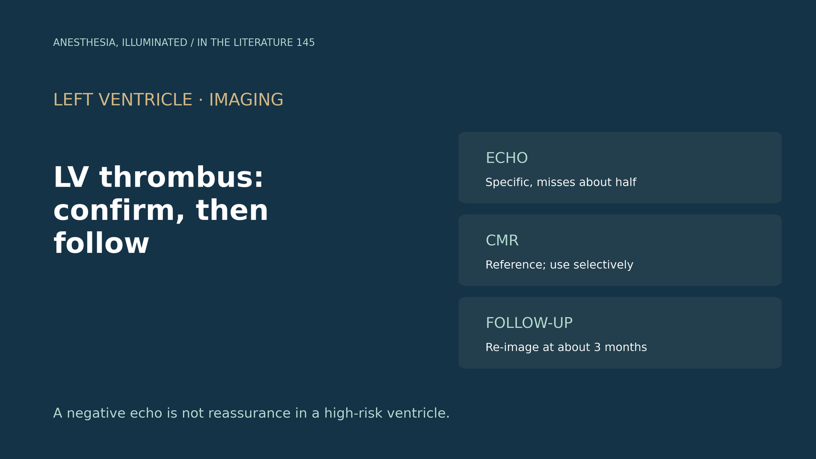
ITL-604713a6abe2
Juan Li, MD · Updated November 17, 2026
Watch the video
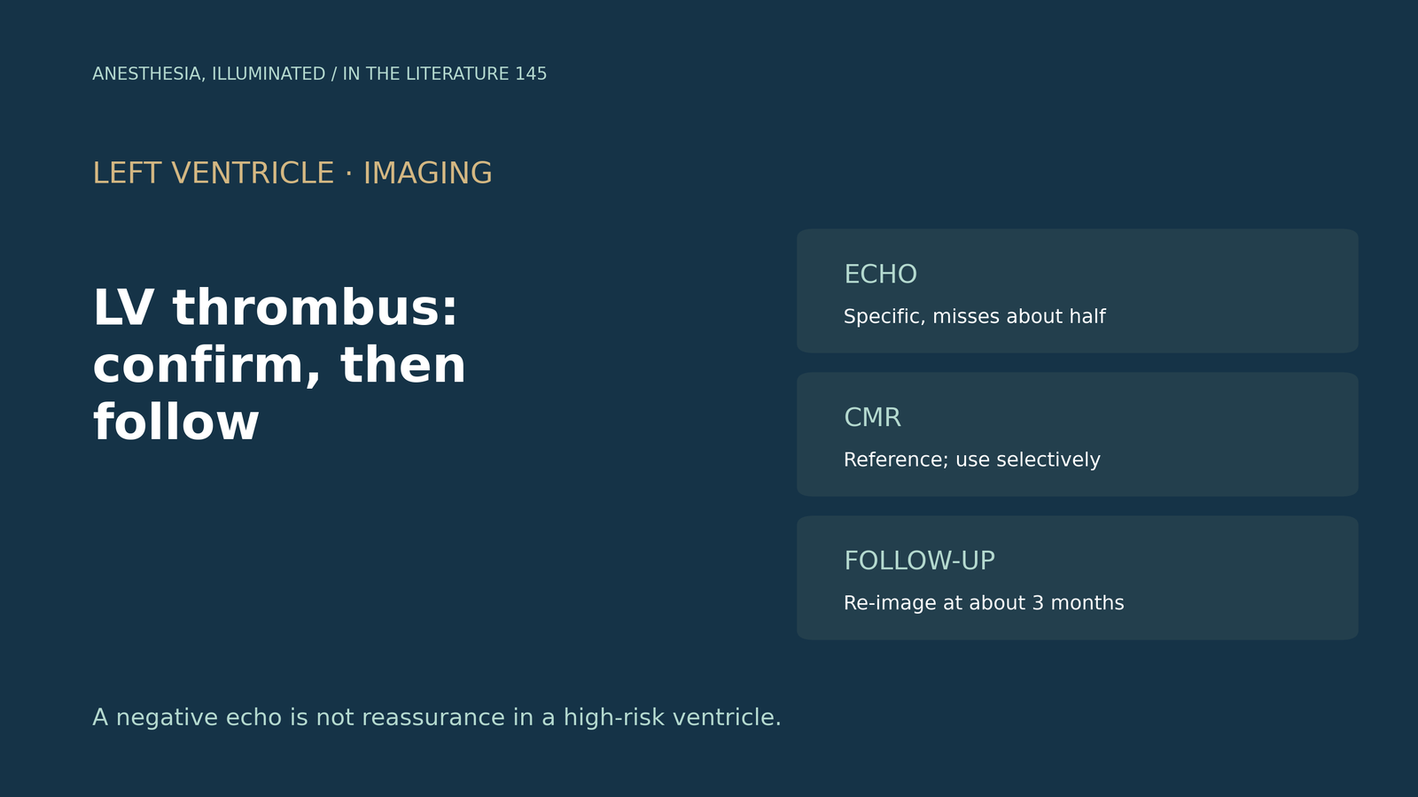
Key points
- Echo rules in, not out. Against cardiac magnetic resonance, non-contrast transthoracic echo had a sensitivity of 47% and a specificity of 98% across 10 studies with 2019 patients.[1]
- Contrast helps less than hoped. Contrast echo had a sensitivity of 58%, a non-significant gain, and still misses about half of thrombi.[1]
- An updated meta-analysis of 18 studies with 3478 participants gave a pooled echo sensitivity of 0.46 and an area under the curve of 0.985 for magnetic resonance versus 0.59 for echo.[2]
- Select patients for magnetic resonance. Apical wall-motion scoring on echo had a sensitivity of 100% (specificity 54%) and could triage who needs it; contrast echo is suggested as an intermediate step.[1], [2]
- Follow up with imaging. In 63 patients, thrombus resolved in 54% within 24 months, embolism occurred in 11%, and repeat imaging at about 3 months is recommended, but stopping therapy should not depend on time alone.[3], [4]
Two weeks after an anterior myocardial infarction, a patient with an apical aneurysm has a normal-looking apex on a limited transthoracic echo before noncardiac surgery. Is that enough to exclude thrombus, and which test should confirm or follow it?[1], [5]
How well echo detects thrombus
Phuah and colleagues pooled 11 studies with 2113 patients who had both echo and magnetic resonance within 7 days. Non-contrast echo had a sensitivity of 47% and a specificity of 98%; contrast echo had a sensitivity of 58% and a specificity of 98%; the addition of contrast was not found to improve diagnostic performance, and echo is likely to miss about half of thrombi.[1] Calderón Martínez and colleagues updated the analysis to 18 studies with 3478 participants: pooled echo sensitivity was 0.46, and magnetic resonance had an area under the curve of 0.985 versus 0.59 for echo, with late gadolinium enhancement magnetic resonance as the usual reference.[2]
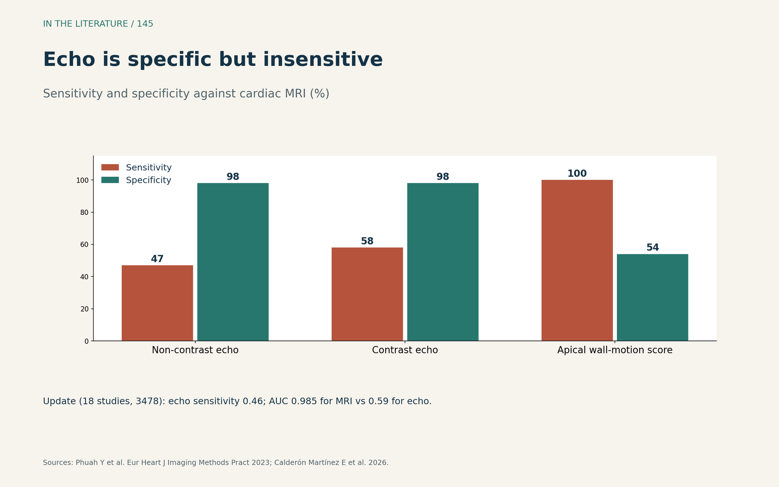
Figure 1. Echo is specific but insensitive. Redrawn from Phuah et al. and Calderón Martínez et al.[1], [2] View larger.

Choosing who needs magnetic resonance
Apical wall-motion scoring on non-contrast echo had a sensitivity of 100% and a specificity of 54% in 2 studies and could identify patients who need magnetic resonance.[1] The updated meta-analysis suggests contrast echo as an intermediate step before referral for magnetic resonance and notes that small, flat thrombi carry lower embolic risk than large, protruding or mobile ones.[2] Hwang and Sohn, commenting on a trial after anterior infarction, argue that routine contrast echo is useful at least for screening patients at risk and for patients with a poor echo window, though the trial was small.[5]
Following the thrombus
Tang and colleagues followed 63 patients with thrombus treated mainly with warfarin and serial echo: contrast was used in 78% at baseline and 86% at follow-up, thrombus resolved in 54% within 24 months, and systemic embolism occurred in 11%.[3] Awashra and colleagues recommend repeat imaging at about 3 months to assess resolution, note that a negative echo does not entirely exclude thrombus in a high-risk patient, and stress that the decision to stop therapy must not be based on time alone.[4]
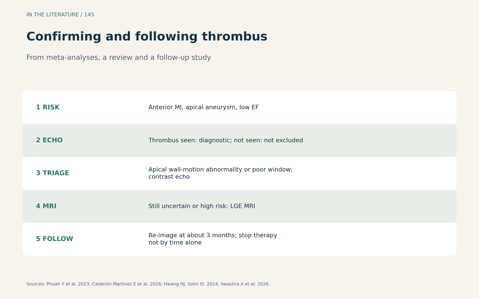
Figure 2. Confirming and following thrombus. Synthesized from Phuah et al., Calderón Martínez et al., Tang et al., Awashra et al. and Hwang and Sohn.[1], [2], [3], [4], [5] View larger.

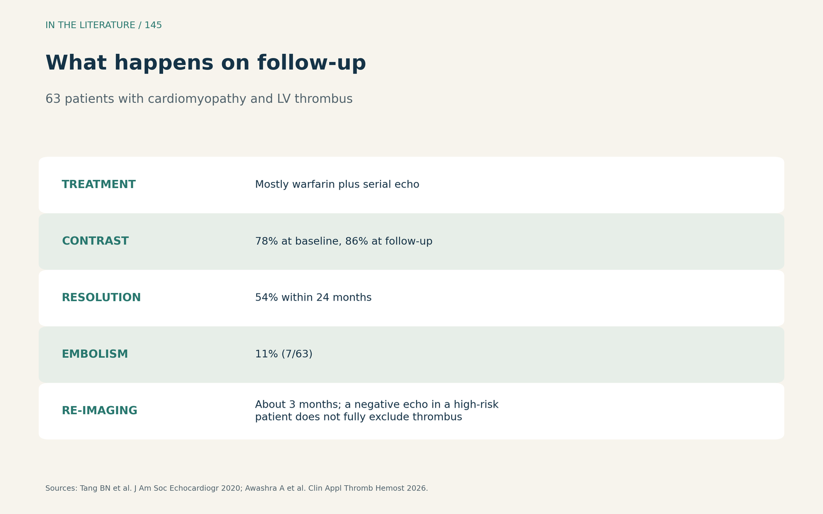
Figure 3. What happens on follow-up. Redrawn from Tang et al. and Awashra et al.[3], [4] View larger.

Three takeaways
- A negative echo is not reassurance. Echo misses about half of thrombi; specificity is high.[1], [2]
- Use magnetic resonance selectively. Apical wall-motion abnormality or a poor window should prompt contrast or magnetic resonance.[1], [5]
- Image again before stopping therapy. Reassess at about 3 months; do not stop anticoagulation by the calendar alone.[3], [4]
References
- Phuah Y, Tan YX, Zaghloul S, et al. A systematic review and meta-analysis of transthoracic echocardiogram vs. cardiac magnetic resonance imaging for the detection of left ventricular thrombus. Eur Heart J Imaging Methods Pract. 2023;1(2):qyad041. doi:10.1093/ehjimp/qyad041
- Calderón Martínez E, Miret Durazo CI, Kumar A, et al. Transthoracic echocardiography vs. cardiac magnetic resonance imaging for the diagnosis of left ventricular thrombus: an updated systematic review and diagnostic accuracy meta-analysis. Eur Heart J Imaging Methods Pract. 2026;4(4):qyag150. doi:10.1093/ehjimp/qyag150
- Tang BN, Ong G, Sandhu P, et al. Natural history of left ventricular thrombi in patients with cardiomyopathy. J Am Soc Echocardiogr. 2020;33(9):1158–1159. doi:10.1016/j.echo.2020.05.024
- Awashra A, Emara A, AbuBaha M, et al. Left ventricular thrombus in oncology patients and beyond: imaging, treatment choices, and duration. Clin Appl Thromb Hemost. 2026;32:10760296261437022. doi:10.1177/10760296261437022
- Hwang HJ, Sohn IS. Efficacy of routine contrast echocardiography for the detection of left ventricular thrombus in patients with anterior ST-elevation myocardial infarction. J Cardiovasc Imaging. 2024;32(1):36. doi:10.1186/s44348-024-00041-2