ITL-82b29bc24946
Juan Li, MD · Updated November 17, 2026
Watch the video
Key points
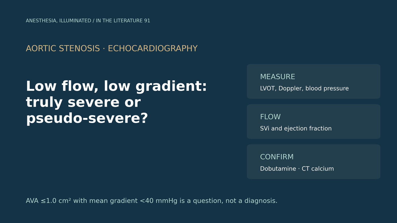
- A valve area of 1.0 cm² or less with a mean gradient below 40 mmHg is a discordant finding. Before calling it low-flow stenosis, exclude measurement error, especially in left ventricular outflow tract (LVOT) diameter, Doppler alignment and blood pressure.[1]
- Classify by stroke volume index (SVi, 35 mL/m²) and ejection fraction: classical low-flow (reduced ejection fraction), paradoxical low-flow (preserved ejection fraction) and normal-flow low-gradient, which usually reflects moderate stenosis.[1], [2]
- In classical low-flow stenosis, dobutamine stress echocardiography separates true severe from pseudo-severe stenosis when flow rises, but standard criteria leave many cases indeterminate. Projected valve area classifies more patients.[3], [4]
- CT aortic valve calcium scoring is flow-independent and uses sex-specific thresholds. It agrees with stress echocardiography in only about 70% of cases, and a low score does not rule out benefit from valve replacement.[2], [5], [6]
- Paradoxical low-flow stenosis is the hardest to confirm: dobutamine adds little in small ventricles, and intervention should follow careful confirmation of severity.[1], [2], [7]
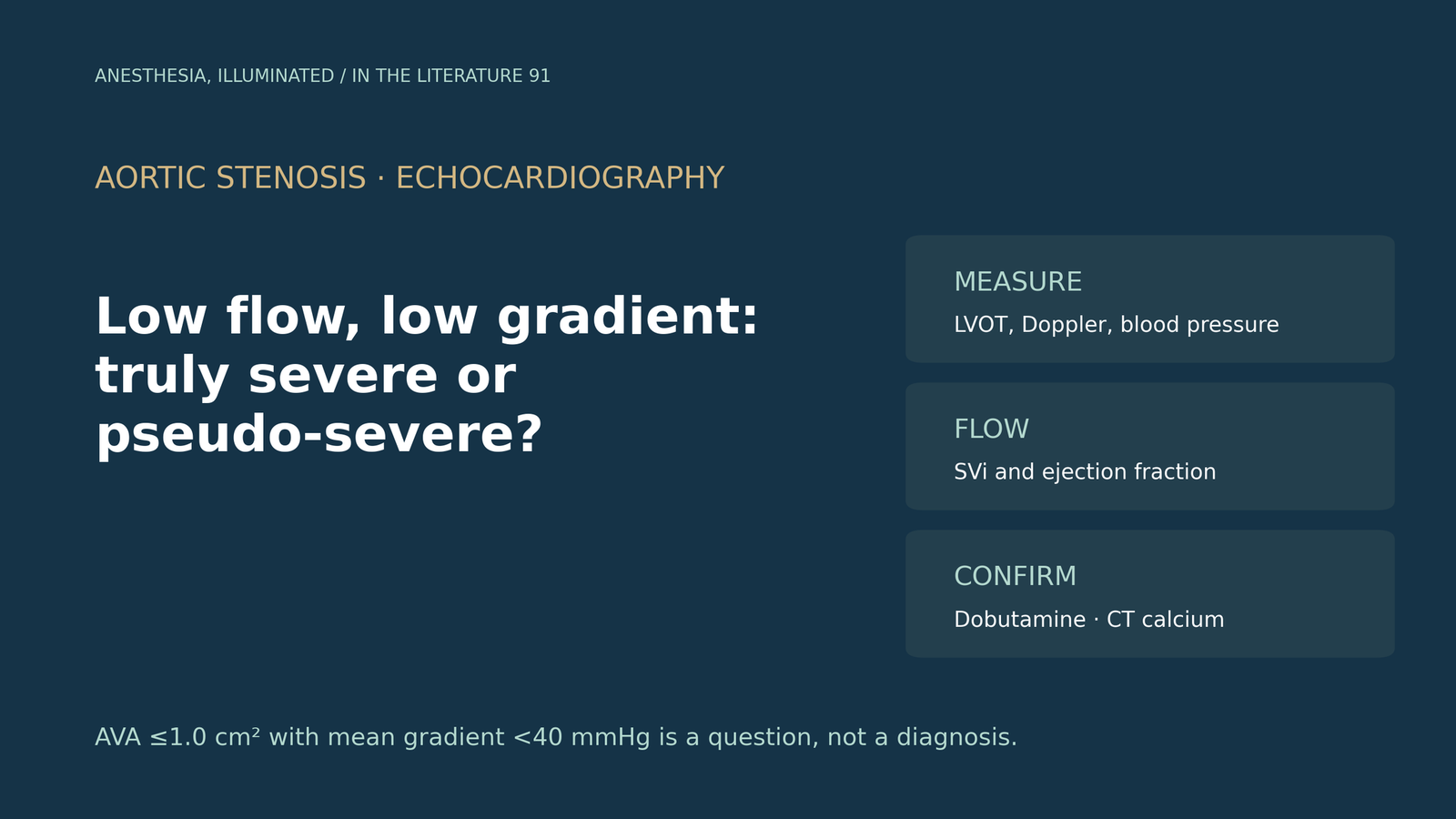
An older patient is referred before surgery with a calculated aortic valve area of 0.8 cm², a mean gradient of 25 mmHg and either reduced or apparently normal ejection fraction. The report says "severe aortic stenosis", but the gradient says "moderate". Is the valve truly severely stenotic, pseudo-severe because the ventricle cannot open it, or simply mismeasured, and which test settles the question?[1], [2]
Start by doubting the numbers
The European Association of Cardiovascular Imaging and American Society of Echocardiography (EACVI/ASE) recommendations stress that the continuity equation is only as good as its inputs. Measurement variability for LVOT diameter ranges from 5% to 8%, and because the diameter is squared it becomes the greatest potential source of error in valve area. Assuming a circular outflow tract underestimates stroke volume and can create apparent "low flow".[1]
Misaligned Doppler underestimates velocity and gradient, so multiple windows are needed. Hypertension can alter velocity and gradient and should be recorded, with blood pressure controlled before the examination when possible. The document advises that severe stenosis should be questioned in particular when peak velocity is below 3.0 m/s and mean gradient below 20 mmHg.[1]
Classify by flow and ejection fraction
The 2025 European Society of Cardiology/European Association for Cardio-Thoracic Surgery (ESC/EACTS) guideline categorizes discordant stenosis by flow, using an SVi threshold of 35 mL/m² while noting that sex-specific thresholds have been proposed. With a mean gradient below 40 mmHg and valve area of 1 cm² or less, it distinguishes three groups:[2]
- Classical low-flow, low-gradient: SVi of 35 mL/m² or less and ejection fraction below 50%.
- Paradoxical low-flow, low-gradient: SVi of 35 mL/m² or less with ejection fraction of 50% or more.
- Normal-flow, low-gradient: SVi above 35 mL/m² with preserved ejection fraction.
The EACVI/ASE document states that patients in the normal-flow group should not be diagnosed with severe stenosis; they usually have moderate disease and a miscalculated valve area.[1]
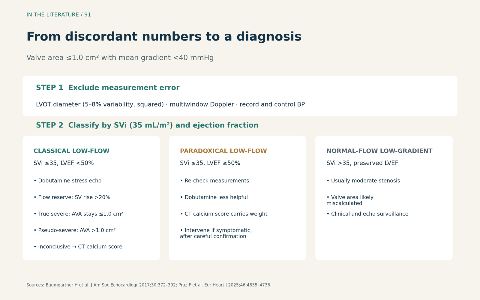
Figure 1. From discordant numbers to a diagnosis. Synthesized from the EACVI/ASE recommendations and the 2025 ESC/EACTS guideline; thresholds as stated in those documents.[1], [2] View larger.

Classical low-flow stenosis: what dobutamine can and cannot show
In classical low-flow stenosis, two conditions look alike at rest. In true severe stenosis, the valve causes the ventricular dysfunction. In pseudo-severe stenosis, moderate stenosis coexists with another cause of ventricular dysfunction, and a weak ventricle cannot open the valve fully.[1]
Low-dose dobutamine stress echocardiography tests the valve at higher flow. A stroke volume increase of more than 20% indicates flow (contractile) reserve. Severe stenosis is suggested when velocity reaches 4 m/s or mean gradient exceeds 30–40 mmHg while valve area does not exceed 1.0 cm² at any flow rate. A final valve area above 1.0 cm² with a gradient below 40 mmHg indicates pseudo-severe stenosis.[1], [5] The 2025 ESC/EACTS guideline recommends intervention when severe stenosis is confirmed by stress echocardiography or calcium scoring, and guideline-directed medical therapy for pseudo-severe stenosis.[2]
The limits show up in practice. Sato and colleagues studied 235 patients with classical low-flow stenosis who underwent dobutamine stress echocardiography. Flow reserve was present in 138 (59%). By standard criteria, 86 (37%) had true severe stenosis, 86 had pseudo-severe stenosis, and severity could not be determined in 63. Absence of flow reserve was not associated with mortality (P = 0.62). After weighting, valve replacement was associated with better survival (hazard ratio 0.40; 95% CI 0.31–0.51). No ischemia was detected during the tests.[3]
Projected valve area, which standardizes valve area to a flow rate of 250 mL/s, could be analyzed in 233 patients and classified 60% as true severe. By projected valve area, valve replacement was associated with better survival in true severe but not pseudo-severe stenosis.[3] In an accompanying editorial, Annabi and colleagues summarized earlier data showing an accuracy of only 48% for a stress mean gradient of 40 mmHg or more and 47% for the combined criterion, versus 70% for projected valve area.[4] These are observational, single-center findings, and projected valve area is not yet a standard guideline criterion.
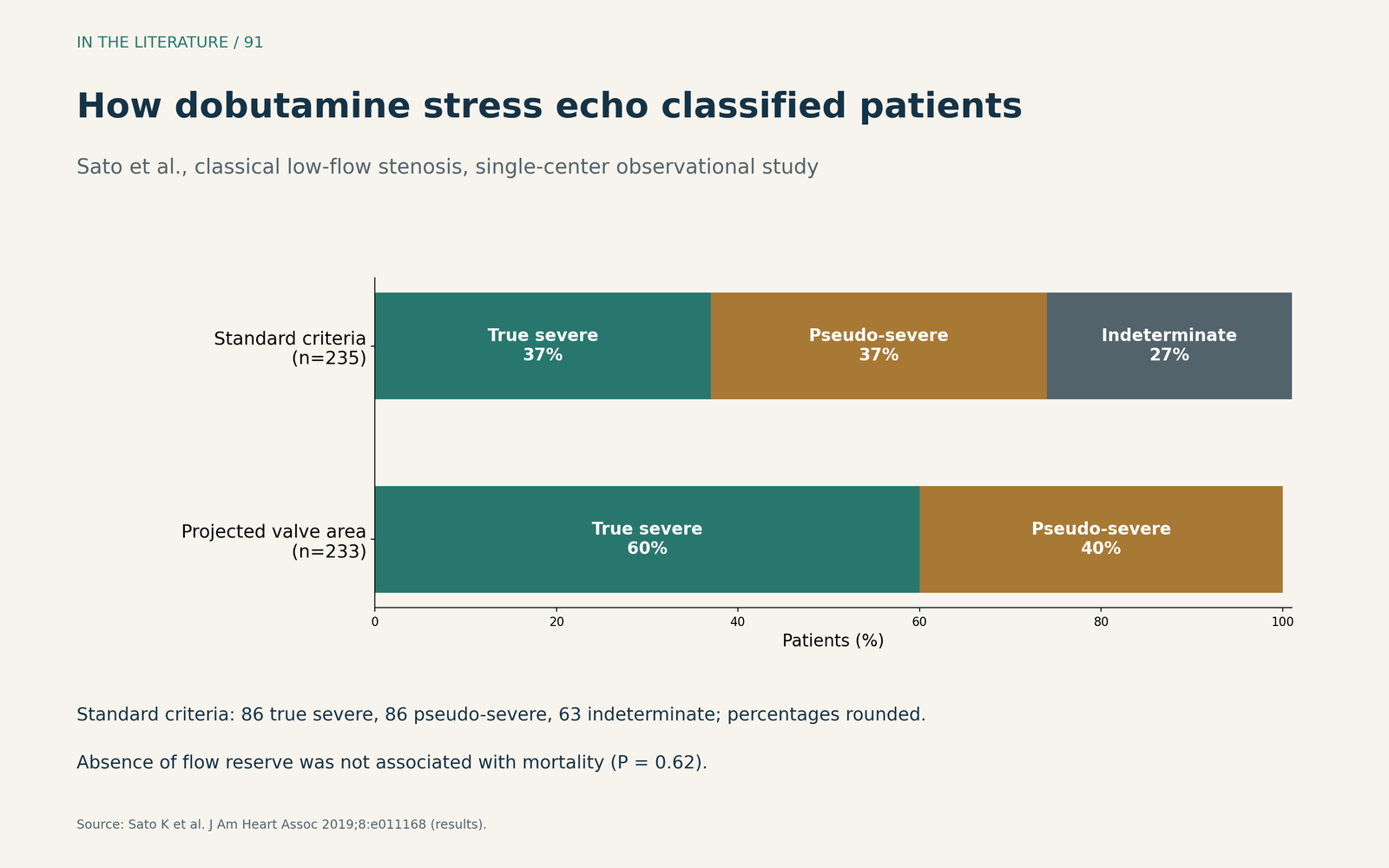
Figure 2. Standard criteria leave many cases indeterminate. Redrawn from the results of Sato et al. (235 patients; projected valve area analyzable in 233).[3] View larger.

Calcium scoring: flow-independent, but not a verdict
The ESC/EACTS guideline states that CT aortic valve calcium scores above 2000 Agatston units (AU) in men and 1200 AU in women indicate severe stenosis with high sensitivity and specificity (85%). Higher thresholds (3000 AU in men, 1600 AU in women) are very specific. Severe stenosis becomes unlikely below 1600 AU in men and 800 AU in women. Caution is needed when stenosis can be severe without heavy calcification, as in bicuspid valves, amyloidosis and fibrotic disease after rheumatic fever, radiation or inflammation.[1], [2] For classical low-flow stenosis with reduced ejection fraction, the guideline describes calcium scoring and stress echocardiography as complementary.[2]
The two tests often disagree. In a letter by Kragholm and colleagues describing 55 patients with classical low-flow stenosis, 26 of 37 (70.3%) classified as true severe by stress echocardiography had a concordant calcium score. Stress echocardiography was inconclusive in 11 (20%).[5]
A low score also does not settle prognosis. Juhász and colleagues found a low calcium score in 40% of patients with low-gradient stenosis, compared with 10% of those with high gradients. In low-gradient stenosis, the calcium score did not predict mortality. Valve intervention was associated with lower mortality even in patients with low-gradient stenosis and a low score (adjusted hazard ratio 0.102; 95% CI 0.028–0.369). This was a retrospective study, so the authors cautioned that no causal conclusions can be drawn.[6]
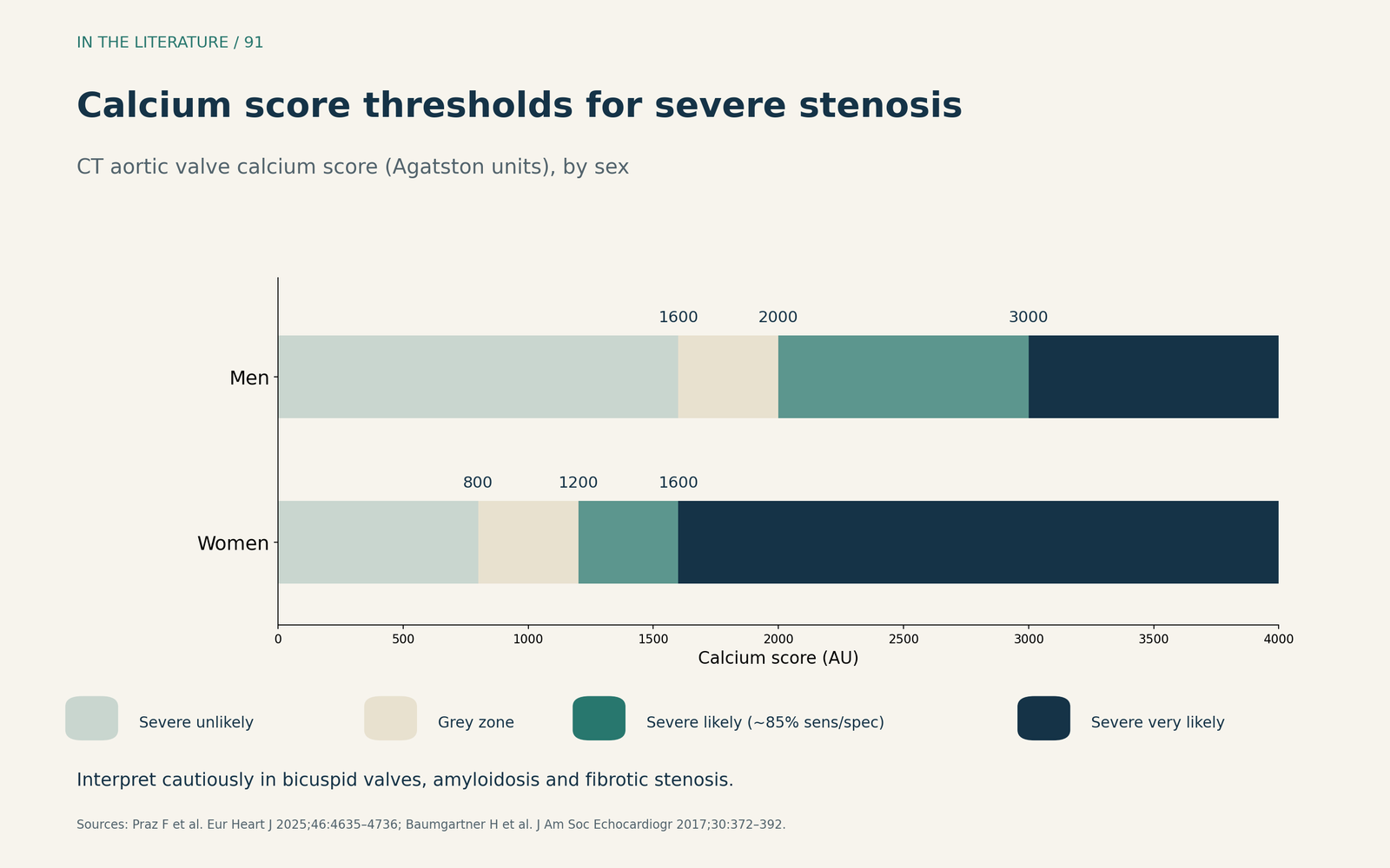
Figure 3. Calcium thresholds differ by sex, with a grey zone between them. Redrawn from the thresholds in the 2025 ESC/EACTS guideline and the EACVI/ASE recommendations.[1], [2] View larger.

Paradoxical low-flow stenosis: the hardest case
With preserved ejection fraction, a small, hypertrophied ventricle produces low stroke volume. The EACVI/ASE recommendations note that dobutamine may be less helpful in these small ventricles, and it is unclear how to distinguish pseudo-severe from true severe stenosis in this setting. Calcium scoring and careful exclusion of measurement error therefore carry more weight.[1]
One proposed refinement addresses preload. Buffle and colleagues note that dobutamine fails to increase SVi in one third of patients. They added passive leg raising to dobutamine in 45 patients, including 25 with paradoxical stenosis. This raised stroke volume and gradient, and the combined protocol tended to reclassify most paradoxical cases as pseudo-severe.[7] The study was small, used no outcome or CT reference standard, and the reclassification was not statistically significant, so it remains investigational.
The ESC/EACTS guideline concludes that intervention improves outcomes compared with medical treatment in paradoxical low-flow stenosis, but should be considered in symptomatic patients only after careful confirmation that the stenosis is severe.[2]
Three takeaways
- Check the measurement before the diagnosis. Re-measure the outflow tract, interrogate from multiple windows and record blood pressure; normal-flow low-gradient stenosis is usually moderate.[1], [2]
- Use dobutamine in classical low-flow stenosis, and read it carefully. Absent flow reserve does not mean no benefit from valve replacement, and projected valve area classifies more patients than standard criteria.[3], [4]
- Treat calcium as one piece of evidence. Sex-specific thresholds help, especially in paradoxical stenosis, but discordant or low scores need an integrated Heart Team judgment.[2], [5]–[7]
References
- Baumgartner H, Hung J, Bermejo J, et al. Recommendations on the echocardiographic assessment of aortic valve stenosis: a focused update from the European Association of Cardiovascular Imaging and the American Society of Echocardiography. J Am Soc Echocardiogr. 2017;30(4):372–392. doi:10.1016/j.echo.2017.02.009
- Praz F, Borger MA, Lanz J, et al. 2025 ESC/EACTS Guidelines for the management of valvular heart disease. Eur Heart J. 2025;46(44):4635–4736. doi:10.1093/eurheartj/ehaf194
- Sato K, Sankaramangalam K, Kandregula K, et al. Contemporary outcomes in low-gradient aortic stenosis patients who underwent dobutamine stress echocardiography. J Am Heart Assoc. 2019;8(6):e011168. doi:10.1161/JAHA.118.011168
- Annabi MS, Clavel MA, Pibarot P. Dobutamine stress echocardiography in low-flow, low-gradient aortic stenosis: flow reserve does not matter anymore. J Am Heart Assoc. 2019;8(6):e012212. doi:10.1161/JAHA.119.012212
- Kragholm K, Abdelkarim O, Sinha A, et al. Aortic valve calcium score in addition to dobutamine stress echocardiography for low-flow, low-gradient aortic stenosis assessment. J Am Soc Echocardiogr. 2023;36(12):1324–1326. doi:10.1016/j.echo.2023.08.009
- Juhász D, Vecsey-Nagy M, Jermendy ÁL, et al. Prognostic and therapeutic implications of a low aortic valve calcium score in patients with low-gradient aortic stenosis. Eur Heart J Cardiovasc Imaging. 2025;26(2):287–298. doi:10.1093/ehjci/jeae276
- Buffle E, Papadis A, Boscolo Berto M, et al. Passive leg raise stress echocardiography in severe paradoxical low-flow, low-gradient aortic stenosis. J Am Soc Echocardiogr. 2022;35(11):1123–1132. doi:10.1016/j.echo.2022.07.005