
ITL-e478d4de6cd7
Watch the video

Key points
- Assess perfusion promptly and repeatedly; the patient's clinical state determines the urgency of the response.[1]
- Low flow can reflect inadequate LV filling, increased afterload or obstruction. A small LV does not, by itself, establish hypovolemia or justify fluid loading.[1], [2]
- Lack of marked LDH elevation does not exclude outflow-graft obstruction; in one small series, most affected patients had LDH below twice the local upper limit.[3]
- Echocardiography is an early assessment tool, but persistent concern about the outflow graft may require CT angiography even when TTE is unrevealing. Recent imaging research supports this distinction without validating a universal alarm algorithm.[2], [4]
A durable LVAD reports low flow, but the alarm alone does not reveal whether the problem is reduced blood delivery to the pump, excessive resistance to forward flow or a mechanical obstruction. The first task is to establish how well the patient is perfusing while the LVAD team helps interpret the device and investigate the cause. This discussion concerns the initial assessment of low-flow alarms; circulatory collapse requires an immediate emergency response.[1], [2]
Read the patient and the controller together
Check mental status, peripheral perfusion, blood pressure and rhythm, and look for congestion or bleeding. Blood-pressure assessment needs to account for the degree of pulsatility; an absent palpable pulse alone is not sufficient evidence of absent circulation. Deteriorating perfusion warrants urgent escalation while assessment continues. A patient who initially appears well still needs reassessment if alarms persist.[1]
Record the alarm pattern and compare speed, power, estimated flow and pulsatility index with the patient's usual values. Displayed pump flow is an estimate derived from device parameters, not a direct measurement of systemic cardiac output. PI and power are also context dependent. A single reading cannot distinguish hypovolemia, RV failure, hypertension and mechanical obstruction.[2]
When device thrombosis or dysfunction is suspected, interrogation and review of longitudinal log files with the LVAD team and manufacturer can add information that a snapshot misses. A power rise may suggest increased rotor resistance; a fall can accompany inflow or outflow obstruction. These patterns guide investigation but do not independently establish the diagnosis.[1]
Organize the differential around filling, pressure and pathway
Filling: is enough blood reaching the LV? Hypovolemia, RV failure and tamponade can all reduce LV filling. Review LV size together with RV function, septal position, venous congestion and pericardial fluid or organized clot. A small LV in a patient with a dilated, congested right heart is a different problem from a small LV after blood loss. Giving fluid simply because the display says “low flow” may worsen congestion; volume replacement should follow evidence of hypovolemia and reassessment.[1], [2]
Pressure: is afterload opposing pump flow? Hypertension can reduce LVAD forward flow. Obtain an interpretable pressure measurement and relate it to the alarm and clinical state. Arrhythmias also belong in the initial assessment because they can disrupt filling and RV performance. Neither an isolated high PI nor an isolated low estimated flow supplies the missing hemodynamic context.[1], [2]
Pathway: is blood encountering an inflow or outflow obstruction? Echo can identify inflow malposition, dynamic contact with the endocardium and some obstructing material. Outflow problems include kinking, twisting, intraluminal material and external compression. The whole graft may not be adequately assessed by TTE; CT provides complementary anatomic information.[2]
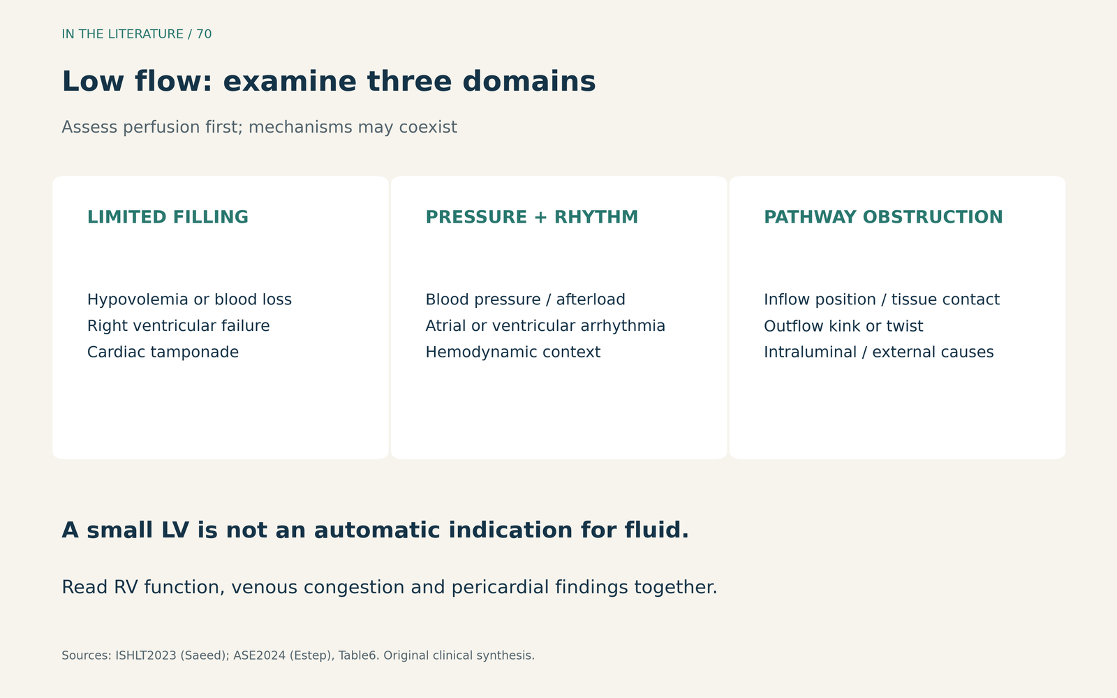
Figure 1. Use a physiologic differential before choosing a treatment. These domains can coexist; a small LV is a finding to explain, not a fluid prescription.[1], [2] View larger.

Do not use mild LDH elevation or unrevealing TTE to close the investigation
Peters and colleagues described 11 recognized outflow-graft obstructions in a retrospective, mixed-device cohort. Ten patients presented with low-flow alarms. Eight had LDH below twice the local upper limit of normal—this is not the same as saying that all had normal LDH. TTE did not directly visualize the obstruction in any patient. Of nine patients who underwent CTA, eight had the obstruction identified and localized; the inconclusive study required angiography.[3]
This selected series does not provide a validated LDH cutoff or a formal estimate of CTA sensitivity. It does show why absence of marked hemolysis and a nondiagnostic TTE can coexist with important graft obstruction. If alarms or signs of impaired support persist after the initial assessment, discuss targeted graft imaging rather than assuming that these findings have excluded a mechanical cause.[1], [2], [3]
What the 2026 imaging study adds
Sato and colleagues compared TTE with CT in 54 patients contributing 62 paired examinations; 19 patients had HeartMate 3 devices. Their endpoint was a CT-defined structural abnormality: bending of at least 90 degrees or lumen stenosis of at least 50%. TTE missed some abnormalities, particularly in the proximal graft. However, only one case was clinically actionable with associated symptoms and device alarms. The study therefore cannot be read as a validation of diagnostic accuracy in patients presenting with acute low-flow alarms.[4]
The useful implication is narrower: TTE and CT answer complementary questions. An unrevealing TTE cannot reliably exclude graft pathology, and an anatomic finding on CT still needs to be related to symptoms, pump trends and hemodynamics. Neither a single Doppler velocity nor a structural abnormality alone establishes which treatment is needed.[2], [4]
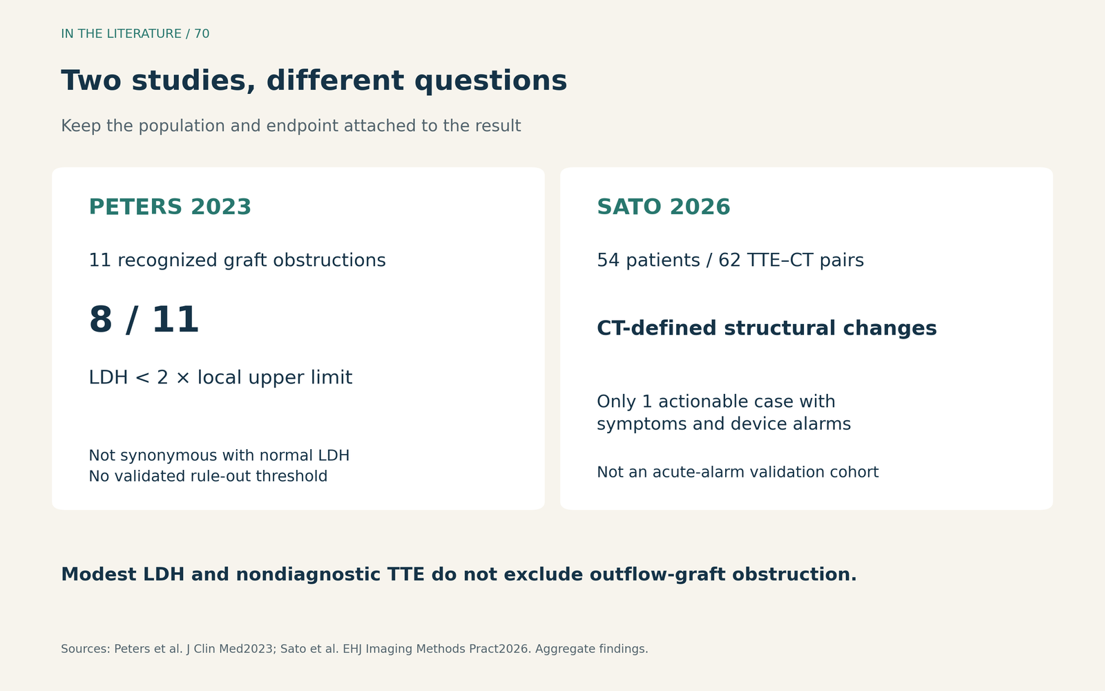
Figure 2. Keep the population and endpoint attached to the finding. These studies support continued investigation when suspicion persists; neither establishes a universal diagnostic rule for all low-flow alarms.[3], [4] View larger.

Let persistence and physiology determine the next test
Use bedside echo to assess filling, RV function, pericardial pathology and LV unloading. Correct an identified reversible cause with the LVAD team, then reassess perfusion and the alarm pattern. Persistent unexplained low flow or evidence of inadequate support warrants further evaluation; CTA is particularly useful when the question concerns outflow-graft anatomy. If imaging remains inconclusive, invasive hemodynamic assessment or angiography may be appropriate for the specific unresolved question.[1], [2]
The sequence depends on stability. A patient with compromised perfusion needs urgent treatment and bedside assessment; transport for CT must be considered in that context. Pump-speed changes and ramp testing require experienced supervision and an explicit diagnostic or therapeutic purpose. Increasing speed simply to improve the displayed number does not resolve an unidentified filling or mechanical problem.[1], [2]
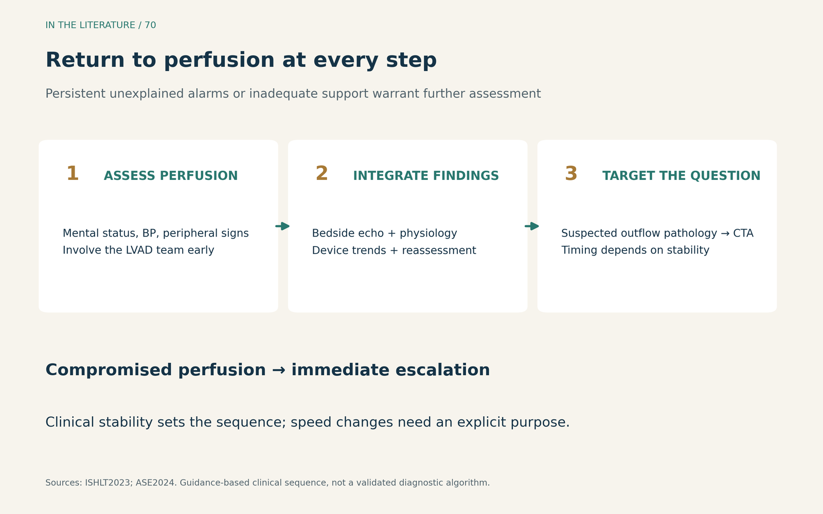
Figure 3. Reassess the patient throughout the investigation. This is an application of the cited guidance, not a separately validated diagnostic algorithm; unstable patients require immediate escalation.[1], [2] View larger.

Three takeaways
- Start with perfusion, then explain the low flow. Examine filling, afterload and the inflow–outflow pathway together with rhythm and device trends.
- Do not let a small LV dictate a fluid bolus. RV failure and tamponade can also limit LV filling; the congestion and imaging findings change the response.
- Keep investigating persistent unexplained alarms. Mild LDH elevation and unrevealing TTE do not exclude graft obstruction; use targeted anatomic imaging and LVAD-team assessment when suspicion remains.[1], [2], [3], [4]
References
- Saeed D, Feldman D, Banayosy AE, et al. The 2023 International Society for Heart and Lung Transplantation Guidelines for Mechanical Circulatory Support: A 10-Year Update. J Heart Lung Transplant. 2023;42(7):e1-e222. doi:10.1016/j.healun.2022.12.004. doi:10.1016/j.healun.2022.12.004
- Estep JD, Nicoara A, Cavalcante J, et al. Recommendations for Multimodality Imaging of Patients With Left Ventricular Assist Devices and Temporary Mechanical Support: Updated Recommendations from the American Society of Echocardiography. J Am Soc Echocardiogr. 2024;37(9):820-871. doi:10.1016/j.echo.2024.06.005. doi:10.1016/j.echo.2024.06.005
- Peters CJ, Zhang RS, Vidula MK, et al. Durable Left Ventricular Assist Device Outflow Graft Obstructions: Clinical Characteristics and Outcomes. J Clin Med. 2023;12(6):2430. doi:10.3390/jcm12062430
- Sato T, Fujino T, Misumi K, et al. Diagnostic accuracy of transthoracic echocardiography to detect structural abnormalities of the outflow graft in patients with left ventricular assist devices. Eur Heart J Imaging Methods Pract. 2026;4(1):qyag017. doi:10.1093/ehjimp/qyag017