
ITL-caf9c896c031
Watch the video

Figure 1. Editorial illustration of a coordinated reassessment; the equipment is conceptual and does not represent a specific LVAD controller.
Key points
- A low-flow alarm can reflect inadequate ventricular filling or altered afterload; it does not identify pump failure by itself.[1]
- New abdominal distension with rising ventilatory pressures and circulatory deterioration during endoscopy should raise concern for tension pneumoperitoneum.[2]
- Gas causing hemodynamic instability or respiratory failure requires emergency decompression; closing a perforation and relieving pressure address different problems.[3]
- In an unresponsive patient with a durable LVAD and impaired perfusion, the 2025 AHA guideline recommends chest compressions; device assessment can proceed concurrently.[4]
During upper GI endoscopy, the LVAD flow display falls and blood pressure deteriorates. Bleeding, anesthetic vasodilation, right ventricular dysfunction and device problems all deserve attention. But if the abdomen is becoming tense and ventilation is becoming difficult, the assessment must extend beyond the controller.[1, 2]
A functioning pump still needs blood to reach it
Continuous-flow LVADs depend on adequate left ventricular filling. Hypovolemia, right ventricular failure or reduced systemic venous return can lower pump flow. Excessive afterload can also reduce flow, so the same alarm can arise from different hemodynamic problems.[1]
Tension pneumoperitoneum provides a particularly relevant mechanism during endoscopy: pressurized intraperitoneal gas can impair venous return and ventilation. In an LVAD patient, reduced filling can then threaten pump output. This connection is a physiological inference from LVAD physiology and endoscopic complication reports; it is not a validated LVAD-specific diagnostic rule.[1, 2]
Treat displayed flow as a trend within the clinical picture. Assess perfusion, blood pressure, rhythm, ventilation and the device together; echocardiography can help distinguish inadequate filling from right ventricular dysfunction or other causes. Reflexively increasing pump speed does not correct the cause of an underfilled ventricle.[1]
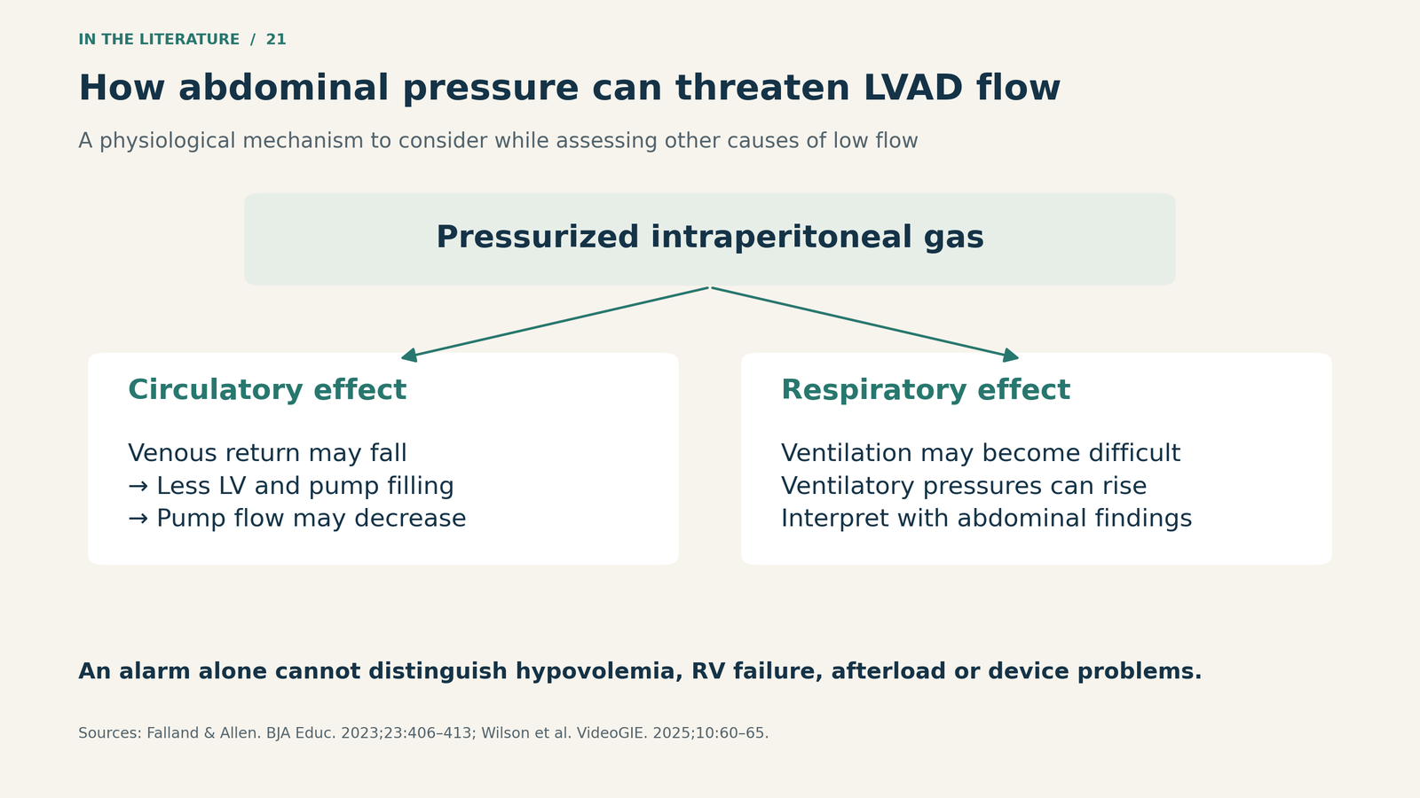
Figure 2. A mechanism to consider, not a diagnostic test. The combination of procedural timing, abdominal findings, ventilation and perfusion is more informative than the alarm alone.[1, 2]

Distension becomes urgent when it affects circulation or breathing
Abdominal gas alone is not synonymous with tension physiology. Wilson and colleagues emphasize progressive abdominal distension or rigidity accompanied by elevated ventilatory pressures, hypotension or shock. Their 2025 technical report illustrates two advanced endoscopic procedures: a colonic submucosal dissection and a gastric tumor resection. In both, needle decompression improved abdominal distension and ventilatory pressures, allowing treatment of the defect and completion of the procedure.[2]
An earlier retrospective series by Chiapponi and colleagues included 11 patients referred with iatrogenic bowel perforation. Three had tension physiology and underwent emergency decompression before surgery, with reported stabilization. These were selected perforation cases, not a denominator for estimating the risk of tension pneumoperitoneum during routine endoscopy.[5]
Neither report establishes an LVAD-specific success rate or a risk-free technique. They support recognizing a potentially reversible pressure problem while arranging definitive management. They also show why the response after decompression must be reassessed, rather than assuming the emergency is over.[2, 5]
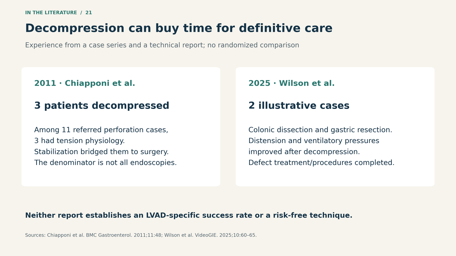
Figure 3. Different clinical settings, not pooled efficacy data. These reports describe rescue treatment; they do not quantify benefit specifically in LVAD recipients.[2, 5]

Relieve the pressure and address its source
The ESGE position statement recommends emergency decompression when gas under tension causes hemodynamic instability or respiratory failure. It separately addresses endoscopic closure, determined by the defect, patient condition and available expertise. Failed closure or clinical deterioration calls for surgical consultation and further treatment.[3]
CT helps evaluate suspected perforation, particularly in a stable patient; it should not turn into a prerequisite that delays emergency treatment of established tension physiology. Bedside ultrasound may help distinguish free intraperitoneal gas from luminal overdistension and assist with site selection when available. It is an adjunct, not a requirement to postpone rescue.[2, 3, 5]
For an LVAD recipient, involve the device team early. The pump and driveline course matter when planning abdominal access. Applying that anatomical precaution to decompression is a clinical planning inference: a generic entry-site diagram cannot account for every patient's implanted hardware or prior surgery.[1, 2]
Assess perfusion while the team addresses the cause
A continuous-flow LVAD patient may lack a palpable pulse despite adequate perfusion, and an automated cuff or pulse oximeter may be unreliable. The AHA assessment therefore incorporates skin findings, capillary refill, measured blood pressure and waveform capnography, alongside responsiveness.[4]
For unresponsiveness with impaired perfusion, chest compressions are a Class 1 recommendation, based on expert opinion. Starting compressions immediately while another rescuer assesses device-related reversible causes is Class 2b, also based on expert opinion. The guideline describes checking alarms, the driveline and power connections while treating non-device causes. These recommendations concern durable LVAD recipients beyond the implantation hospitalization; the peri-implantation setting has separate guidance.[4]
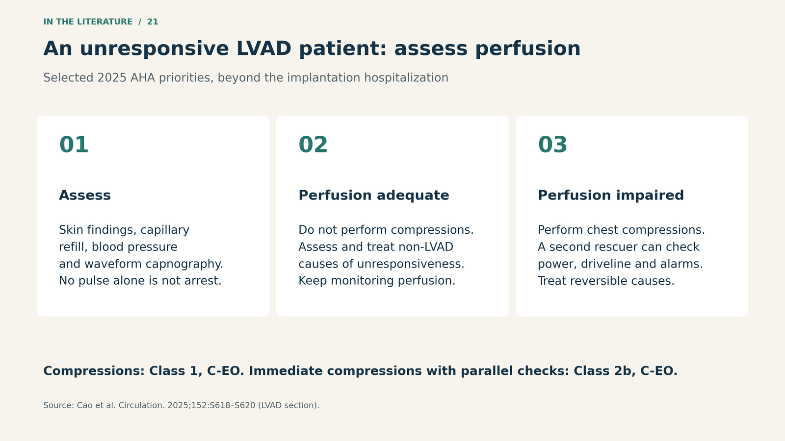
Figure 4. Selected priorities from the 2025 AHA LVAD guidance, not a replacement for the complete resuscitation algorithm. Absence of a palpable pulse alone does not establish cardiac arrest.[4]

Three takeaways
- Interpret the alarm through physiology. Check filling, right ventricular function, afterload and device function rather than assuming one cause.
- Examine the abdomen when ventilation and circulation worsen together. Tension pneumoperitoneum requires pressure relief and a plan for the underlying defect.
- Do not let device troubleshooting delay indicated resuscitation. Assess perfusion and coordinate parallel tasks with the endoscopy, anesthesia and LVAD teams.
References
- Falland R, Allen SJ. Perioperative management of patients with a ventricular assist device undergoing non-cardiac surgery. BJA Educ. 2023;23(10):406–413. doi:10.1016/j.bjae.2023.06.003.
- Wilson NJ, Karna R, Roldan GA, Osorio Cintron R, Leaf A, Guerrero Vinsard D, et al. Percutaneous needle decompression for tension pneumoperitoneum during GI endoscopy: a step-by-step guide. VideoGIE. 2025;10(1):60–65. doi:10.1016/j.vgie.2024.09.003.
- Paspatis GA, Arvanitakis M, Dumonceau JM, Barthet M, Saunders B, Turino SY, et al. Diagnosis and management of iatrogenic endoscopic perforations: European Society of Gastrointestinal Endoscopy (ESGE) Position Statement – Update 2020. Endoscopy. 2020;52(9):792–810. doi:10.1055/a-1222-3191.
- Cao D, Arens AM, Chow SL, Easter SR, Hoffman RS, Lagina AT III, et al. Part 10: Adult and Pediatric Special Circumstances of Resuscitation: 2025 American Heart Association Guidelines for Cardiopulmonary Resuscitation and Emergency Cardiovascular Care. Circulation. 2025;152(16 suppl 2):S578–S672. doi:10.1161/CIR.0000000000001380.
- Chiapponi C, Stocker U, Körner M, Ladurner R. Emergency percutaneous needle decompression for tension pneumoperitoneum. BMC Gastroenterol. 2011;11:48. doi:10.1186/1471-230X-11-48.