ITL-5103cc63b6de
Juan Li, MD · Updated November 18, 2026
Watch the video
Key points
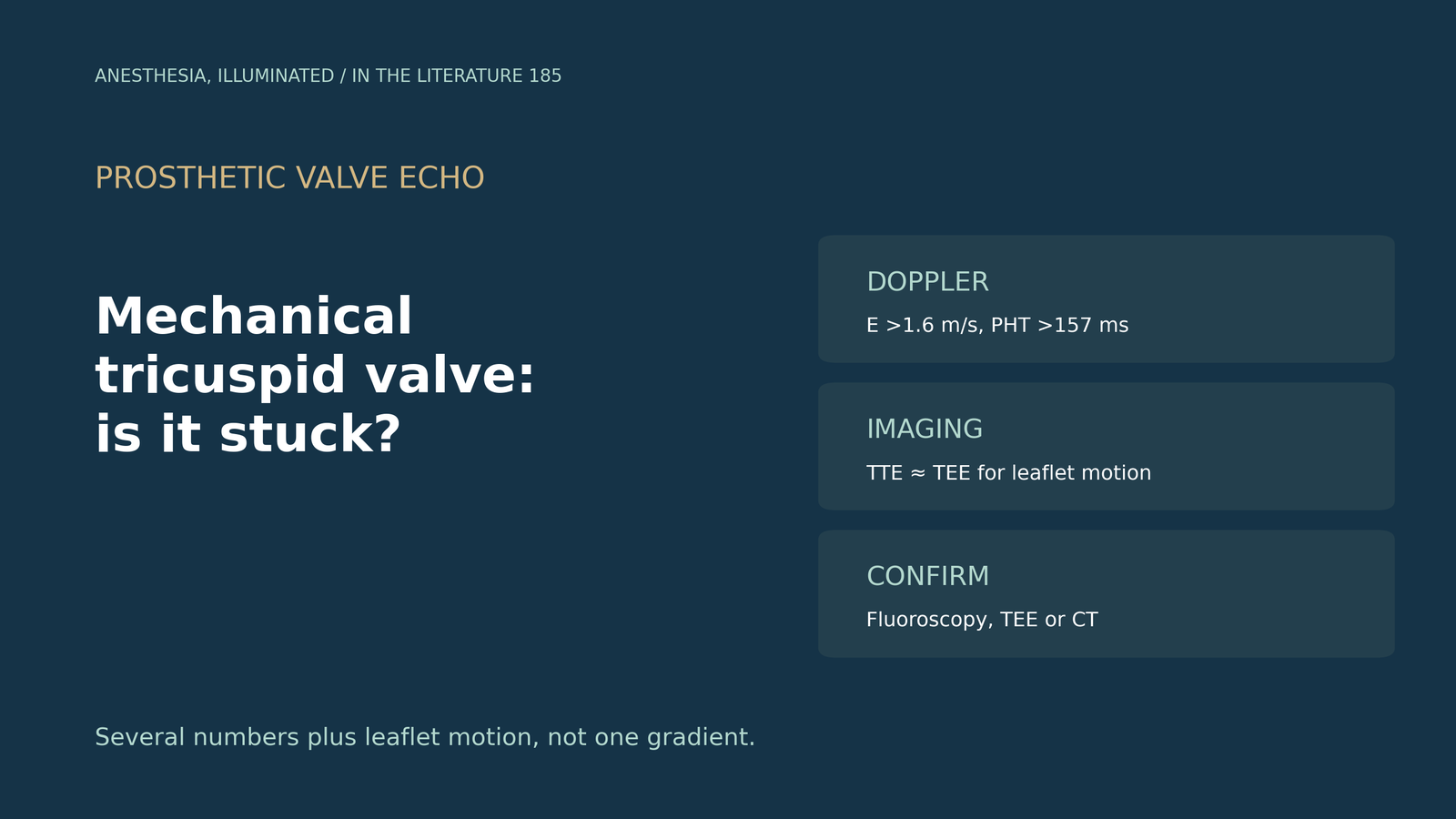
- Mechanical tricuspid valves are common and prone to thrombosis. They make up 39% to 54% of tricuspid prostheses, and thrombosis is more frequent in the tricuspid or mitral than the aortic position.[1], [2]
- TTE is the starting point. Transthoracic and transesophageal echo detected abnormal leaflet motion similarly (90% vs 88%), but TTE saw thrombus in only 14% of episodes.[1]
- Use several Doppler numbers. E velocity above 1.6 m/s, mean gradient above 5 mm Hg and pressure half-time above 157 ms identified dysfunction, with areas under the curve above 0.95.[1]
- Confirm leaflet motion. With suspected dysfunction, TEE, CT or fluoroscopy is recommended even if TTE is normal, and fluoroscopy or cine-CT should assess leaflet motion.[2]
- Read the whole picture. Rising gradients, increased regurgitation and CT attenuation help separate thrombus from pannus, and thrombosis can occur with a relatively low gradient.[3], [4]
A patient with a mechanical tricuspid prosthesis and a history of irregular anticoagulation presents with fatigue and leg swelling, and the gradient across the valve looks higher than before. Which echo findings indicate restricted leaflet motion, and how should it be confirmed?[1], [2]
A multiparametric echo approach
Fadel and colleagues compared 21 episodes of mechanical tricuspid leaflet dysfunction with 56 controls. Most dysfunction episodes involved both leaflets. TTE and TEE detected abnormal leaflet motion similarly (90% vs 88%), but TTE detected thrombus in only 14% of episodes, and flow propagation abnormalities on color Doppler were absent in all controls. Dysfunction cutoffs were E velocity above 1.6 m/s, mean gradient above 5 mm Hg and pressure half-time above 157 ms; pressure half-time and velocity time integral cutoffs of 157 ms and 42 cm performed similarly, with areas under the curve above 0.95. The half-time cutoff was lower than the guideline value (157 vs 230 ms), and the continuity method did not give an accurate effective orifice area. The authors call TTE the modality of choice for initial assessment.[1]
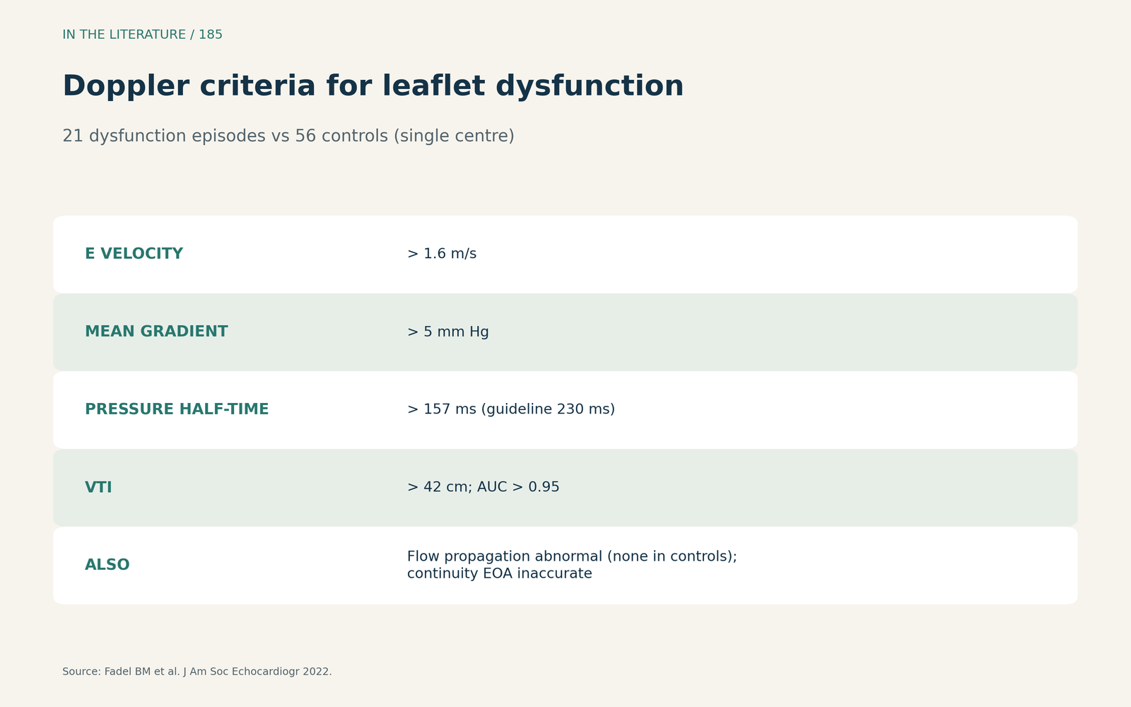
Figure 1. Several numbers, not one. Redrawn from Fadel et al.[1] View larger.

Confirming leaflet motion
The 2020 ACC/AHA guideline notes higher thrombosis rates for mechanical valves in the mitral or tricuspid position than in the aortic position. When prosthetic dysfunction is suspected clinically, TEE, gated CT or fluoroscopy is recommended even if TTE does not show it (Class 1), and in mechanical valve stenosis fluoroscopy or cine-CT is recommended to assess leaflet motion (Class 1). High gradients with normal leaflet motion and no thrombus point to prosthesis–patient mismatch or pannus.[2]
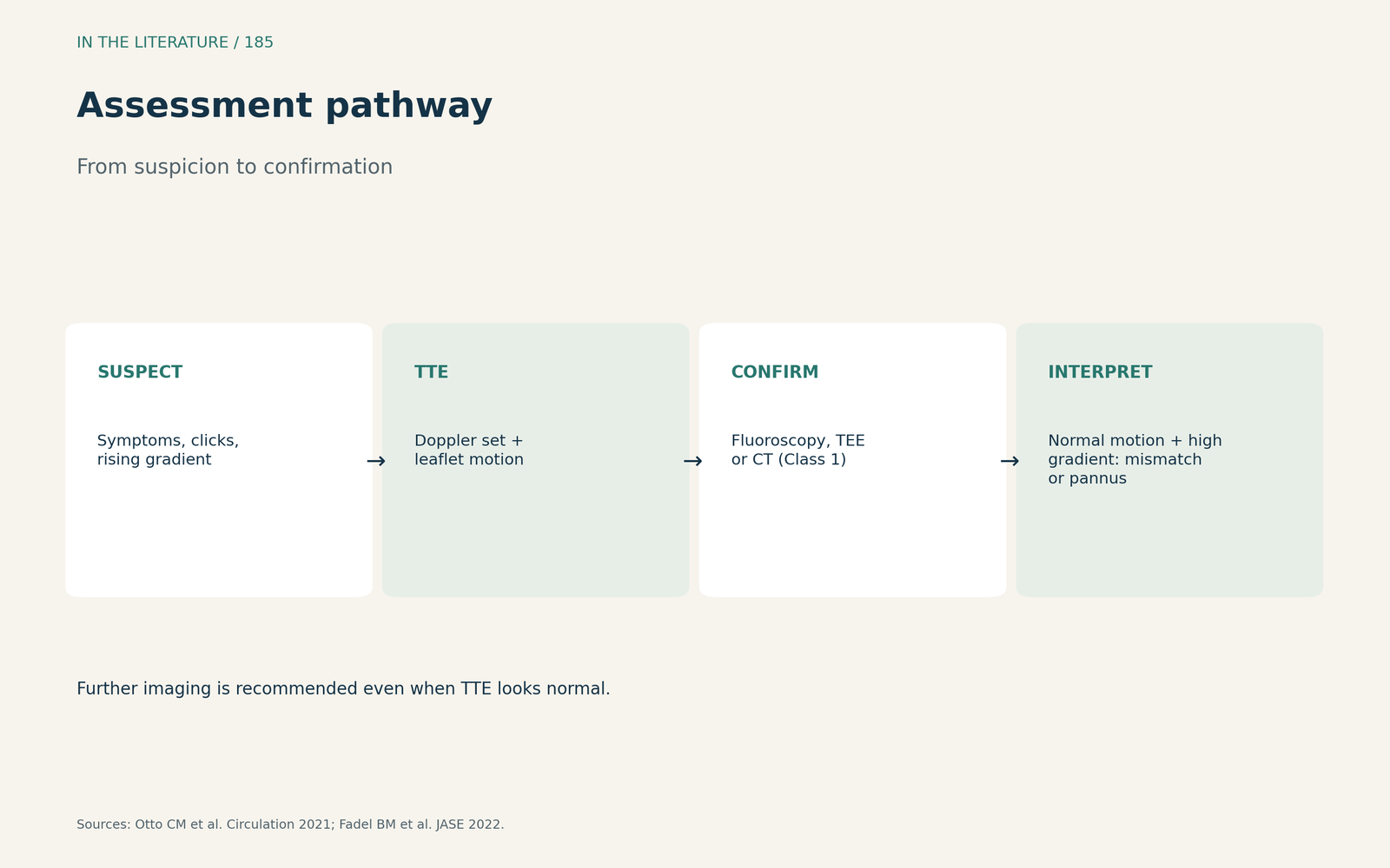
Figure 2. From suspicion to confirmation. Synthesized from Otto et al. and Fadel et al.[2], [1] View larger.

Lessons from recent cases
Afzal and colleagues describe a mechanical tricuspid valve whose gradients rose from 7 and 4 to 12 and 9 mm Hg (peak and mean). They cite tricuspid velocity above 1.9 m/s and mean gradient above 6 mm Hg as suggesting obstruction, use cine fluoroscopy to assess disc motion in real time, and note that CT attenuation above 145 Hounsfield units favours pannus; the patient received ultraslow t-PA, 25 mg over 25 hours.[3] In Shingu's series of right-sided mechanical valve thrombosis, increased transvalvular velocity on routine echo prompted suspicion, cinefluoroscopy showed reduced leaflet mobility in all cases, increased regurgitation was a clue, and one thrombosis was found despite a relatively low gradient.[4]
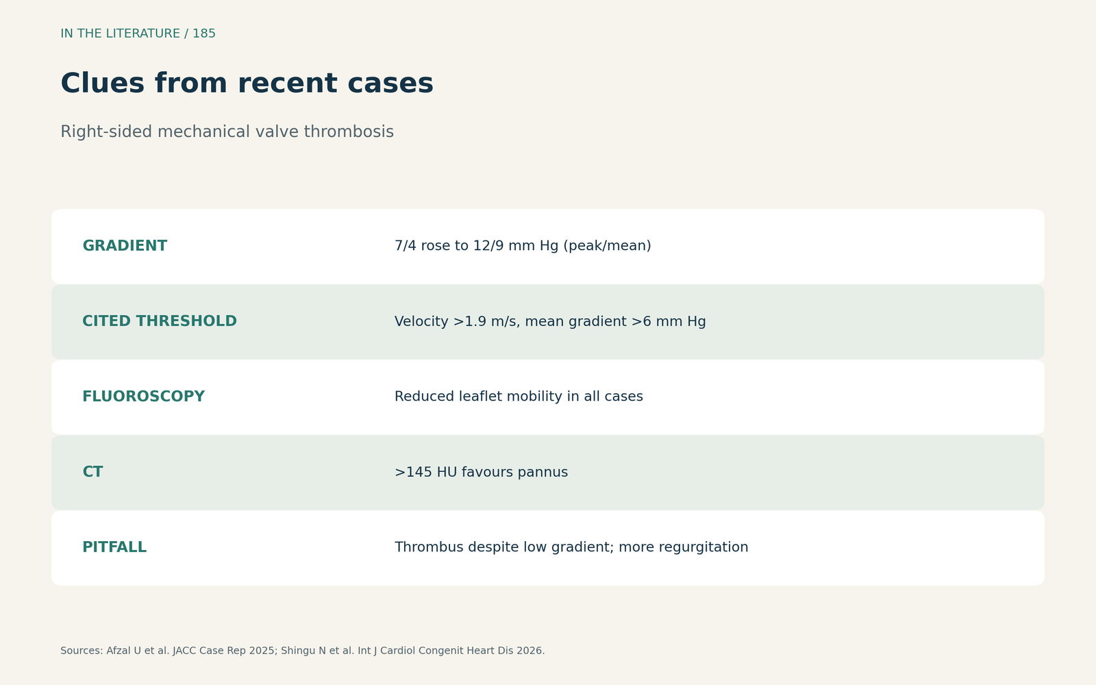
Figure 3. Clues beyond the gradient. Synthesized from Afzal et al. and Shingu et al.[3], [4] View larger.

Three takeaways
- Combine Doppler measures and compare with the patient's baseline.[1], [3]
- Confirm leaflet motion with fluoroscopy, TEE or CT when dysfunction is suspected, even if TTE looks normal.[2]
- A low gradient does not exclude thrombosis; watch regurgitation and clinical context.[4]
References
- Fadel BM, Alassas K, Clavel MA, et al. Multiparametric approach for the assessment of mechanical prosthetic tricuspid leaflet function. J Am Soc Echocardiogr. 2022;35(2):206–216. doi:10.1016/j.echo.2021.09.001
- Otto CM, Nishimura RA, Bonow RO, et al. 2020 ACC/AHA guideline for the management of patients with valvular heart disease: a report of the American College of Cardiology/American Heart Association Joint Committee on Clinical Practice Guidelines. Circulation. 2021;143(5):e72–e227. doi:10.1161/CIR.0000000000000923
- Afzal U, Lau J, Yap J, et al. Ultraslow thrombolysis of mechanical tricuspid prosthetic thrombosis. JACC Case Rep. 2025;30(19):104211. doi:10.1016/j.jaccas.2025.104211
- Shingu N, Suenaga T, Ishikita A, et al. Feasibility and safety of a standardized low-dose thrombolytic therapy for right-sided mechanical prosthetic valve thrombosis in adult congenital heart disease. Int J Cardiol Congenit Heart Dis. 2026;25:100707. doi:10.1016/j.ijcchd.2026.100707