
ITL-cbda3d1d415d
Juan Li, MD · Updated November 17, 2026
Watch the video

Key points
- Confirm the actual posterior leaflet attachment throughout the cardiac cycle; a single systolic gap can mislead.1,2
- “True” and “pseudo” describe proposed imaging patterns. They do not, by themselves, establish dangerous or benign disease.2,3
- When mitral valve prolapse (MVP) coexists, assess symptoms and rhythm: EHRA recommends a standard 24-hour Holter in patients with MVP.1
- MVP with unexplained syncope or nonsustained ventricular tachycardia (NSVT) warrants cardiac magnetic resonance (CMR) assessment under the EHRA consensus.1
- MAD alone does not establish an indication for an implantable cardioverter-defibrillator (ICD). Treatment requires the wider clinical and arrhythmic context.1
An echocardiography report identifies mitral annular disjunction (MAD). The next useful question is not simply how many millimeters were measured. It is whether the image identifies the actual leaflet attachment, and whether this patient has symptoms, ventricular arrhythmias or additional structural findings that change management. Separate those two questions before turning an imaging label into a treatment decision.1,2
First confirm what is separating
MAD describes separation between the mitral leaflet–atrial wall junction and the underlying ventricular myocardium. Follow the posterior leaflet to its true hinge point in a long-axis cine sequence, including diastole and systole. The EHRA consensus specifically calls for dynamic assessment throughout the cardiac cycle.1
Faletra and colleagues describe how redundant posterior leaflet tissue can lie against the atrial wall in systole, creating the appearance of disjunction even when the actual attachment is normally located. Their proposed “true-MAD” pattern has an atrially displaced attachment in both phases; “pseudo-MAD” reflects the misleading systolic appearance. This morphological proposal requires further validation, including its relationship to arrhythmic MVP.2
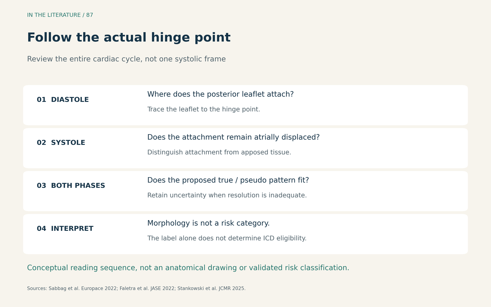
Figure 1. A conceptual image-reading sequence, not an anatomical drawing or validated risk classification. Inadequate resolution should prompt further assessment rather than a confident benign label. Sources: Sabbag et al. 2022; Faletra et al. 2022; Stankowski et al. 2025.1–3 Enlarge

The importance of definitions is visible in Stankowski and colleagues’ 2025 CMR study: among 290 clinically referred patients, 100 had study-defined true-MAD and 45 had pseudo-MAD. The study used a separation threshold of at least 1 mm across specified long-axis views. These counts are not general-population prevalence estimates. Interobserver reproducibility for extent was imperfect, with an intraclass correlation coefficient of 0.67. Crucially, there was no ventricular-arrhythmia analysis or clinical follow-up. The study helps explain imaging variability; it does not validate either label as a prognostic category.3
Then ask which clinical findings change the next step
For a patient with MVP, the EHRA consensus supports structured assessment with history, a 12-lead ECG, ambulatory rhythm monitoring and comprehensive echocardiography. Establish whether there has been unexplained syncope, document ventricular ectopy or NSVT, and assess MR, ventricular function and other relevant valve findings. MAD is one part of this assessment, not a substitute for it.1
The recommendation for a standard 24-hour Holter applies to MVP. It should not be silently expanded into a universal rule for every incidental isolated MAD finding. Longer monitoring may be useful when symptoms remain unexplained or the initial recording does not adequately characterize suspected arrhythmias; the choice depends on the clinical context.1
CMR becomes particularly useful when echocardiography is inadequate or when MVP accompanies unexplained syncope or NSVT. It can characterize ventricular structure and function, MAD and late gadolinium enhancement. The location of fibrosis matters: the consensus gives particular relevance to enhancement involving the mitral apparatus, such as the papillary muscles or periannular region. This is not a mandate to obtain CMR solely because a small gap was measured.1
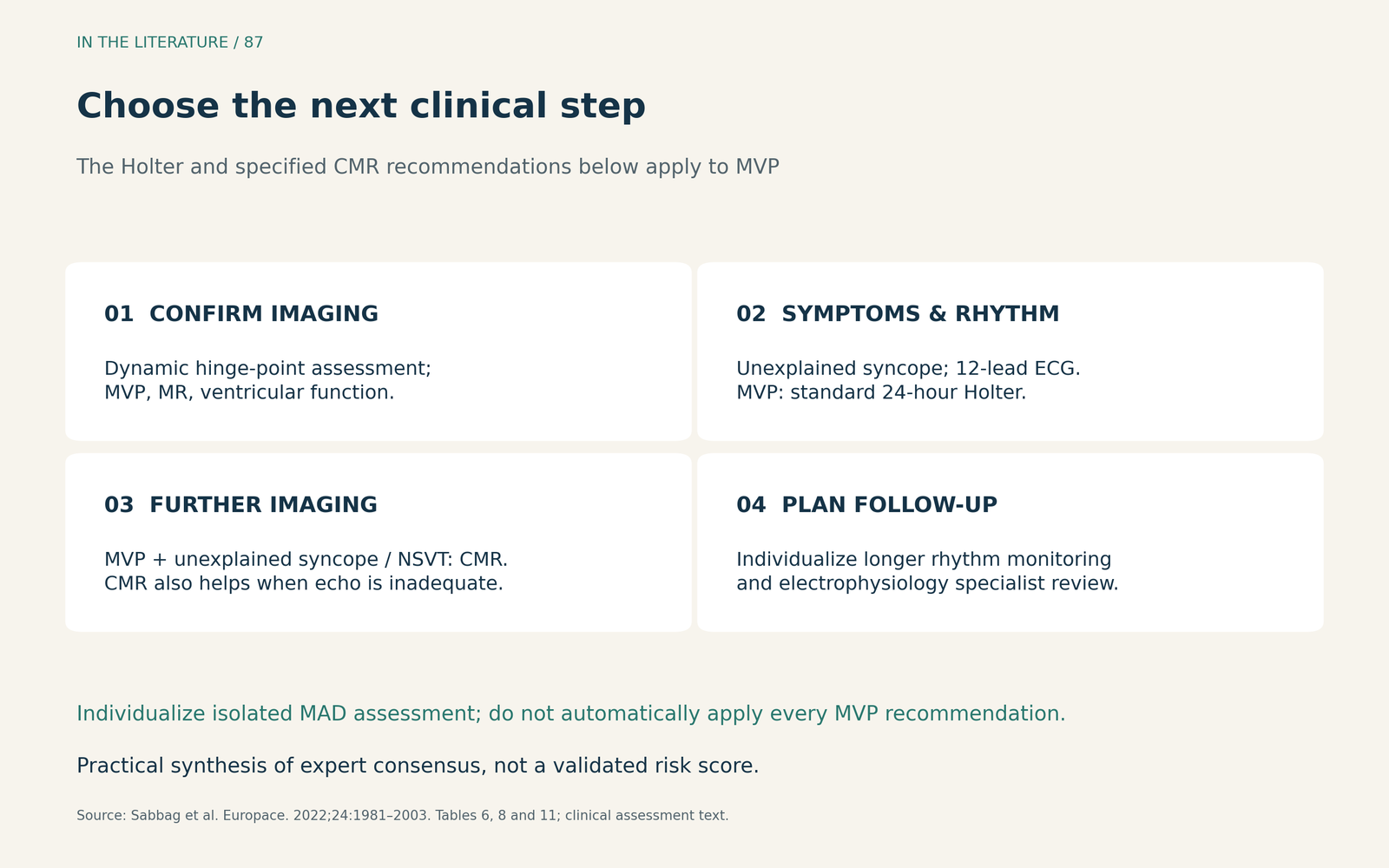
Figure 2. Practical synthesis of EHRA expert consensus. The Holter and specified CMR recommendations shown here are for patients with MVP; isolated MAD requires individualized assessment. This is not a validated risk score.1 Enlarge

What the newest monitoring study adds
Ezzeddine and colleagues’ 2026 retrospective report included 56 of 635 patients with imaging-detected MAD who were selected for an implantable loop recorder. Over a median 21.5 months, 12/56 had ventricular arrhythmias detected: 11 NSVT and one PVC-triggered ventricular fibrillation. Seven subsequently received an ICD, for heterogeneous clinical indications.4
These findings support considering prolonged surveillance in selected patients whose risk remains uncertain. They do not establish the event rate for all incidental MAD, or prove that loop recorders prevent sudden death. Device selection was discretionary, the cohort was small and there was no controlled comparison of monitoring strategies.4
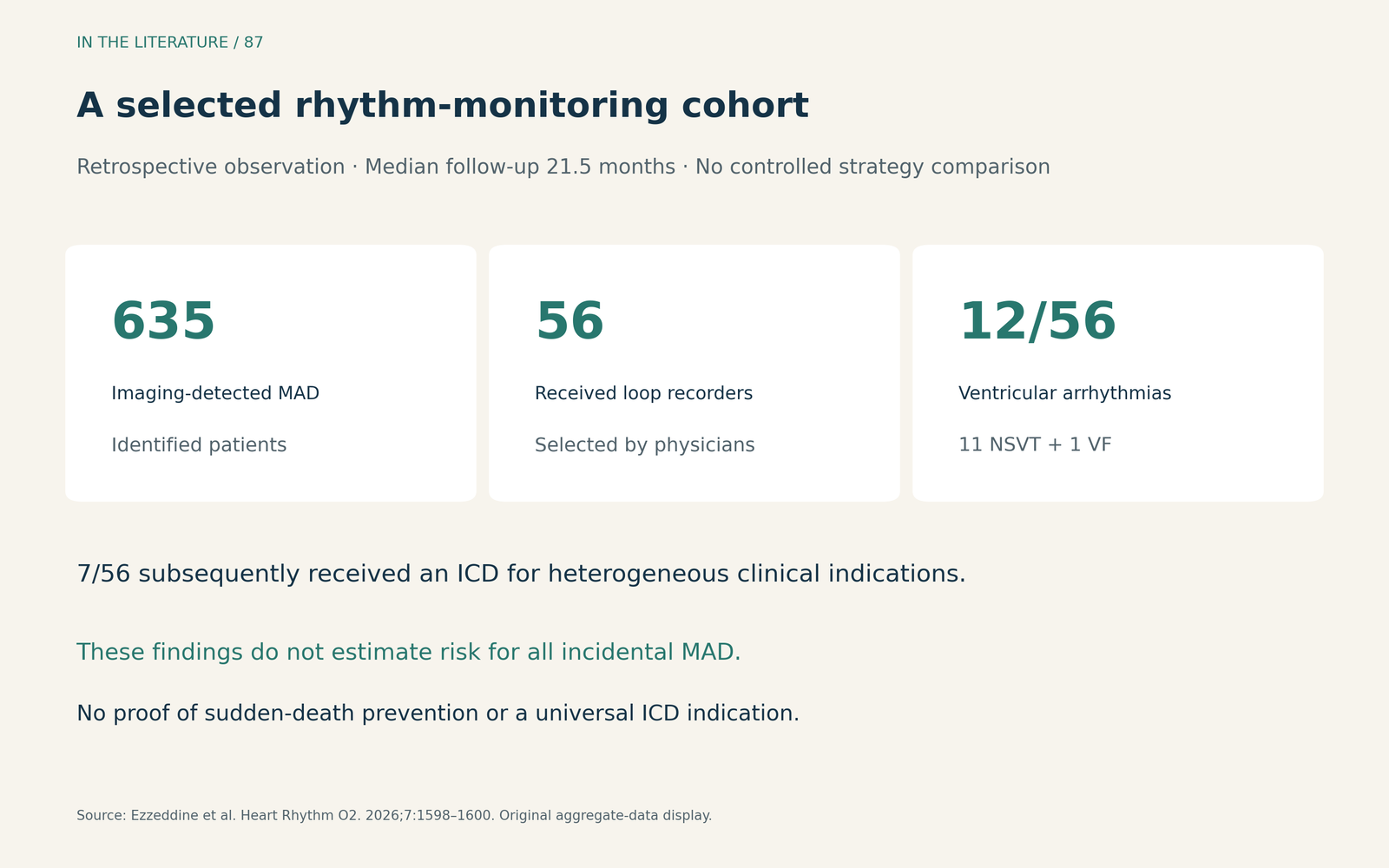
Figure 3. Original display of aggregate results from Ezzeddine et al., Heart Rhythm O2 2026. The monitored patients were selected; the remaining patients do not form a controlled comparison. ICD implantation was an observed management decision, not a universal MAD indication.4 Enlarge

Keep the ICD decision separate from the image label
Established primary- and secondary-prevention indications still apply. For arrhythmic MVP, EHRA also describes selected situations in which an ICD may be reasonable: one option combines a high-risk feature with at least two phenotypic risk features. MAD is only one phenotypic feature. This is expert consensus, and no randomized trial has demonstrated primary-prevention ICD benefit in a specific MVP subset.1
For the clinician receiving the echo report, the practical output is therefore a documented plan: confirm the dynamic finding; state whether MVP and important MR coexist; record symptoms and rhythm findings; and identify whether additional imaging, monitoring or electrophysiology assessment is needed. Avoid treating a distance measurement as an independent device indication.1
Three takeaways
- Confirm the hinge point dynamically. Review diastole and systole before accepting a MAD label.
- Assess the patient’s rhythm and structural context. In MVP, use the consensus-based Holter and CMR indications; do not extrapolate them indiscriminately to isolated MAD.
- Do not equate morphology with treatment. True/pseudo labels and MAD distance alone do not determine ICD eligibility; selected monitoring evidence requires careful interpretation.
References
- Sabbag A, Essayagh B, Barrera JDR, et al. EHRA expert consensus statement on arrhythmic mitral valve prolapse and mitral annular disjunction complex in collaboration with the ESC Council on valvular heart disease and the European Association of Cardiovascular Imaging endorsed by the Heart Rhythm Society, by the Asia Pacific Heart Rhythm Society, and by the Latin American Heart Rhythm Society. Europace. 2022;24(12):1981–2003. doi:10.1093/europace/euac125
- Faletra FF, Leo LA, Paiocchi VL, et al. Morphology of Mitral Annular Disjunction in Mitral Valve Prolapse. J Am Soc Echocardiogr. 2022;35(2):176–186. doi:10.1016/j.echo.2021.09.002
- Stankowski K, Catapano F, Donia D, et al. True- and pseudo-mitral annular disjunction in patients undergoing cardiovascular magnetic resonance. J Cardiovasc Magn Reson. 2025;27(1):101413. doi:10.1016/j.jocmr.2024.101413
- Ezzeddine FM, Zhang K, Siontis K, et al. Long-term continuous rhythm monitoring in patients with mitral annular disjunction. Heart Rhythm O2. 2026;7(8):1598–1600. doi:10.1016/j.hroo.2026.04.015