
ITL-a1106bea1448
Juan Li, MD · Updated November 17, 2026
Watch the video

Key points
- Identify the viewpoint and anatomical landmarks before assigning a leaflet segment; rotating an image changes its screen position, not its identity.1,2
- Combine systematic multiplane imaging with targeted orthogonal views and three-dimensional assessment to establish the location and extent of disease.1,2
- Deliberately inspect both commissures: a conspicuous central lesion does not establish that the rest of the valve is normal.1,3
- Use regurgitant jet origin and direction to corroborate morphology; jet direction alone cannot reliably name a scallop.1,2
- Give the repair team a description of the affected regions, mechanism and associated lesions, rather than only a label such as “posterior leaflet prolapse.”1
An eccentric mitral regurgitant jet makes the abnormality obvious. The harder question is whether the lesion is confined to a posterior scallop, extends toward a commissure, or includes another leaflet region. That distinction is what the surgeon needs before planning a repair. The practical goal of transesophageal echocardiography (TEE) is therefore a reproducible anatomical description, supported by more than one view.1
Establish orientation before naming a segment
In Carpentier nomenclature, the posterior leaflet has P1, P2 and P3 scallops; the anterior leaflet has corresponding A1, A2 and A3 segments, rather than three anatomically separate scallops. P1 is adjacent to the anterolateral commissure and left atrial appendage; P3 is toward the posteromedial commissure, with P2 between them.2
The ASE/SCA/STS surgery-based TEE guideline by Nicoara and colleagues recommends orienting the three-dimensional view with the aortic valve at twelve o’clock. The left atrial appendage identifies the lateral commissural side, while the interatrial septum identifies the medial side. State whether the valve is viewed from the atrium or ventricle: “screen left” is not a reliable anatomical label when the volume has been rotated or the viewing perspective changed.1
A left atrial en-face view is useful for communicating with the surgeon, but retain the landmarks that make its orientation intelligible. A narrowly cropped volume may display the lesion clearly while losing the structures needed to name it correctly.1,2
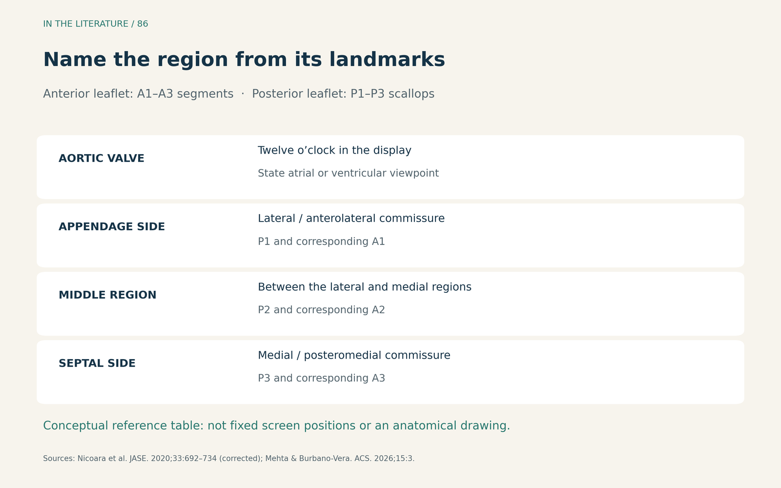
Figure 1. Name the region from its anatomical relationships. The anterior regions are segments; the posterior regions are scallops. The table does not prescribe fixed screen positions. Sources: Nicoara et al., JASE 2020; Mehta and Burbano-Vera, Annals of Cardiothoracic Surgery 2026.1,2 Enlarge

Confirm the lesion across the coaptation line
Start with a comprehensive examination, then interrogate the suspected region. In simultaneous biplane imaging, place the cursor over the structure of interest in the reference view and inspect the orthogonal plane. The point is to confirm what the imaging plane actually crosses, not to assume that a familiar probe angle always displays the same segment.1
The commissural-biplane approach uses a true bicommissural view, then sweeps the orthogonal plane through the lateral, middle and medial regions. Grayscale and color images are complementary. This is an addition to the conventional multiplane examination, not a reason to abandon it.3
Three-dimensional multiplanar reconstruction (MPR) extends that approach. Mehta and Burbano-Vera’s 2026 technical review describes moving the planes along the systolic coaptation line to identify the location and extent of prolapse or flail. An en-face impression can then be checked against a correctly aligned tomographic plane. These are practical imaging methods; the review does not establish a new diagnostic accuracy percentage or prove improved repair outcomes.2
Do not stop when P2 looks abnormal. In Abazid and colleagues’ study, P2 was involved in 63 of 102 surgical patients, but other posterior regions, anterior regions and commissures were also affected. Those segment counts overlap; they are not separate groups of patients.3
Why the commissures deserve a separate check
Abazid and colleagues prospectively studied 102 patients undergoing mitral surgery. The cohort was predominantly primary MR, but was not restricted to degenerative prolapse. They compared conventional multiplane imaging with a commissural-biplane approach. Three-dimensional TEE was the main reference, with surgical findings providing complementary information when images were nondiagnostic or when evaluating clefts or perforations.3
Among the ten patients with commissural abnormalities, the commissural-biplane approach identified 8/10, compared with 3/10 using conventional multiplane imaging. That is a useful reason to deliberately add a commissural sweep. It is not evidence that a single approach is sufficient for every valve.3
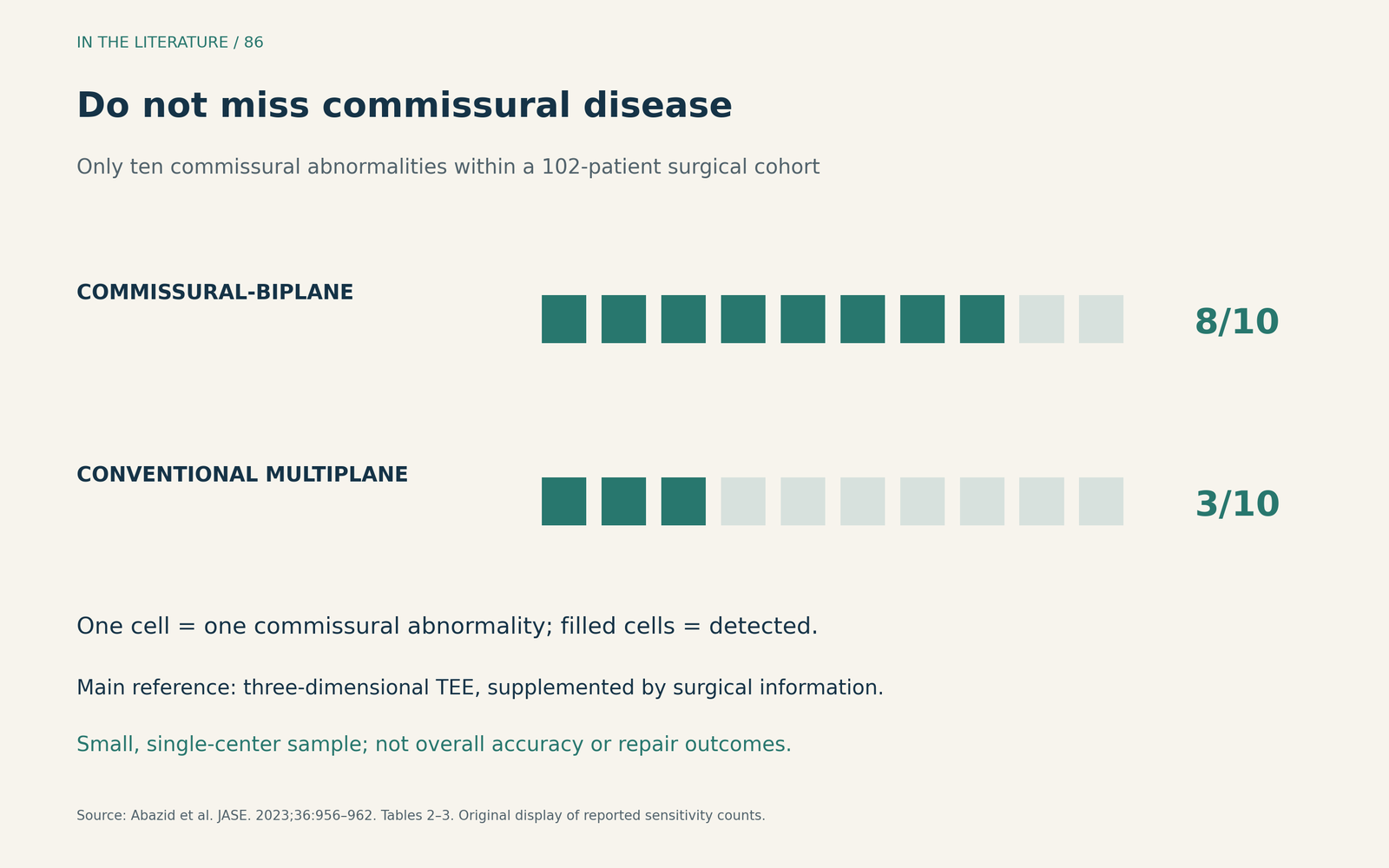
Figure 2. Reported detection among ten patients with commissural abnormalities, within a 102-patient surgical cohort. The reference was mainly three-dimensional TEE, supplemented by surgical information. These are sensitivity counts, not overall accuracy or repair outcomes. Source: Abazid et al., JASE 2023, Tables 2–3.3 Enlarge

The denominator is small, the study was performed at one center, and the authors acknowledge the absence of a clear definition of commissural prolapse. Some published accuracy and specificity entries also contain arithmetic inconsistencies; the detection counts above agree with the reported sensitivities. The clinically defensible conclusion is complementary imaging, not a universal performance guarantee.3
Use the jet as a clue, then report the anatomy
Regurgitant flow typically points away from a region with excessive leaflet motion. Nevertheless, an anteriorly directed jet does not, by itself, prove P2 prolapse. The 2026 review describes P1 lesions that can direct flow posteriorly, and mixed restricted and excessive motion can change the dominant jet direction. Follow the jet back to its origin and match it to the directly imaged lesion.1,2
For the repair discussion, describe the affected scallops or segments, whether the abnormal motion is prolapse or flail, how far it extends, and whether a commissure or additional region is involved. Include the subvalvular abnormalities and leaflet thickening or calcification that may matter to the repair. Inspect suspected clefts in diastole and correlate them with morphology and flow; an ordinary posterior indentation should not automatically acquire a pathological label.1,2
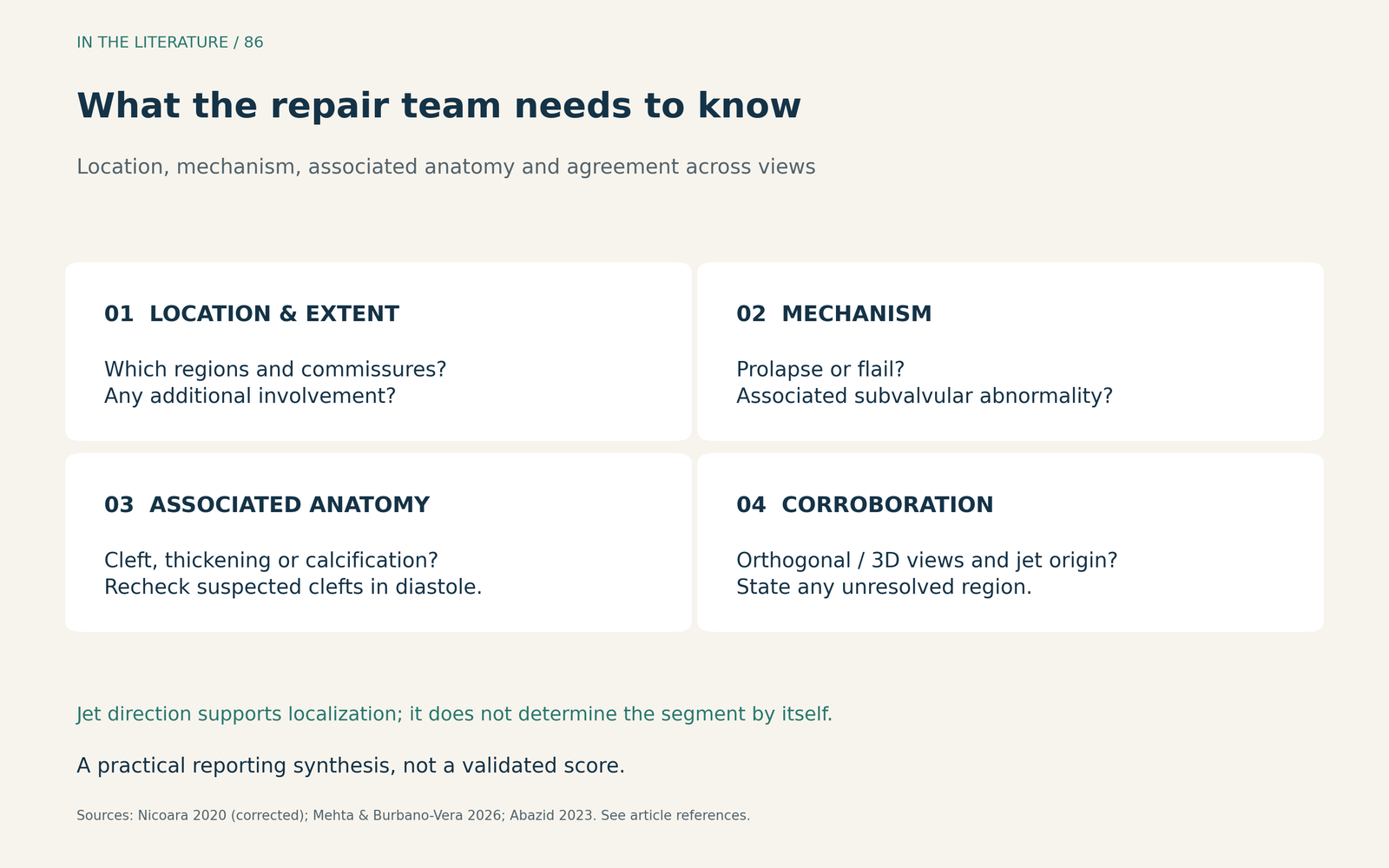
Figure 3. A practical reporting synthesis from the cited imaging guidance, not a validated score. Identify what is established and what remains uncertain; reconcile disagreement with targeted views and the surgical team. Sources: Nicoara et al. 2020; Mehta and Burbano-Vera 2026; Abazid et al. 2023.1–3 Enlarge

If two views disagree, the useful next step is to revisit orientation, plane alignment and volume quality before committing to a segment label. Communicate the unresolved region explicitly. “Localization remains uncertain near the commissure” is more actionable than a confident label unsupported by the images. This is a practical application of comprehensive imaging and team communication, rather than a separate diagnostic rule.1,2
Three takeaways
- Orient before you label. Use the aortic valve, left atrial appendage and interatrial septum, and specify the viewing perspective.
- Map beyond the obvious lesion. Confirm the region in orthogonal or reconstructed planes and deliberately examine the commissures and remaining leaflet regions.
- Report a repair-relevant lesion description. Combine location, extent, abnormal motion and associated anatomy; use jet direction as supporting evidence and state residual uncertainty.
References
- Nicoara A, Skubas N, Ad N, et al. Guidelines for the Use of Transesophageal Echocardiography to Assist with Surgical Decision-Making in the Operating Room: A Surgery-Based Approach: From the American Society of Echocardiography in Collaboration with the Society of Cardiovascular Anesthesiologists and the Society of Thoracic Surgeons. J Am Soc Echocardiogr. 2020;33(6):692–734. Correction: 2020;33(11):1426. doi:10.1016/j.echo.2020.03.002 Correction
- Mehta AR, Burbano-Vera N. Intraoperative transesophageal echocardiography: the roadmap for successful mitral valve repair. Ann Cardiothorac Surg. 2026;15(1):3. doi:10.21037/acs-2025-dmv-10
- Abazid RM, Frost A, Manian U, et al. Diagnostic Accuracy of Transesophageal Echocardiographic Commissural-Biplane Approach in Identifying Mitral Valve Anatomy. J Am Soc Echocardiogr. 2023;36(9):956–962. doi:10.1016/j.echo.2023.04.008