ITL-9aabc50ee93e
Juan Li, MD · Updated November 18, 2026
Watch the video
Key points
- TEE defines the lesion. Intraoperative TEE establishes the anatomic basis of primary MR, and 3D TEE better identifies leaflet pathology and the affected scallop.[1], [2], [3]
- Look for failure-prone anatomy. SAM risk markers include a basal septum above 15 mm, an anterior-to-posterior leaflet ratio below 1.3 and C-sept below 2.5 cm; tenting marks chronic secondary MR.[1], [3]
- Durability drives the decision. Early repair is reasonable when the chance of durable success exceeds 95% with mortality below 1%, while rheumatic repairs carry 50% to 60% reoperation at 20 years.[2]
- Check the result under real loading. A successful repair should leave no more than mild MR at normal filling pressure, and a mean gradient above 6 mm Hg suggests iatrogenic stenosis.[1], [3]
- Repair is worth the effort. Twenty-year survival was 46% after repair versus 23% after replacement, and converting a failed repair did not worsen long-term mortality in one cohort.[4], [5]
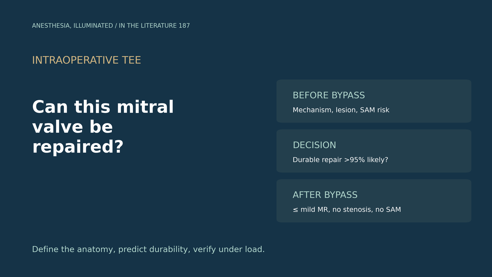
Before planned surgery for degenerative mitral regurgitation, the anaesthesiologist performs the pre-bypass TEE while the surgeon asks: "Is this valve repairable, and what should I watch for?" Which TEE findings support repair, and how is the repair judged afterward?[1], [2]
Before bypass: anatomy and failure-prone features
The ASE/SCA/STS guideline states that TEE is best suited to describe mitral anatomy in detail and that SAM risk should be assessed before repair; a basal septum thicker than 15 mm independently predicts SAM. In secondary MR, tenting serves as a surrogate of chronicity, and functional MR may appear less severe under general anaesthesia.[1] The 2020 ACC/AHA guideline gives intraoperative TEE the role of establishing the anatomic basis of primary MR, with the final repair-or-replace decision made in the operating room. Early repair is reasonable when durable success exceeds 95% with mortality below 1%, whereas rheumatic repair carries reoperation rates of 50% to 60% at 20 years.[2] Neema and Nagarjuna add that 3D TEE better identifies leaflet pathology and the affected scallop, and list SAM risk markers including an AML/PML ratio below 1.3 and C-sept below 2.5 cm; SAM occurs in 1% to 16% of repairs.[3]
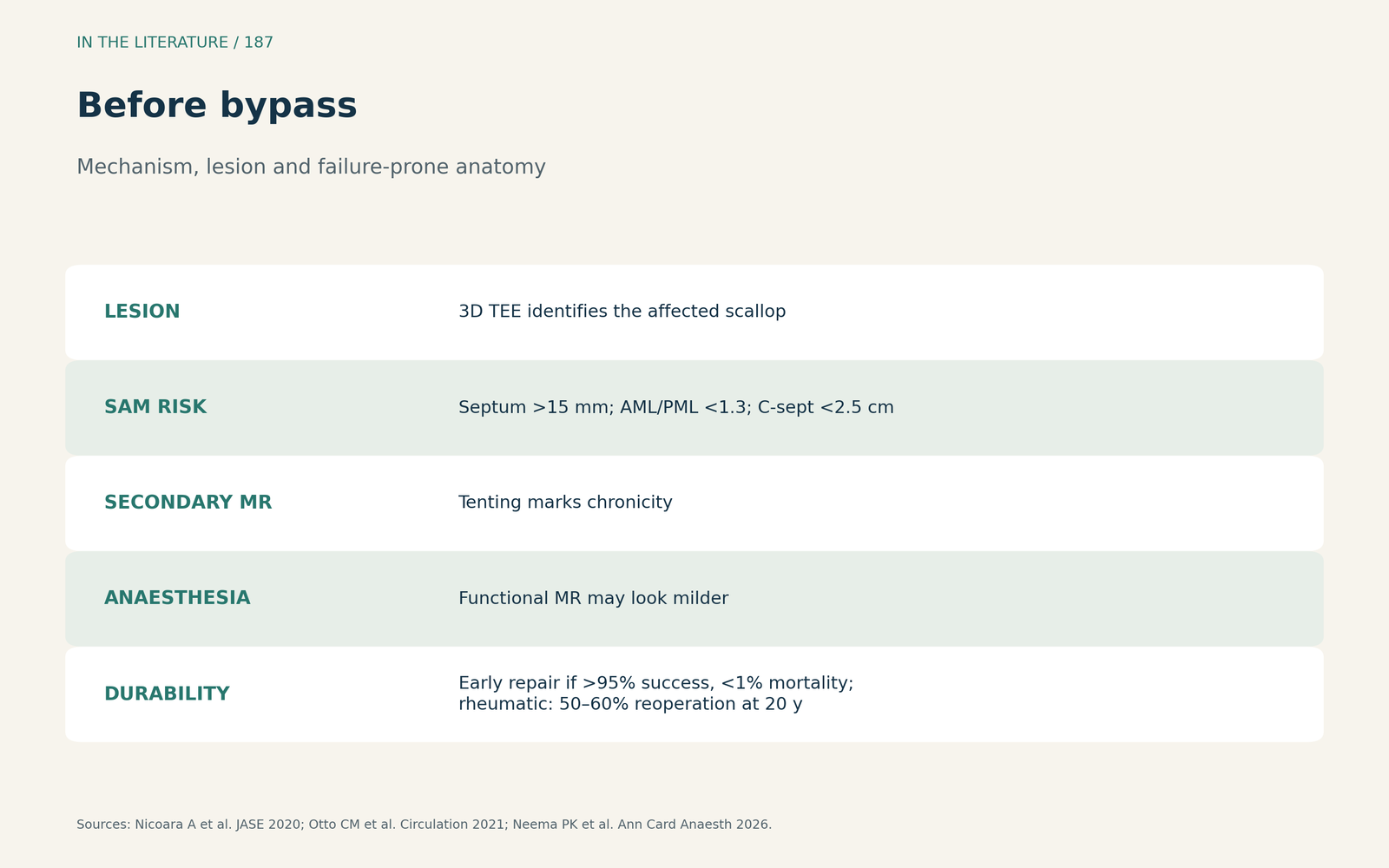
Figure 1. Mechanism, lesion and failure-prone anatomy. Synthesized from Nicoara et al., Otto et al. and Neema et al.[1], [2], [3] View larger.

After bypass: judging the repair
A successful repair should leave no more than mild MR, and greater than mild MR should prompt further investigation; iatrogenic stenosis criteria include a mean gradient above 6 mm Hg.[1] The ACC/AHA guideline notes that more than trivial MR after repair usually leads to revision.[2] Residual MR should be checked at normal filling pressure, and coaptation height should be 5 to 8 mm at A2-P2.[3]
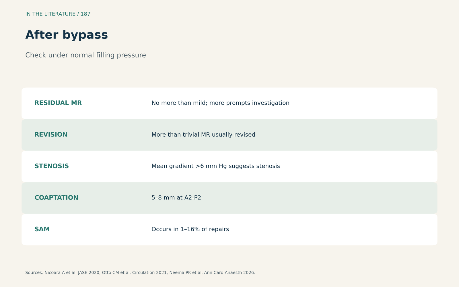
Figure 2. Leak, gradient, SAM. Synthesized from Nicoara et al., Otto et al. and Neema et al.[1], [2], [3] View larger.

Why repairability matters
In the MIDA registry of flail degenerative MR, 20-year survival was 46% after repair versus 23% after replacement, and 20-year freedom from 3+ MR after repair was 88%; the authors still regard a low likelihood of durable repair as a legitimate reason to replace.[4] Ogami and colleagues studied 133 patients (8%) in whom repair was attempted and converted to replacement; a failed repair may require another cross-clamp and arrest, yet conversion was not associated with long-term mortality, and the authors favour attempting repair in borderline repairable anatomy.[5] In secondary disease the calculus differs: in severe ischaemic MR, the CTSN trial (Goldstein et al., N Engl J Med 2016) randomized 251 patients to repair or replacement, and recurrence over 2 years was higher with repair; echocardiographic measures such as tenting area and coaptation distance predict recurrence after restrictive annuloplasty.[6]
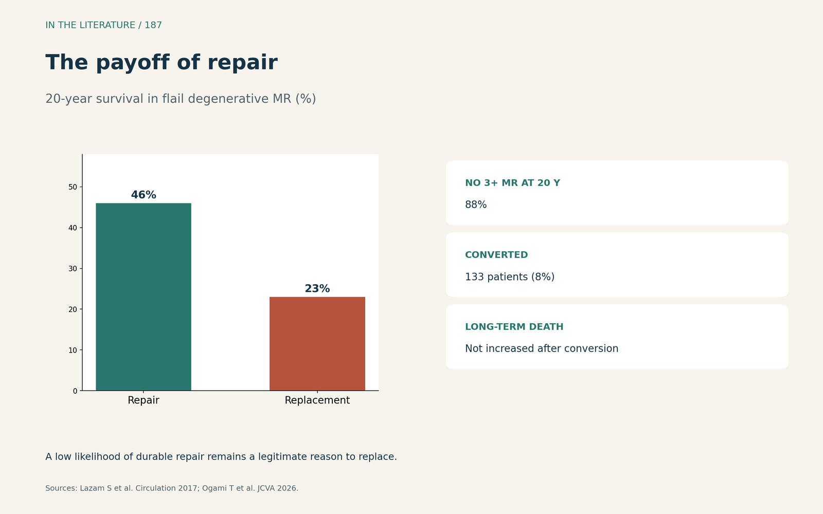
Figure 3. The payoff of a durable repair. Redrawn from Lazam et al. and Ogami et al.[4], [5] View larger.

Three takeaways
- Describe the mechanism, the lesion and the failure-prone anatomy before bypass.[1], [3]
- Judge repair under normal loading: no more than mild MR, no stenosis, no SAM.[1], [2]
- A durable repair is worth pursuing, but repairability thresholds remain expert consensus.[2], [4], [5]
References
- Nicoara A, Skubas N, Ad N, et al. Guidelines for the use of transesophageal echocardiography to assist with surgical decision-making in the operating room: a surgery-based approach. J Am Soc Echocardiogr. 2020;33(6):692–734. doi:10.1016/j.echo.2020.03.002
- Otto CM, Nishimura RA, Bonow RO, et al. 2020 ACC/AHA guideline for the management of patients with valvular heart disease: a report of the American College of Cardiology/American Heart Association Joint Committee on Clinical Practice Guidelines. Circulation. 2021;143(5):e72–e227. doi:10.1161/CIR.0000000000000923
- Neema PK, Nagarjuna P. A comprehensive echocardiographic guide for mitral valve repair: a narrative review. Ann Card Anaesth. 2026;29(1):22–34. doi:10.4103/aca.aca_184_25
- Lazam S, Vanoverschelde JL, Tribouilloy C, et al. Twenty-year outcome after mitral repair versus replacement for severe degenerative mitral regurgitation: analysis of a large, prospective, multicenter, international registry. Circulation. 2017;135(5):410–422. doi:10.1161/CIRCULATIONAHA.116.023340
- Ogami T, Chetkof E, Bonatti JO, et al. Outcomes of patients undergoing mitral valve replacement after failed attempt at mitral valve repair. J Cardiothorac Vasc Anesth. Published online 2026. doi:10.1053/j.jvca.2026.08.146
- Goldstein D, Moskowitz AJ, Gelijns AC, et al. Two-year outcomes of surgical treatment of severe ischemic mitral regurgitation. N Engl J Med. 2016;374(4):344–353. doi:10.1056/NEJMoa1512913