
ITL-e8b58fa96c68
Juan Li, MD · Updated November 17, 2026
Watch the video

Key points
- In rheumatic stenosis, well-aligned planimetry is the preferred anatomical measurement; three-dimensional multiplanar imaging can help find the correct orifice plane. 1
- Calcific stenosis requires a different approach: acoustic shadowing and complex obstruction make planimetry less reliable. 2
- Pressure half-time (PHT) reflects pressure equalization as well as valve narrowing; abnormal chamber compliance can undermine its agreement with anatomical area. 1, 3
- A continuity-equation area is credible only when its flow assumptions and component measurements are credible. 1
- CT can supplement difficult echocardiography, but recent correlation data do not establish interchangeable measurements or better clinical outcomes. 4
A mitral valve looks tight, yet PHT suggests a substantially larger opening than planimetry. Choosing the smaller number may overcall obstruction; choosing the larger may dismiss it. The useful question is which measurement is valid in this valve, at this heart rate, under these loading conditions?
First identify where the obstruction is
Rheumatic disease usually combines commissural fusion with leaflet-tip restriction. For planimetry, identify the smallest opening at the leaflet tips, align the short-axis plane, and adjust gain before tracing. A plane above the tips or an oblique cut can enlarge the apparent orifice; excessive gain can shrink it. Three-dimensional multiplanar reconstruction helps correct alignment, but still requires a usable dataset. 1
Mitral annular calcification (MAC) is a different substrate. Calcium can extend into the leaflet bases and subvalvular apparatus, while shadowing obscures borders. The 2025 ESC/EACTS guideline specifically cautions that planimetry is less reliable here and supports liberal use of transesophageal echocardiography (TEE). Do not transfer the rheumatic leaflet-tip technique automatically to an irregular calcific channel. 1, 2
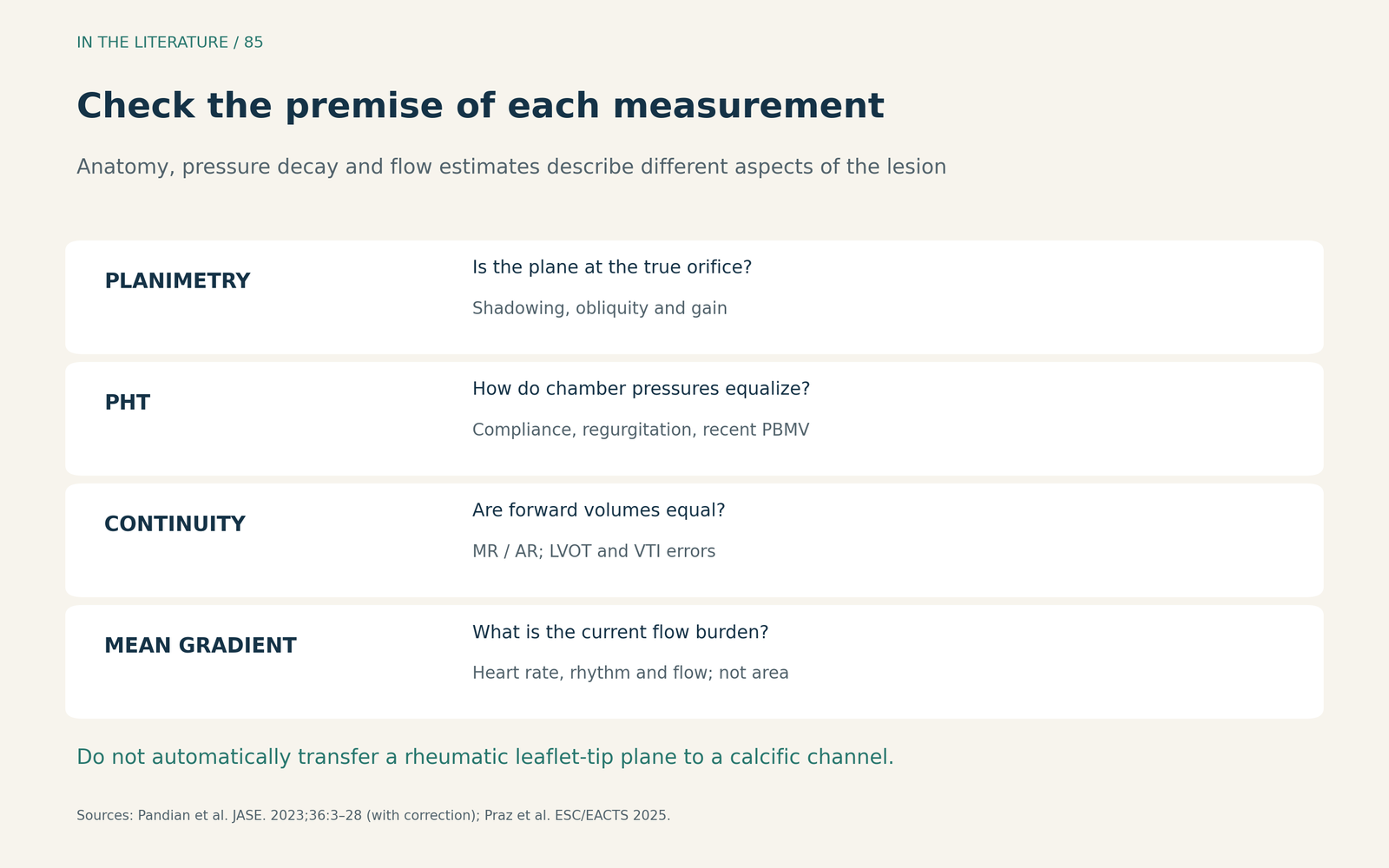
Figure 1. A practical comparison of measurements. PHT and continuity-equation estimates depend on different assumptions; the mean gradient describes hemodynamic burden rather than area. Adapted as an original clinical summary from ASE 2023, with its correction, and ESC/EACTS 2025. 1, 2

Ask what the Doppler estimate is measuring
The familiar equation is MVA = 220/PHT, with area in cm² and PHT in milliseconds. A longer PHT produces a smaller calculated area. Its physiological premise can fail when atrial or ventricular compliance changes, with associated regurgitation, or immediately after balloon commissurotomy. In a biphasic deceleration envelope, the early steep segment is not the appropriate slope. 1
Li and colleagues studied 244 patients with rheumatic stenosis: 110 had both TTE and three-dimensional TEE measurements, while 134 formed a TTE-only validation cohort. In the TEE comparison, PHT-derived area agreed less well with three-dimensional area when net atrioventricular compliance was low. Compliance was estimated noninvasively, and TTE and TEE were not necessarily simultaneous. This supports investigating a mismatch rather than letting PHT overrule credible anatomy. The study does not validate a rule for MAC-related stenosis. 3
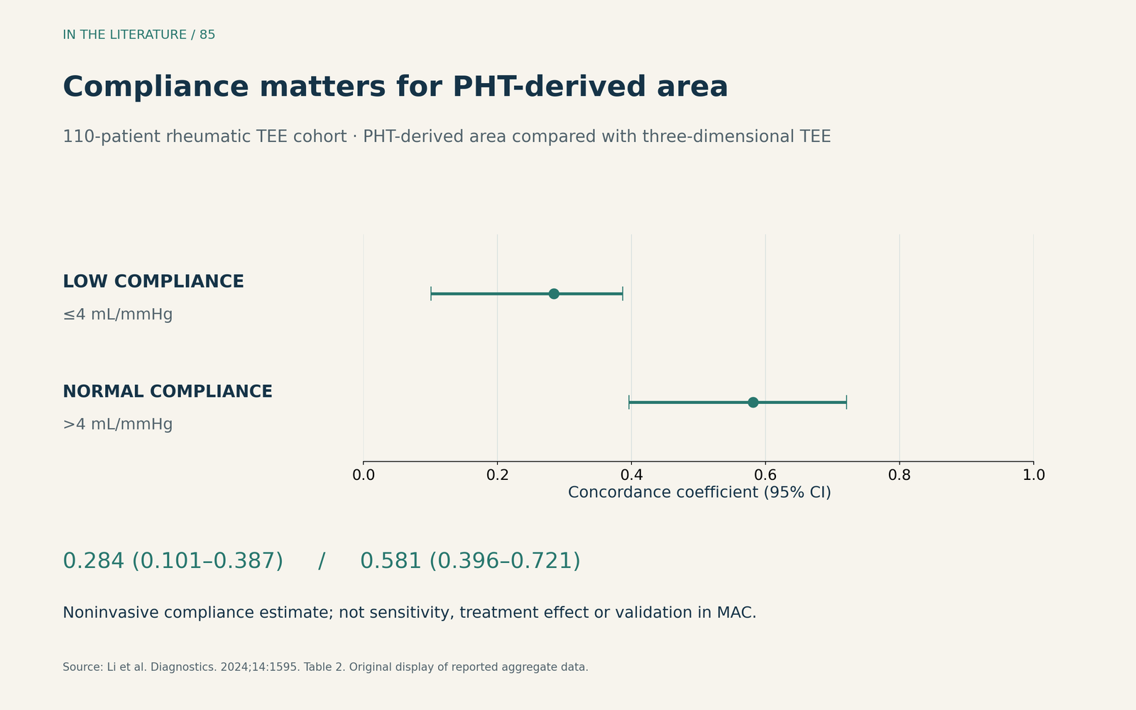
Figure 2. Reported concordance coefficients for PHT-derived versus three-dimensional TEE area: 0.284 (95% CI 0.101–0.387) with low compliance and 0.581 (0.396–0.721) with normal compliance. The comparison comes from the 110-patient TEE cohort. These are agreement statistics, not sensitivity, clinical event rates or treatment effects. Low compliance was defined as ≤4 mL/mmHg. Original display of Table 2 data. 3

The continuity equation divides forward stroke volume by mitral inflow VTI. Significant MR or AR breaks the simple equality of flow used with the LV outflow tract. Errors in outflow-tract measurement also propagate into the result. It can provide a useful cross-check when its assumptions hold, rather than an automatic tie-breaker. 1
Use the gradient to understand the consequences
A high mean gradient can reflect fast heart rate or increased flow as well as obstruction; a low gradient alone does not establish a mild lesion. Record heart rate and rhythm, assess associated regurgitation, and interpret pulmonary pressure and chamber findings alongside area. 1, 2
For rheumatic disease, ESC/EACTS 2025 links an area ≤1.5 cm² to clinically severe stenosis in conjunction with clinical factors. The number should not become a universal treatment trigger. If symptoms remain discordant with resting findings, exercise echocardiography can show the accompanying changes in gradient and pulmonary pressure. In MAC-related disease, the mean gradient remains informative even when anatomical measurements are difficult. 2
When another image can change the interpretation
Steinhoff and colleagues' 2026 retrospective study assessed CT planimetry in 85 patients: 72 had native valves and 13 had bioprostheses or previous repair. Echo planimetry was available in only 41. CT and echo area were moderately correlated; excellent CT interobserver agreement came from duplicate readings in just 23 patients. Reproducibility does not establish diagnostic truth, and this mixed cohort does not make CT a universal replacement for echo. 4
The practical opportunity is targeted: when echo remains inconclusive, a suitable gated CT—particularly one already obtained for procedural planning—may clarify the geometry. CT also characterizes the distribution of calcium when intervention for MAC-related disease is being considered. 2, 4
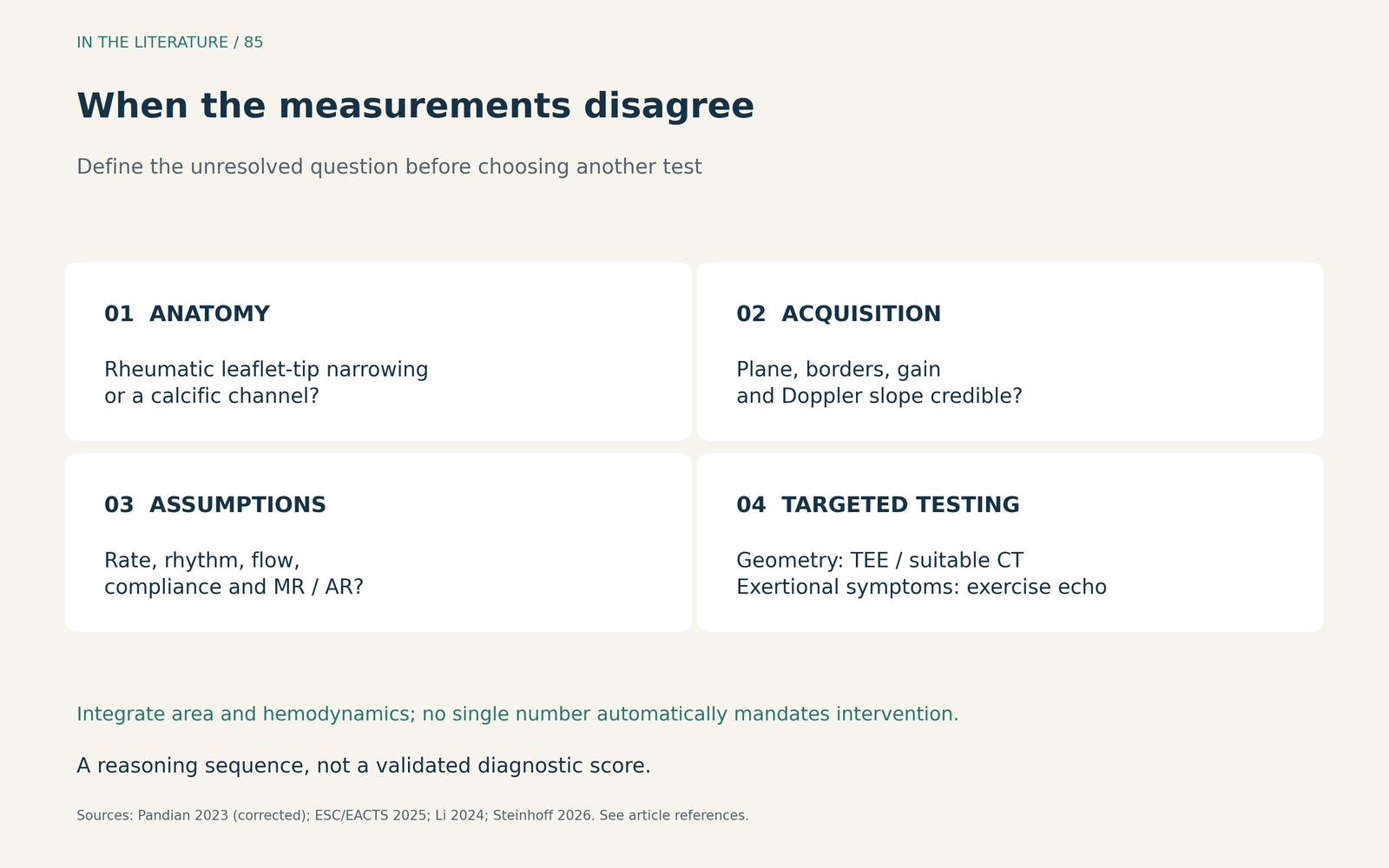
Figure 3. A clinical reasoning sequence, not a validated diagnostic score. Additional testing should answer the unresolved question: geometry, measurement validity or exertional hemodynamics. 1–4

Three takeaways
- Start with the lesion. Rheumatic leaflet-tip obstruction and calcific annular disease require different interpretation of planimetry.
- Explain disagreement before selecting a number. Recheck the plane, Doppler slope, flow assumptions, heart rate and associated regurgitation.
- Report an integrated conclusion. State the most credible area estimate, the hemodynamic burden and the specific uncertainty that additional imaging or exercise testing could resolve.
References
- Pandian NG, Kim JK, Arias-Godinez JA, et al. Recommendations for the Use of Echocardiography in the Evaluation of Rheumatic Heart Disease: A Report from the American Society of Echocardiography. J Am Soc Echocardiogr. 2023;36(1):3–28. Correction: 2023;36(4):445. doi:10.1016/j.echo.2022.10.009 Correction
- Praz F, Borger MA, Lanz J, et al. 2025 ESC/EACTS Guidelines for the management of valvular heart disease. Eur J Cardiothorac Surg. 2025;67(8):ezaf276. doi:10.1093/ejcts/ezaf276
- Li T, Leow R, Chan MW, et al. Impact of Net Atrioventricular Compliance on Mitral Valve Area Assessment—A Perspective Considering Three-Dimensional Mitral Valve Area by Transesophageal Echocardiography. Diagnostics (Basel). 2024;14(15):1595. doi:10.3390/diagnostics14151595
- Steinhoff P, Schmitt R, Hazard D, et al. Diagnostic performance of computed tomographic angiography-based planimetry for the assessment of mitral stenosis. Int J Cardiovasc Imaging. Published online September 17, 2026. doi:10.1007/s10554-026-03803-9. doi:10.1007/s10554-026-03803-9