ITL-655a4bd73e07
Juan Li, MD · Updated November 17, 2026
Watch the video
Key points
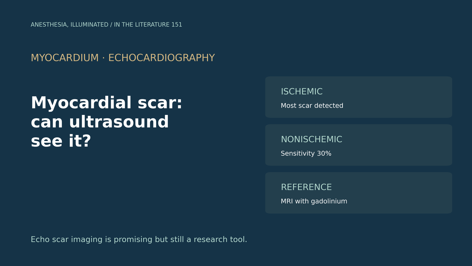
- Ischemic scar is visible. Power modulation echo detected scar in all 71 patients with prior infarction, with a per-segment sensitivity of 82% and specificity of 97% against magnetic resonance.[1]
- Nonischemic scar is mostly missed. In unselected patients, agreement with magnetic resonance was only moderate (kappa 0.465), and sensitivity for nonischemic scar was 30%.[1]
- The method has practical limits: it comes from a single vendor and probe, scar volume analysis is time-consuming, anterior and apical segments are poorly seen, and results depend on vendor processing.[2], [3]
- Strain is indirect. Reduced strain suggests fibrosis but is not necessarily a sign of it; late gadolinium enhancement remains the gold standard.[4]
- Echo scar carries prognostic information but is not ready to be used alone; adding it raised the area under the curve for defibrillator shocks from 0.734 to 0.782.[5]
A patient with a prior inferior infarction and ischemic cardiomyopathy comes for noncardiac surgery; magnetic resonance is unavailable, and a transthoracic echo shows an akinetic, bright inferior wall. Can ultrasound tell whether this is scar, and how much?[1], [4]
How echo can show scar
Collagen-rich scar reflects ultrasound differently from normal muscle and behaves more nonlinearly. Power modulation exploits this by cancelling the linear signal of normal myocardium and leaving the nonlinear scar signal.[2] Papachristidis and colleagues compared three-dimensional power modulation echo with magnetic resonance. In 71 patients with prior infarction, echo detected scar in all, with a per-segment sensitivity of 82% and specificity of 97%; sensitivity was higher in inferior and posterior segments, and echo scar volumes were smaller than on magnetic resonance.[1]
In an unselected cohort, sensitivity was 62%, specificity 89% and accuracy 72%, with moderate agreement (kappa 0.465). Echo was diagnostic in 30 of 33 patients with ischemic scar (91%) but found scar in only 30% of those with nonischemic scar.[1]
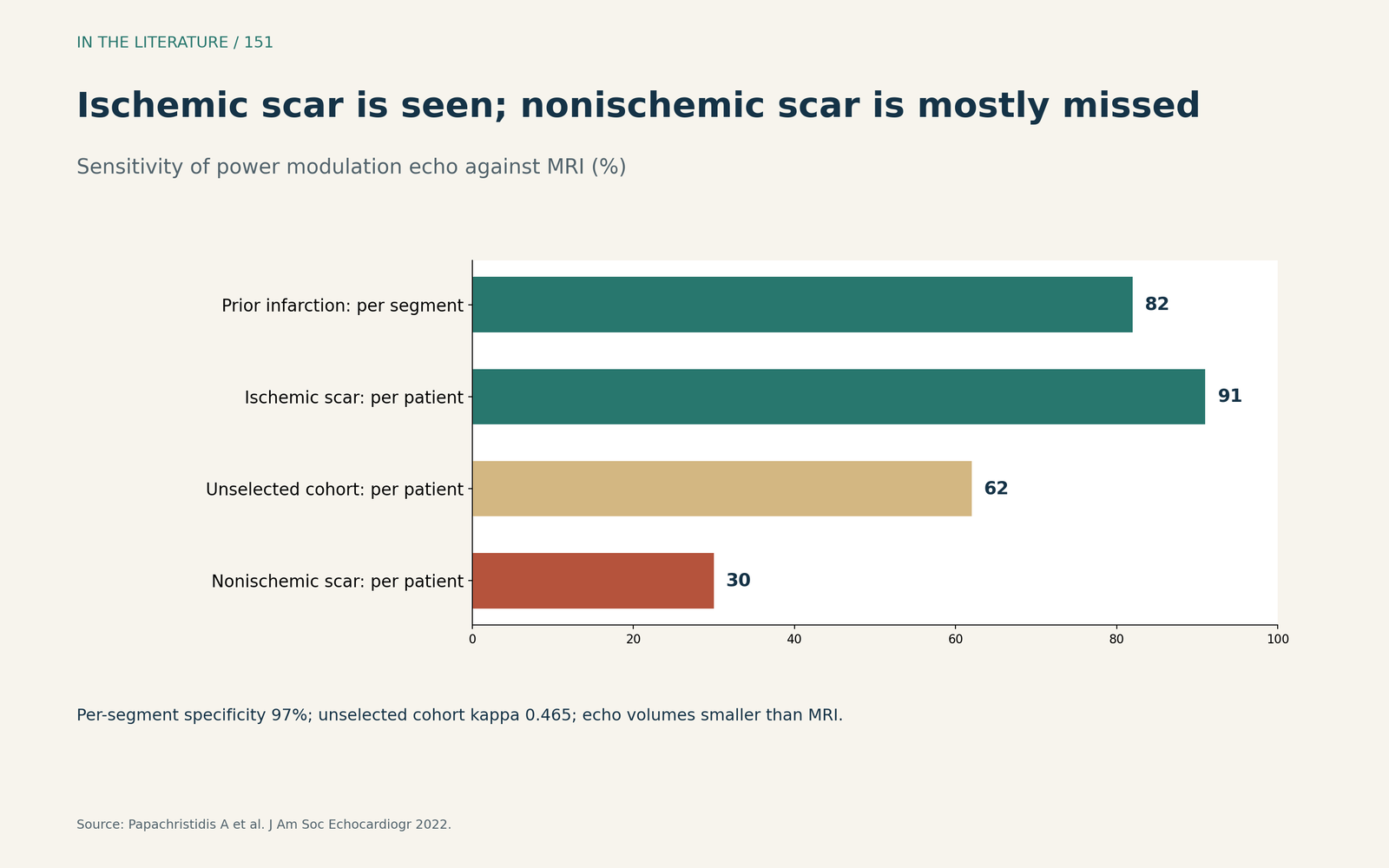
Figure 1. Ischemic scar is seen; nonischemic scar is mostly missed. Redrawn from Papachristidis et al.[1] View larger.

Why it is not routine yet
Appadurai and Thomas note that scar volume calculation was time-consuming, that the data came from a single vendor, model and probe, that anterior, anterolateral and apical segments were poorly detected, and that incremental value over wall motion assessment is unproven.[2] In a phantom and pilot study, agreement with magnetic resonance was best at a mechanical index of 0.8, and results cannot be translated automatically to other systems.[3]
Strain is the more widely available ultrasound tool, but Gao and colleagues emphasize that it is an indirect marker: echo is the most common way to measure strain, yet reduced strain in an individual patient is not necessarily a sign of fibrosis, and late gadolinium enhancement remains the gold standard.[4]
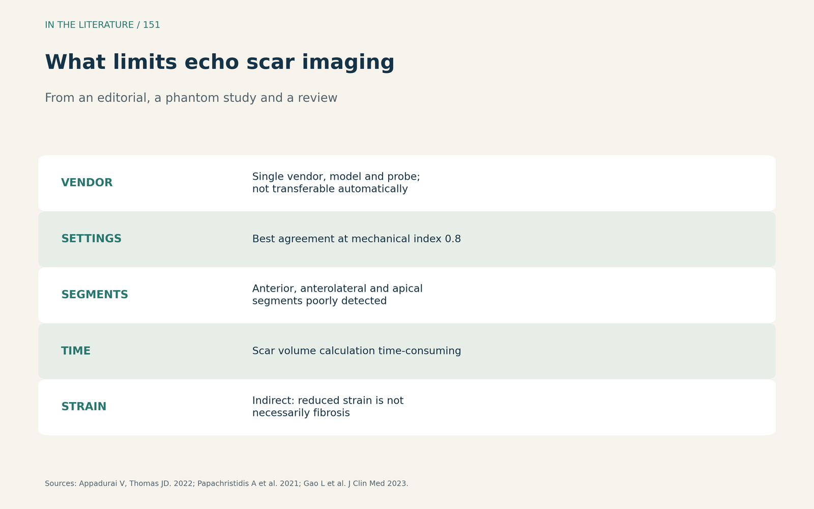
Figure 2. What limits echo scar imaging. Synthesized from Appadurai and Thomas, Papachristidis et al. and Gao et al.[2], [3], [4] View larger.

Does echo scar matter?
Gaibazzi and colleagues used a pulse-cancellation method in a case-control study: echo scar was present in 80% of patients with appropriate defibrillator shocks and 52% of controls, and adding it raised the area under the curve from 0.734 to 0.782, but they state it is not ready to be used in isolation.[5] In pigs, a contrast nanodroplet method correlated with magnetic resonance infarct area (r 0.81), but safety studies are needed before clinical use.[6]
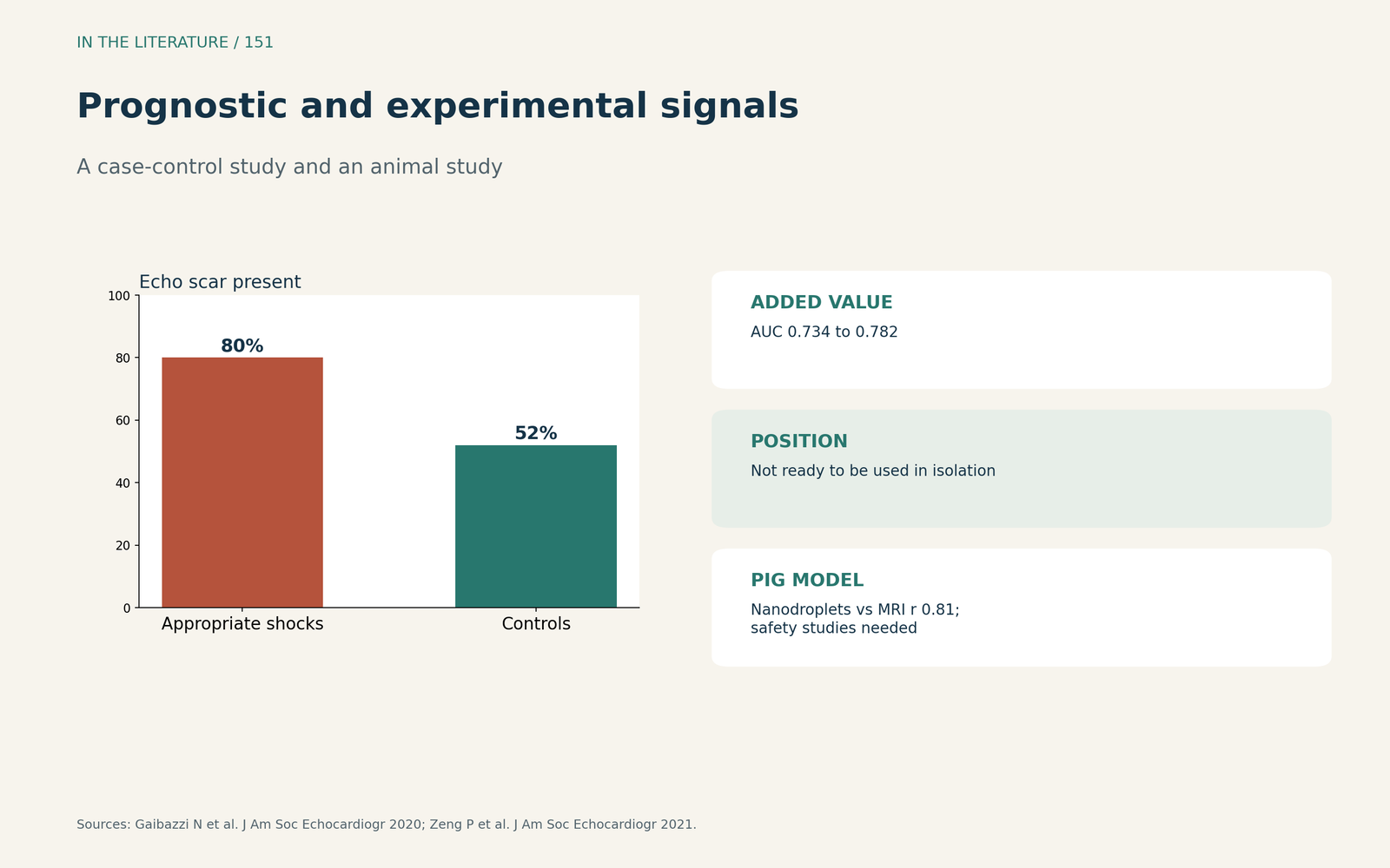
Figure 3. Prognostic and experimental signals. Redrawn from Gaibazzi et al. and Zeng et al.[5], [6] View larger.

Three takeaways
- Echo can see ischemic scar. With specialized processing, detection against magnetic resonance is good, especially in inferior and posterior walls.[1]
- Do not rely on it for nonischemic scar. Sensitivity was only 30%, and strain is nonspecific.[1], [4]
- Magnetic resonance remains the reference. Echo scar methods are single-vendor research tools.[2], [5]
References
- Papachristidis A, Theodoropoulos KC, Marvaki A, et al. Power modulation echocardiography to detect and quantify myocardial scar. J Am Soc Echocardiogr. 2022;35(11):1146–1155. doi:10.1016/j.echo.2022.06.009
- Appadurai V, Thomas JD. Detecting scar in echocardiography: has the power shifted? J Am Soc Echocardiogr. 2022;35(11):1156–1158. doi:10.1016/j.echo.2022.09.001
- Papachristidis A, Queirós S, Theodoropoulos KC, et al. The impact of vendor-specific ultrasound beam-forming and processing techniques on the visualization of in vitro experimental "scar": implications for myocardial scar imaging using echocardiography. J Am Soc Echocardiogr. 2021;34(10):1095–1105.e6. doi:10.1016/j.echo.2021.05.016
- Gao L, Zhang L, Zhang Z, et al. Clinical utility of strain imaging in assessment of myocardial fibrosis. J Clin Med. 2023;12(3):743. doi:10.3390/jcm12030743
- Gaibazzi N, Suma S, Lorenzoni V, et al. Myocardial scar by pulse-cancellation echocardiography is independently associated with appropriate defibrillator intervention for primary prevention after myocardial infarction. J Am Soc Echocardiogr. 2020;33(9):1123–1131. doi:10.1016/j.echo.2020.04.020
- Zeng P, Qian L, Lof J, et al. Delayed echo enhancement imaging to quantify myocardial infarct size. J Am Soc Echocardiogr. 2021;34(8):898–909. doi:10.1016/j.echo.2021.02.019