
ITL-24dbd45e338f
Juan Li · In the Literature · Updated November 16, 2026
Watch the video

Key points
- Establish where regurgitation originates before calling it paravalvular leak; an eccentric intravalvular jet can be misleading.
- Grade severity from several concordant findings. The apparent color-jet area or circumferential extent alone is insufficient.
- Record the examination's timing and hemodynamic context; measurements obtained immediately after deployment and weeks later are not interchangeable.
- When findings disagree, first check acquisition and physiology, then choose additional imaging for the unresolved question.
- CMR can help quantify regurgitation in a stable patient with uncertain echocardiographic findings; diagnostic agreement does not prove that an imaging strategy improves outcomes.
Start at the origin of the jet
After TAVR deployment, a color jet appears beside the prosthesis. One view looks reassuring; another suggests a larger leak. Before deciding on its grade, answer a simpler question: where does the blood cross the valve or its surrounding seal?
Prosthetic shadowing and eccentric flow make a single view unreliable. Sweep through multiple planes, identify each jet's origin and distinguish intravalvular from paravalvular regurgitation. TTE and TEE have complementary blind spots; additional views should address the region that remains uncertain. The ASE prosthetic-valve guideline and Hoffmeister and colleagues' focused review both support this approach. [1, 2]
This distinction matters when measuring circumferential extent. An eccentric jet can travel around the annulus after emerging from a much smaller orifice. Trace its origin and vena contracta—the narrowest jet region—rather than treating the entire downstream color arc as the leak's circumference. When several separate jets are present, the nonregurgitant gaps between them do not become part of the leaking arc. [1, 2]
Build a grade from findings that agree
The 2024 ASE guideline integrates jet characteristics and vena-contracta measurements with Doppler evidence, quantitative regurgitation measures when feasible, and the ventricular response. Circumferential extent contributes to that assessment; its own table footnote cautions against using it alone. [1]
For practical reporting, separate four observations: the mechanism and location, the jet origin and extent, the hemodynamic evidence, and the effect on the ventricle. Report the overall grade together with the findings that support it. A color image that looks dramatic is not a substitute for that explanation. Likewise, a normal-sized ventricle immediately after implantation cannot exclude an important acute leak: chamber remodeling reflects time as well as regurgitant burden. [1, 2]
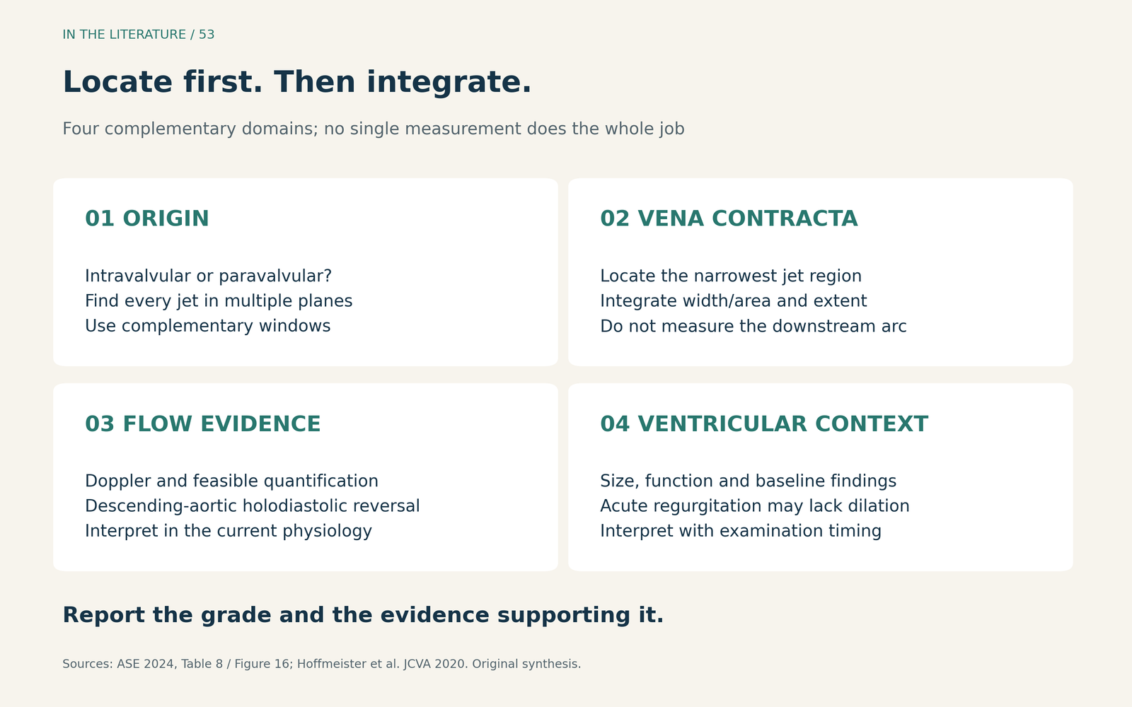
Figure 1. An original clinical summary of the integrated assessment. The displayed color jet is distinguished from its vena contracta; a single reassuring domain does not overrule discordant evidence. [1, 2] View figure

When the measurements disagree, resolve the reason
First revisit image quality, the plane of measurement and the patient's physiology. A different loading state or a poorly aligned acquisition may explain part of the discrepancy. The ASE approach is to investigate these explanations before forcing inconsistent findings into a confident grade. [1]
The next examination should answer a specific question. Additional TTE or TEE can clarify an obscured jet and its mechanism. CT can contribute anatomical information. CMR can quantify regurgitant flow when echocardiography remains inadequate or conflicts with the clinical picture. These tests have different strengths; CMR is not an immediate rescue investigation for an unstable patient in the procedure room. [1]
Also distinguish total aortic regurgitation from its paravalvular component. A flow-based regurgitant fraction does not, by itself, locate the leak. It must be interpreted alongside the anatomical examination. State when the measurement was obtained and under what conditions rather than silently treating every modality's number as the same quantity. [1]
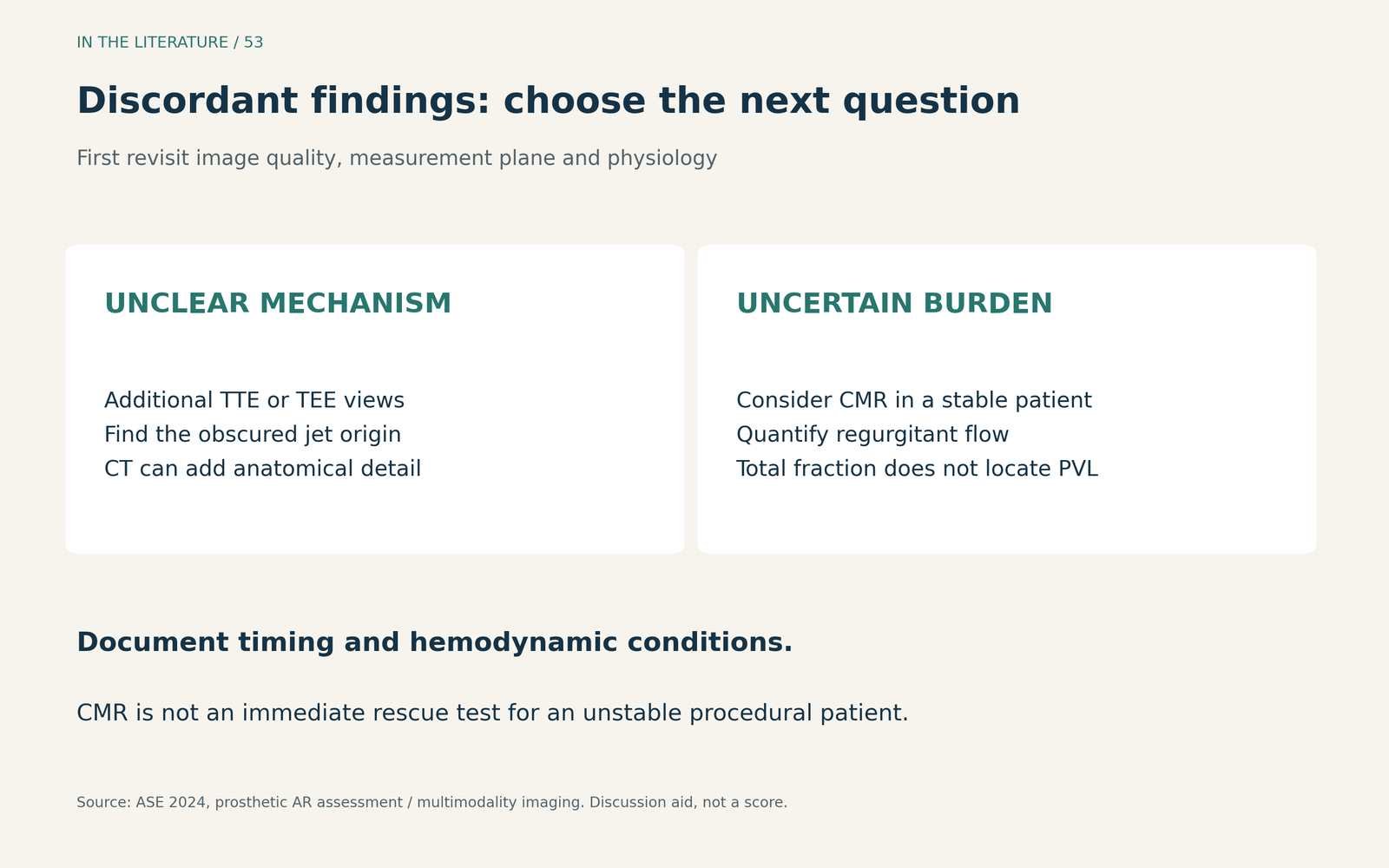
Figure 2. A discussion aid, not a validated decision score. Imaging is selected according to the unresolved question and the patient's stability; the diagram does not require every patient to undergo every modality. [1] View figure

Quantitative measurements add information, with conditions
In the prospective APPOSE substudy, Rooijakkers and colleagues compared intraprocedural diastolic delta—the end-diastolic aortic pressure minus left ventricular end-diastolic pressure—and angiographic videodensitometry with subsequent CMR. Only 51 of the 77 patients in the primary APPOSE analysis had analyzable final aortograms. CMR was performed 40 ± 15 days after TAVR, so these were not measurements at the same physiological moment. [3]
Combining the two intraprocedural measures produced an AUC of 0.90 (95% CI, 0.81–0.99), compared with 0.82 and 0.80 for the individual measures, for detecting the study's definition of relevant regurgitation: CMR regurgitant fraction greater than 20%. That definition is not a universal threshold for severe PVL. This small, single-center Portico cohort supports complementary diagnostic information; it does not establish an automatic postdilatation trigger or demonstrate better outcomes from the combined strategy. [3]
The 2026 COMPARE-TAVI 1 imaging substudy offers a useful counterweight to the assumption that TTE always underestimates regurgitation. Only the published abstract was available for this discussion. Among 327 participants examined 4–6 weeks after TAVI, TTE and CMR grades agreed in 281; TTE graded 31 higher and 15 lower than CMR. The parent trial randomized valve platforms, not imaging strategies. These findings concern aortic regurgitation after TAVI and do not establish a clinical-outcome advantage for routine CMR. [4]
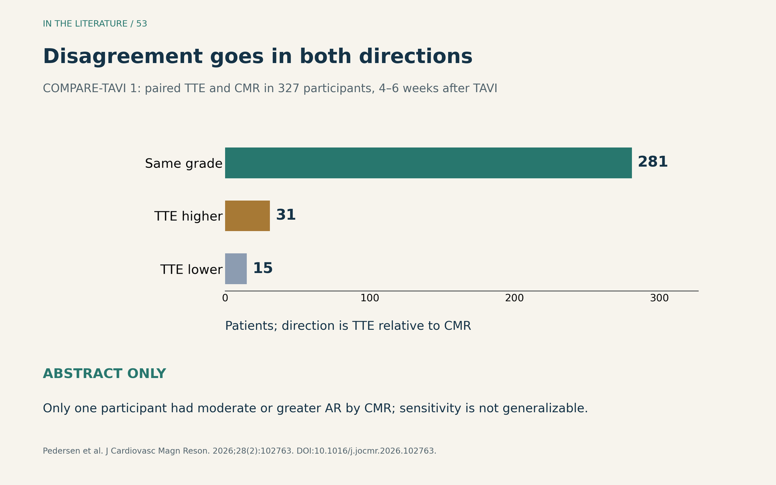
Figure 3. Original display of the aggregate counts reported in the COMPARE-TAVI 1 abstract. Direction is TTE relative to CMR, at 4–6 weeks after TAVI. Only one participant had moderate or greater AR by CMR, so the study's sensitivity estimate should not be generalized from that very small denominator. Abstract only. [4] View figure

Why getting the grade right matters
Warraich and colleagues' retrospective cohort of 3,600 TAVI recipients classified PVL at 30 days, with median follow-up of 3.0 years. Moderate-to-severe PVL was associated with higher adjusted mortality than none/trace PVL: HR 1.80 (95% CI, 1.31–2.46). The mild-PVL estimate was HR 1.01 (95% CI, 0.89–1.15). These were clinical TTE grades without standardized laboratory review. [5]
The practical lesson is to document the grade and its supporting evidence carefully. This cohort does not show that every mild leak is harmless, prove that correcting a leak improves survival, or supply an immediate postdeployment intervention threshold. Its 30-day assessment also should not be relabeled as the initial procedural result. [5]
Three takeaways
- Locate before grading. Identify the true jet origin, separate intravalvular from paravalvular flow, and measure the vena contracta rather than the downstream color arc.
- Make the evidence agree—or explain why it does not. Integrate complementary findings, document timing and physiology, and investigate a discrepancy before assigning a confident grade.
- Use another modality for a defined question. Clarify anatomy when the mechanism is uncertain and consider CMR quantification in a stable patient when the regurgitant burden remains unresolved.
References
- Zoghbi WA, Jone PN, Chamsi-Pasha MA, et al. Guidelines for the Evaluation of Prosthetic Valve Function With Cardiovascular Imaging: A Report From the American Society of Echocardiography Developed in Collaboration With the Society for Cardiovascular Magnetic Resonance and the Society of Cardiovascular Computed Tomography. J Am Soc Echocardiogr. 2024;37(1):2–63. doi:10.1016/j.echo.2023.10.004.
- Hoffmeister KJ, Henderson ZT, Hussey PT, et al. Guidelines for the Evaluation of Valvular Regurgitation After Percutaneous Valve Repair or Replacement: A Focused Review for the Cardiac Anesthesiologist. J Cardiothorac Vasc Anesth. 2020;34(10):2740–2753. doi:10.1053/j.jvca.2019.11.040.
- Rooijakkers MJP, Elkoumy A, Stens NA, et al. Periprocedural Assessment of Paravalvular Regurgitation After Transcatheter Aortic Valve Replacement Using Diastolic Delta and Videodensitometry. J Am Heart Assoc. 2024;13(18):e035587. doi:10.1161/JAHA.124.035587.
- Pedersen AAU, Pedersen ALD, Mogensen NSB, et al. Cardiovascular magnetic resonance reclassification of aortic regurgitation after transcatheter aortic valve implantation: A COMPARE-TAVI 1 substudy. J Cardiovasc Magn Reson. 2026;28(2):102763. doi:10.1016/j.jocmr.2026.102763. Abstract only.
- Warraich N, Brown JA, Ashwat E, et al. Paravalvular Leak After Transcatheter Aortic Valve Implantation: Results From 3600 Patients. Ann Thorac Surg. 2025;119(5):1037–1044. doi:10.1016/j.athoracsur.2025.01.012.