ITL-961a5a5381ab
Juan Li, MD · Updated November 17, 2026
Watch the video
Key points
- Echocardiography, not surface landmarks, should choose the route. Echo guidance has shifted practice from the subxiphoid to the apical approach, because the distance from skin to fluid is usually shortest there, and guided procedures succeed in more than 95% of cases.[1], [2]
- Image the patient supine before the procedure, enter where the collection is largest and closest to the transducer, confirm position with agitated saline, drain until the effusion has nearly disappeared and repeat echocardiography within 24 hours.[2], [1], [3]
- Surgical drainage is preferred for purulent pericarditis, bleeding into the pericardium and loculated or complex effusions; penetrating trauma with tamponade needs immediate thoracotomy.[4], [2]
- Anticoagulation and low platelets were not associated with more bleeding in a large echo-guided series, although holding anticoagulation for elective procedures is prudent.[3]
- Needle drainage is less invasive but less durable. In a national US cohort it carried 14.6 times the adjusted odds of reintervention, and in malignant effusions it failed more often than a surgical window.[5], [6]
An anesthesiologist is asked to help with a large effusion in a hypotensive patient on anticoagulation. Which route is safest, how should the needle be guided and confirmed, and when is a surgical window the better choice?[1], [4]
Choosing the route with echocardiography
The 2015 American Society of Echocardiography (ASE) guideline on echocardiographic monitoring notes that echo guidance has resulted in more apical, rather than subxiphoid, pericardiocentesis, mainly because the distance from the skin to the effusion is smallest at that location. Guided procedures have a success rate above 95% in a large single-center series.[1] The 2013 ASE pericardial recommendations describe the procedure as usually approached from the para-apical region, with a 97% success rate, and advise imaging with the patient supine when planning pericardiocentesis.[2]
In the Mayo Clinic series of 1,127 echo-guided procedures reported by Ryu and colleagues, the operator chose the access site where the fluid was closest to the transducer and the collection largest.[3]
Confirming position and finishing the procedure
Agitated saline injected through the needle confirms that the tip is in the pericardial space before the guidewire and catheter are advanced. Drainage continues until the effusion has nearly disappeared, and repeat echocardiography within 24 hours checks for reaccumulation.[1] The ESC guideline prefers needle pericardiocentesis with echocardiographic or fluoroscopic guidance and suggests a triage system to guide its timing.[4]
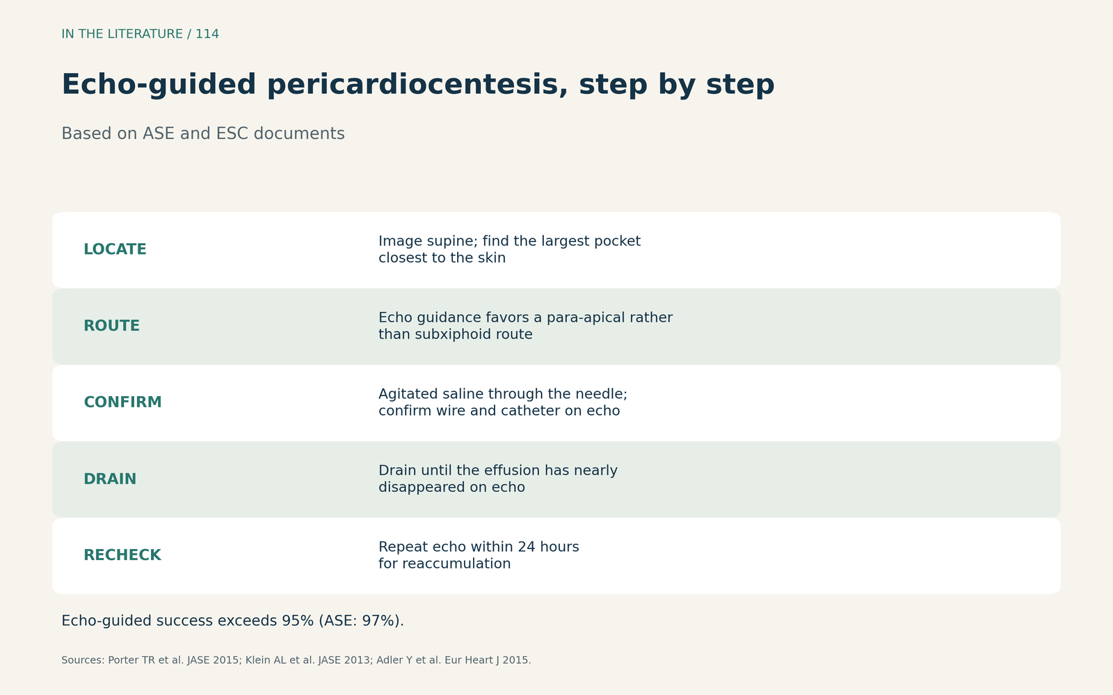
Figure 1. Echo-guided pericardiocentesis, step by step. Synthesized from the ASE monitoring guideline, the ASE pericardial recommendations and the ESC guideline.[1], [2], [4] View larger.

Bleeding risk
In Ryu and colleagues' series, neither platelet count nor anticoagulation status was associated with bleeding complications. Serious bleeds were rare and included internal mammary vein laceration causing mediastinal hematoma and tamponade. The authors still consider it prudent, when feasible, to hold anticoagulation for entirely elective procedures.[3]
When surgery is better
The ESC guideline recommends surgical drainage for purulent pericarditis or urgent bleeding into the pericardium, and immediate thoracotomy for tamponade due to penetrating trauma.[4] The ASE recommends surgical drainage when subacute tamponade is caused by a loculated or complex effusion.[2]
For malignant effusions, the ESC notes a high recurrence rate (40–70%) and that surgical pericardiotomy does not improve clinical outcomes over pericardiocentesis.[4] A 2026 meta-analysis of 6 retrospective studies by Nahle and colleagues found more procedure failure after pericardiocentesis (odds ratio 2.99; 95% CI 1.10–8.14), but lower all-cause mortality (odds ratio 0.68; 95% CI 0.46–0.99) and no significant difference in bleeding; selection bias is likely.[6]
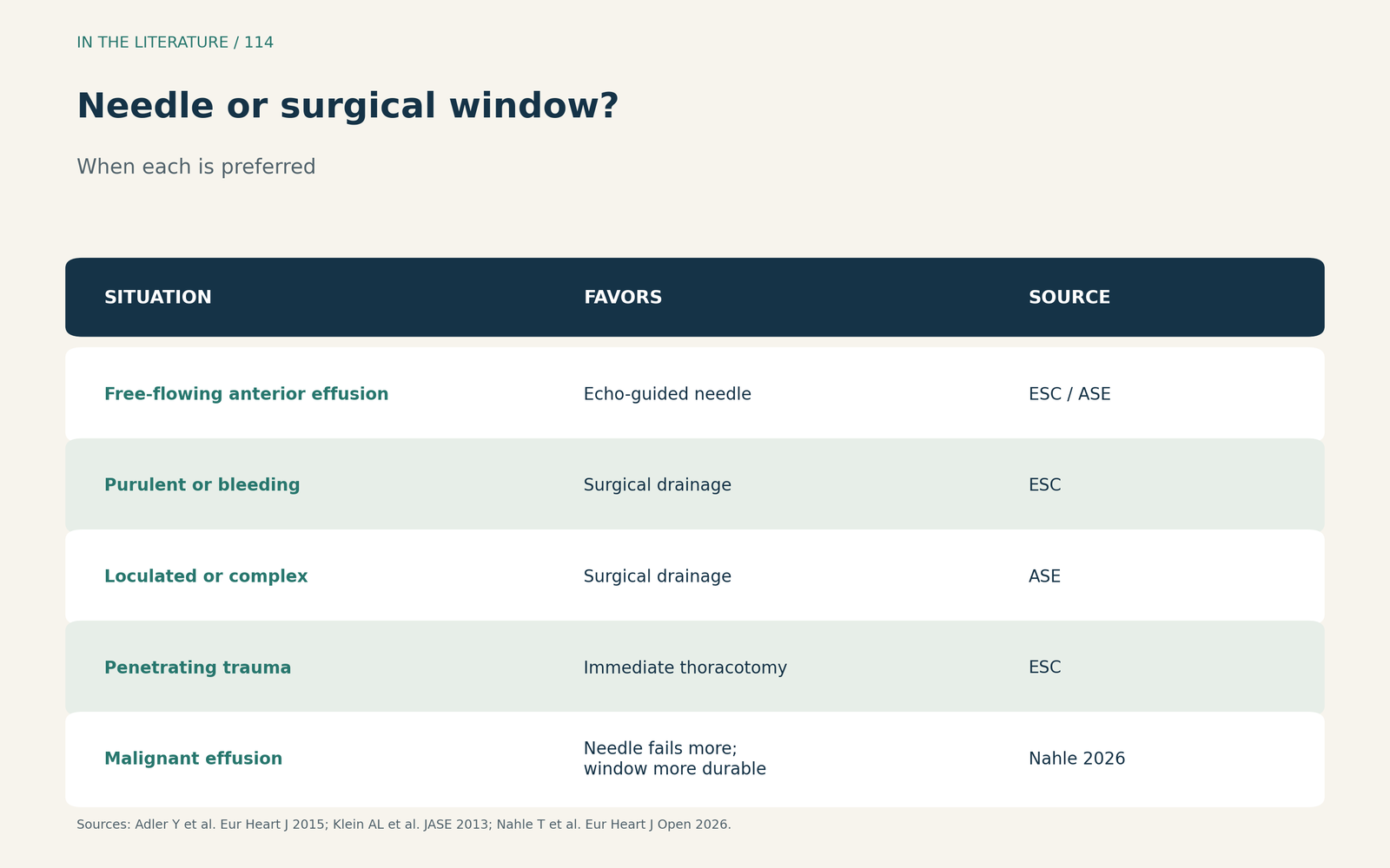
Figure 2. Needle or surgical window? Synthesized from the ESC and ASE documents and Nahle et al.[2], [4], [6] View larger.

National outcomes
In a US national readmissions cohort, Pan and colleagues found higher unadjusted in-hospital mortality after pericardiocentesis than after surgical drainage (10.2% vs 7.31%), although patients receiving pericardiocentesis more often had tamponade (68.0% vs 62.8%). Pericardiocentesis was associated with much greater odds of reintervention (adjusted odds ratio 14.6), and 86.7% of reinterventions were conversions to surgical drainage, suggesting that needle drainage often serves as a bridge.[5]
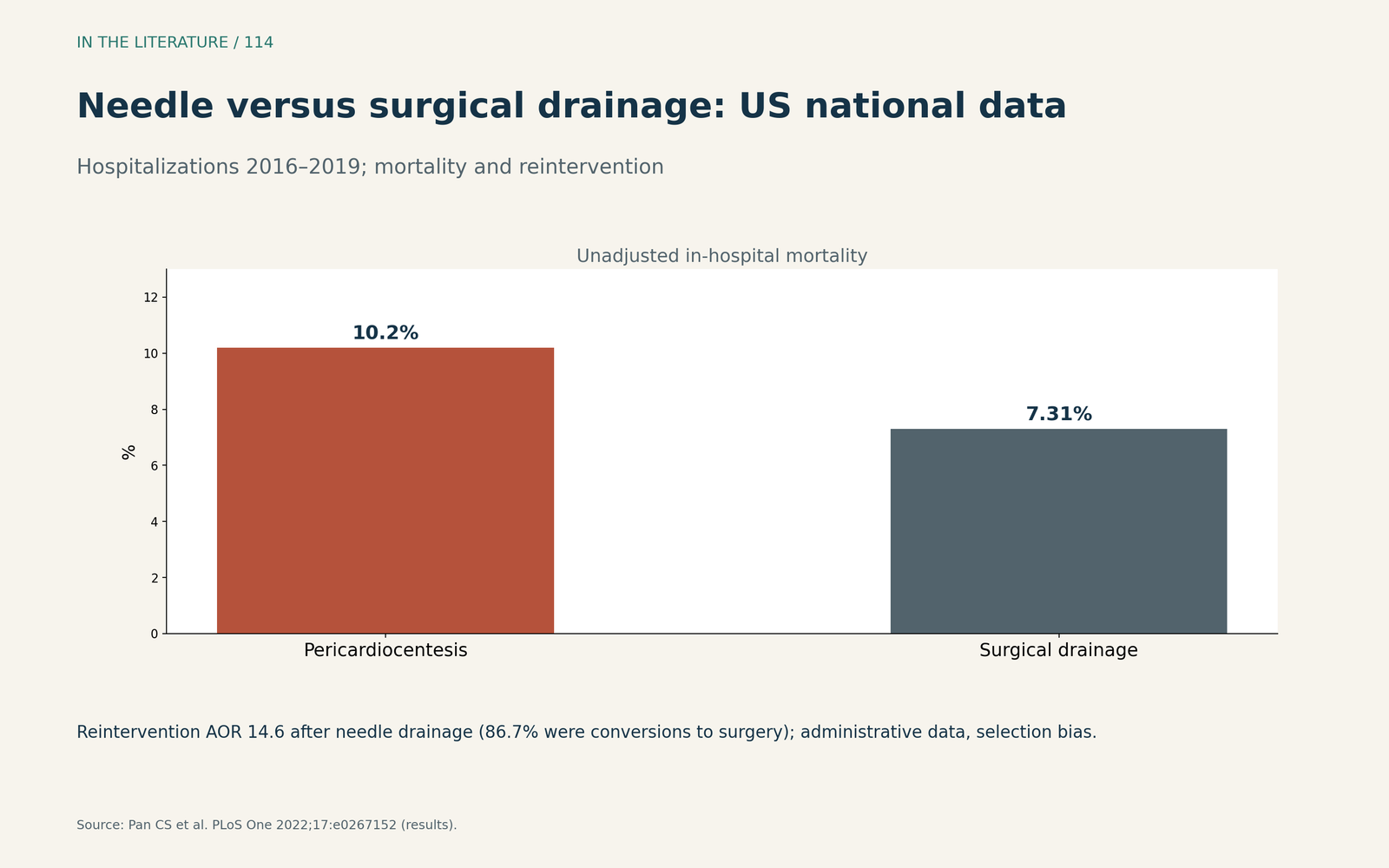
Figure 3. Needle versus surgical drainage in US national data. Redrawn from the results of Pan et al.; administrative data with likely selection bias.[5] View larger.

Three takeaways
- Let echocardiography pick the route. Image supine and enter where the fluid is largest and closest to the skin, often para-apical.[1], [2]
- Confirm, drain and recheck. Use agitated saline, drain to near-empty and repeat echocardiography within 24 hours.[1], [3]
- Know when to choose surgery. Purulent, bleeding, loculated or penetrating-trauma effusions need surgery, and needle drainage of malignant effusions is more likely to need repeat intervention.[4], [5], [6]
References
- Porter TR, Shillcutt SK, Adams MS, et al. Guidelines for the use of echocardiography as a monitor for therapeutic intervention in adults: a report from the American Society of Echocardiography. J Am Soc Echocardiogr. 2015;28(1):40–56. doi:10.1016/j.echo.2014.09.009
- Klein AL, Abbara S, Agler DA, et al. American Society of Echocardiography clinical recommendations for multimodality cardiovascular imaging of patients with pericardial disease: endorsed by the Society for Cardiovascular Magnetic Resonance and Society of Cardiovascular Computed Tomography. J Am Soc Echocardiogr. 2013;26(9):965–1012.e15. doi:10.1016/j.echo.2013.06.023
- Ryu AJ, Kane GC, Pislaru SV, et al. Bleeding complications of ultrasound-guided pericardiocentesis in the presence of coagulopathy or thrombocytopenia. J Am Soc Echocardiogr. 2020;33(3):399–401. doi:10.1016/j.echo.2019.11.006
- Adler Y, Charron P, Imazio M, et al. 2015 ESC guidelines for the diagnosis and management of pericardial diseases. Eur Heart J. 2015;36(42):2921–2964. doi:10.1093/eurheartj/ehv318
- Pan CS, Mabeza RM, Tran Z, et al. Pericardiocentesis or surgical drainage: a national comparison of clinical outcomes and resource use. PLoS One. 2022;17(4):e0267152. doi:10.1371/journal.pone.0267152
- Nahle T, Bhave A, Abou Zeid K, et al. Pericardiocentesis vs. surgical pericardial window for first occurrence of malignancy-related pericardial effusion: a meta-analysis of retrospective studies. Eur Heart J Open. 2026;6(2):oeag018. doi:10.1093/ehjopen/oeag018