ITL-08f2e12667d9
Juan Li, MD · Updated November 17, 2026
Watch the video
Key points
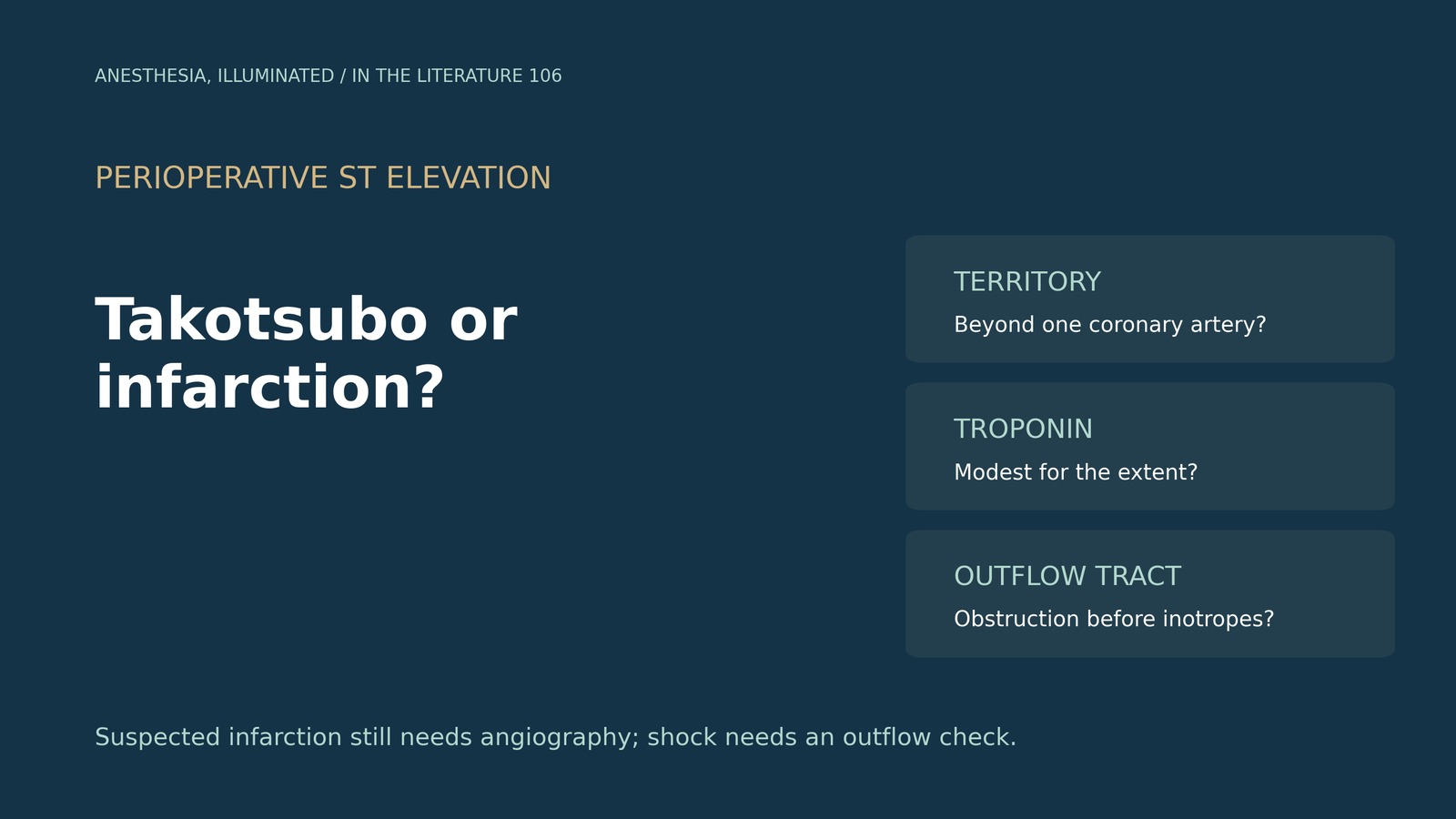
- Takotsubo syndrome (TTS) can mimic acute myocardial infarction on the ECG, troponin and echocardiogram. ST elevation with suspected infarction still requires urgent coronary angiography.[1], [2]
- The most useful bedside clue is a circumferential wall-motion abnormality extending beyond a single coronary territory, with a troponin rise that is modest for the extent of dysfunction.[2], [3]
- Clinical scores and natriuretic peptide to troponin ratios help estimate probability, but no single feature excludes infarction; coexisting coronary disease does not rule out TTS.[1], [3]
- Surgery, anesthesia and catecholamines are recognized triggers; in one review of perioperative cases most presented with heart failure rather than an acute coronary picture.[4]
- The distinction matters most in shock: dynamic left ventricular outflow tract (LVOT) obstruction is common, and inotropes and intra-aortic balloon pumping can worsen it.[1], [5], [6]
After induction or in recovery, the patient becomes hypotensive. New ST elevation appears in the anterior leads, troponin rises, and transesophageal echocardiography shows a hypokinetic apex with hyperdynamic basal segments. Is this an infarct that needs reperfusion, or Takotsubo syndrome, which needs a very different hemodynamic approach?[1], [4]
Why the two look alike
The International Takotsubo Expert Consensus (Part II) notes that ST elevation in TTS can resemble anterior ST-elevation myocardial infarction (STEMI) closely enough that urgent coronary angiography with left ventriculography is required to tell them apart with certainty. Some ECG patterns differ on average. ST elevation in TTS centers on V2–V5 with II and aVR, whereas anterior STEMI centers on V1–V4 with I and aVL. ST depression occurs in fewer than 10% of TTS patients but in over 30% of acute coronary syndromes.[1]
Sonaglioni and colleagues pooled studies comparing TTS with anterior STEMI due to left anterior descending artery occlusion. ST elevation was present in about 48% of TTS patients versus 98% of STEMI patients, and peak troponin I was much lower in TTS. Global longitudinal strain did not separate the groups, and the authors concluded that strain parameters cannot currently be recommended as standalone discriminators.[3]
What points toward Takotsubo
Wall-motion territory. The European Association of Cardiovascular Imaging and Japanese Society of Echocardiography consensus describes the hallmark of TTS as a circumferential pattern of dysfunction extending beyond the territory of a single coronary artery. Typical apical ballooning involves apical segments of all walls, often with basal hypercontraction. Midventricular, basal (reverse) and focal variants also occur.[2]
Biomarkers. Troponin is often similar to that in acute coronary syndrome on admission, but peak values are substantially lower, whereas natriuretic peptides rise markedly and peak about 24–48 hours after symptom onset.[1] Pillitteri and colleagues cite a natriuretic peptide to troponin ratio above 159 as discriminating TTS from infarction with high sensitivity and specificity in one study.[6] Such ratios come from small cohorts and depend on assay and timing; they support, rather than replace, coronary imaging.
Clinical probability. The InterTAK Diagnostic Score combines female sex, emotional or physical trigger, absence of ST depression, psychiatric or neurologic disorders and QTc prolongation into a score of up to 100 points; a score above 70 indicates a high probability of TTS.[1] In a perioperative patient, the physical trigger is often present by definition, which limits how much the score adds.
Coronary and tissue imaging. Coronary angiography remains the reference test when infarction is plausible. Coronary CT angiography is an alternative only in stable patients with low suspicion of acute coronary syndrome, and should not delay invasive angiography when chest pain and acute ECG changes persist.[2] Cardiac magnetic resonance shows edema in the dysfunctional segments without the late gadolinium enhancement of infarction when a threshold of 5 standard deviations is used.[2] Coexisting coronary disease does not exclude TTS; the question is whether the coronary anatomy explains the full extent of dysfunction.[2]
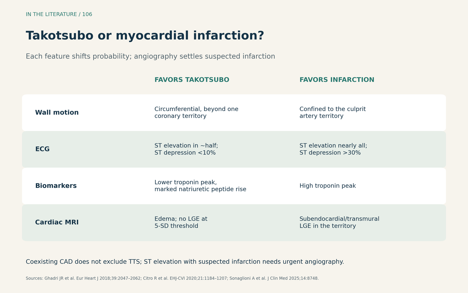
Figure 1. Takotsubo or infarction: comparing the findings. Synthesized from the International Expert Consensus, the EACVI/JSE imaging consensus and the meta-analysis by Sonaglioni and colleagues. Each feature shifts probability; angiography settles suspected infarction.[1]–[3] View larger.

The perioperative setting
Hessel reviewed 131 published perioperative cases and estimated an incidence of about 1 in 6,700 operations. Thirty-seven percent occurred during anesthesia or surgery and 58% after surgery. Most patients presented with heart failure or shock rather than a typical acute coronary syndrome, and the ECG was abnormal in 92%.[4] These are reported cases, so they describe presentation rather than true incidence.
Recognized perioperative triggers include the physical stress of surgery, induction of anesthesia and exogenous catecholamines.[4], [6] In the operating room, echocardiography can show the characteristic wall-motion pattern and reveal LVOT obstruction before a vasoactive strategy is chosen.[2], [5]
Why the distinction changes treatment
Treatment for acute infarction aims at reperfusion and frequently uses inotropic support. In TTS, the hyperdynamic basal segments can create dynamic LVOT obstruction, sometimes with systolic anterior motion of the mitral valve. Di Vece and colleagues define it as an intraventricular gradient of at least 25 mmHg and report a prevalence between 7% and 25%.[5]
When LVOT obstruction is present, the reviews advise stopping positive inotropes, giving careful fluid under hemodynamic monitoring and avoiding intra-aortic balloon pumping. The Di Vece algorithm adds short-acting beta-blockade and, if shock persists, Impella or venoarterial ECMO.[5], [6] The consensus document notes that catecholamine-treated TTS patients have had high reported mortality, and that nitroglycerin should be avoided with LVOT obstruction.[1] When inotropic support is needed without obstruction, the reviews favor levosimendan over catecholamines; this rests on observational data and expert opinion, not randomized trials.[5], [6]
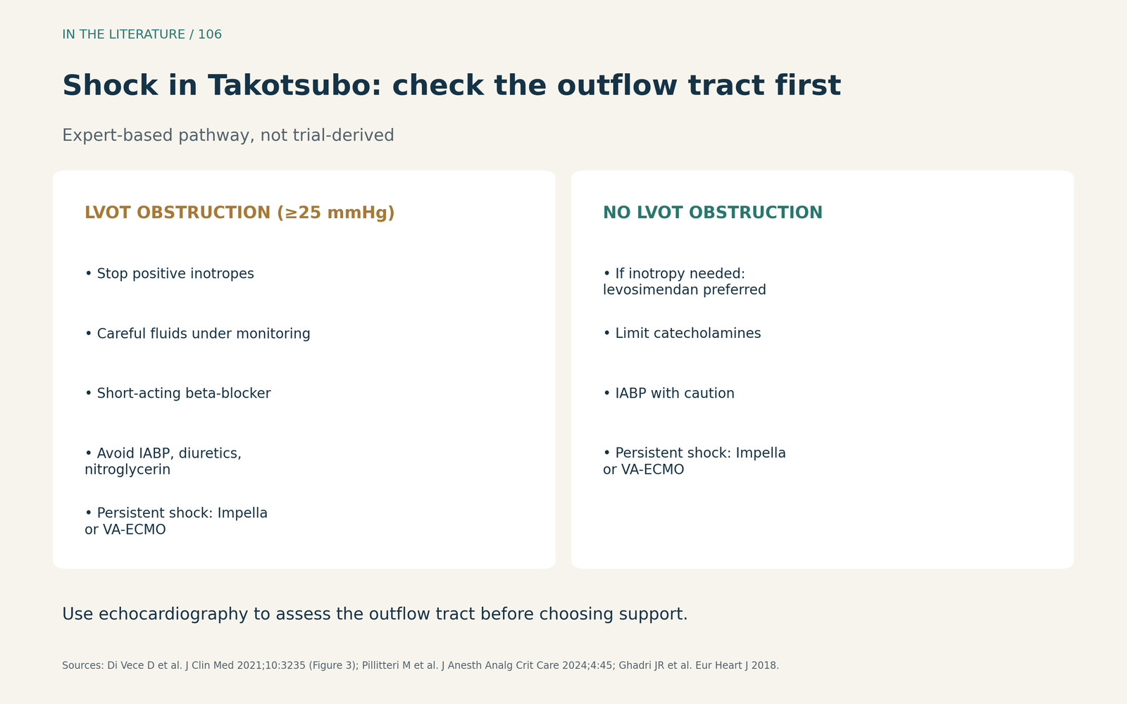
Figure 2. Managing shock depends on LVOT obstruction. Redrawn as text from the algorithm of Di Vece and colleagues and the perioperative review by Pillitteri and colleagues; expert-based, not trial-derived.[5], [6] View larger.

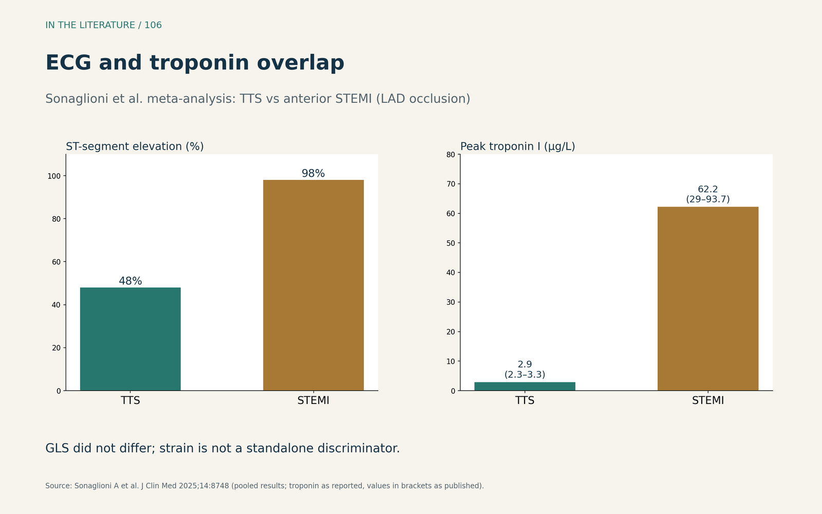
Figure 3. How often the ECG and troponin overlap. Redrawn from the results of the meta-analysis by Sonaglioni et al. comparing TTS with anterior STEMI due to left anterior descending occlusion.[3] View larger.

Three takeaways
- Do not let a pattern replace angiography. ST elevation with suspected infarction still needs urgent coronary angiography; ECG, scores and biomarker ratios only shift probability.[1], [2]
- Look at the territory and the troponin together. Circumferential dysfunction beyond one coronary distribution with a modest troponin rise favors TTS, even when some coronary disease is present.[2], [3]
- Check the outflow tract before treating shock. In TTS, inotropes and balloon pumping can worsen LVOT obstruction; fluids, beta-blockade and early mechanical support are the reviewed alternatives.[5], [6]
References
- Ghadri JR, Wittstein IS, Prasad A, et al. International expert consensus document on takotsubo syndrome (Part II): diagnostic workup, outcome, and management. Eur Heart J. 2018;39(22):2047–2062. doi:10.1093/eurheartj/ehy077
- Citro R, Okura H, Ghadri JR, et al. Multimodality imaging in takotsubo syndrome: a joint consensus document of the European Association of Cardiovascular Imaging (EACVI) and the Japanese Society of Echocardiography (JSE). Eur Heart J Cardiovasc Imaging. 2020;21(11):1184–1207. doi:10.1093/ehjci/jeaa149
- Sonaglioni A, Nicolosi GL, Lombardo M, et al. Comparative meta-analysis of left ventricular mechanics in takotsubo syndrome and anterior STEMI due to left anterior descending artery occlusion. J Clin Med. 2025;14(24):8748. doi:10.3390/jcm14248748
- Hessel EA. Takotsubo cardiomyopathy and its relevance to anesthesiology: a narrative review. Can J Anaesth. 2016;63(9):1059–1074. doi:10.1007/s12630-016-0680-4
- Di Vece D, Silverio A, Bellino M, et al. Dynamic left intraventricular obstruction phenotype in takotsubo syndrome. J Clin Med. 2021;10(15):3235. doi:10.3390/jcm10153235
- Pillitteri M, Brogi E, Piagnani C, et al. Perioperative management of Takotsubo cardiomyopathy: an overview. J Anesth Analg Crit Care. 2024;4(1):45. doi:10.1186/s44158-024-00178-y