
ITL-773cc0cecf86
Juan Li, MD · Updated November 18, 2026
Key points
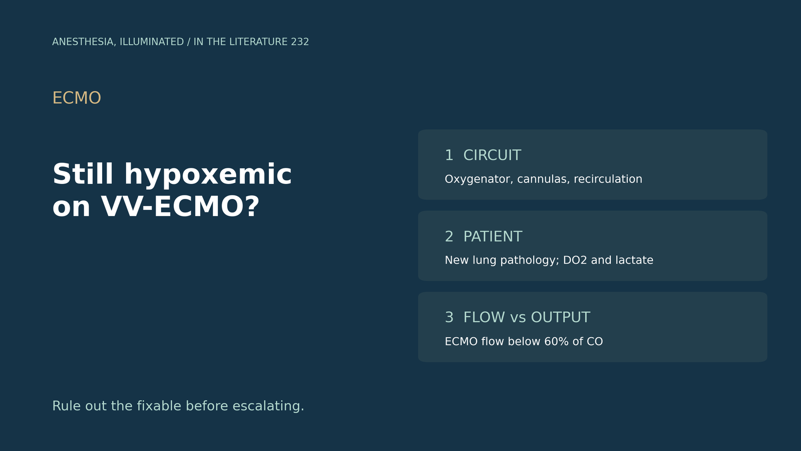
- Circuit first. A rising pre-oxygenator saturation with falling arterial saturation indicates recirculation, and cannula malposition can significantly increase it.[1]
- Then the patient. Mucus plugging, pneumonia, pneumothorax and pulmonary embolism should be sought when saturation falls.[2]
- Saturation is not delivery. PaO2 and SaO2 cannot estimate the oxygen present when circuit and native flows mix.[3]
- Flow ratio. If ECMO flow is below 60% of cardiac output, arterial saturation will predictably be below 90%.[4]
- Lowering cardiac output to raise saturation may reduce oxygen delivery.[3], [5]
A patient on venovenous ECMO for severe ARDS has an arterial saturation in the low 80s despite full circuit flow. What should be checked before escalating support?[2], [5]
Step 1: the circuit and recirculation
Abrams et al. (ASAIO J 2015) describe the bedside sign: a rising pre-oxygenator saturation with falling arterial saturation indicates recirculation, which is most apparent when pre-oxygenator saturation exceeds arterial saturation.[1] Cannula malposition can significantly increase recirculation, and the femoral drainage cannula tip should be positioned near the atrial-IVC junction.[1] Gagliardi and Gagliardi (Medicina 2024) report that recirculation ranges from 2% to 57%, that about 10 to 30% is empirically acceptable without a standardized cut-off, and that cannulas should be at least 10 to 13 cm apart.[6] Recirculation has been shown to increase with ECMO flow.[7] Whether to correct it depends on whether systemic oxygen requirements are being met.[1] For the membrane itself, Shoni et al. (J Cardiothorac Vasc Anesth 2023) advised oxygenator exchange for post-oxygenator PaO2 below 200 mmHg without planned decannulation within 24 hours.[4]
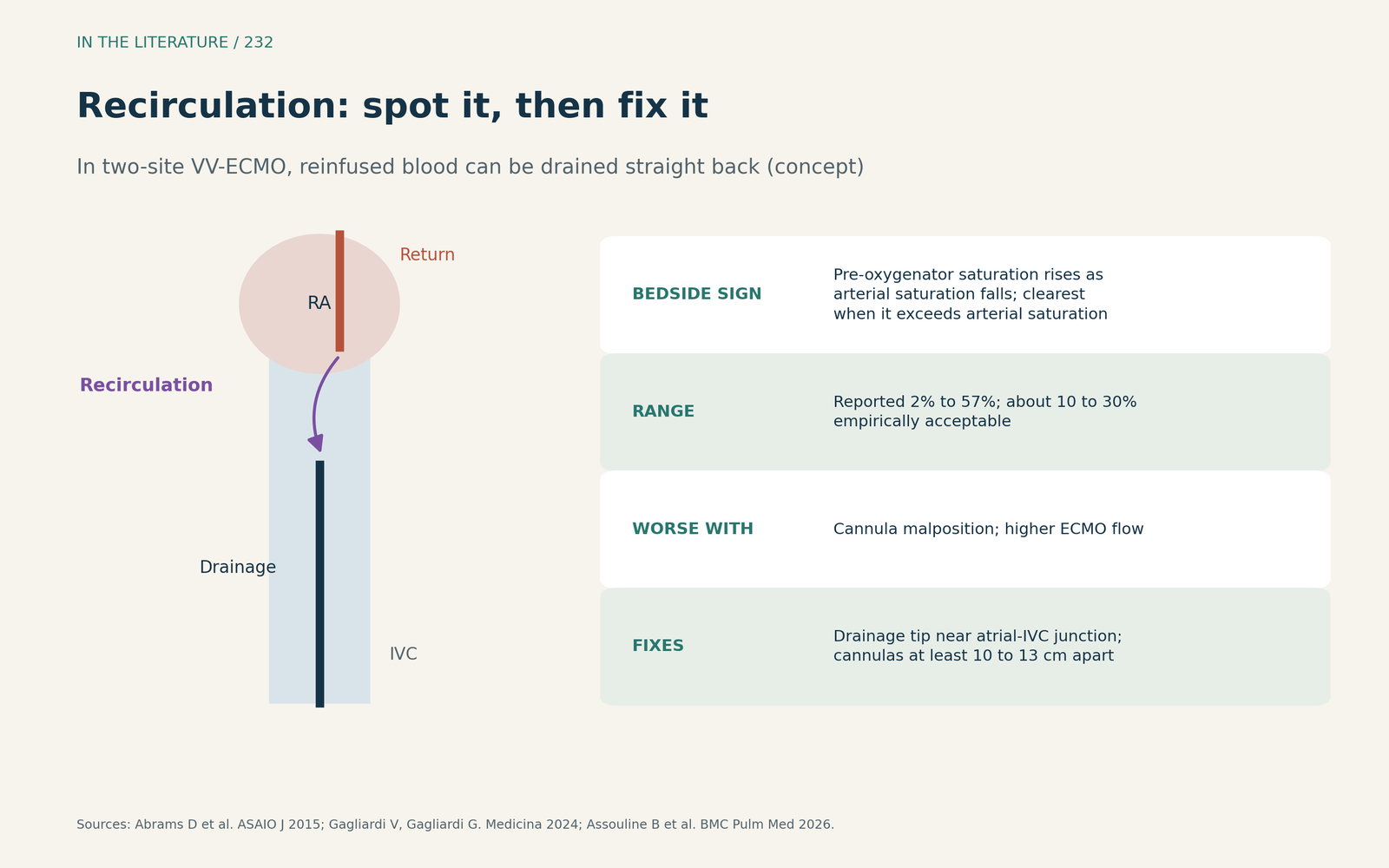
Figure 1. Recirculation: how to spot it and what changes it. Concept redrawn from Abrams et al.; values from Gagliardi and Gagliardi and Assouline et al.[1], [6], [7] View larger.

Step 2: new pathology and oxygen delivery
Ortoleva and Chweich (J Cardiothorac Vasc Anesth 2022) recommend looking for mucus plugging, pneumonia, pneumothorax and pulmonary embolism when saturation falls.[2] Kopanczyk et al. (J Cardiothorac Vasc Anesth 2022) note that PaO2 and SaO2 cannot estimate the oxygen present when flows mix; inadequate oxygenation usually corresponds to a DO2/VO2 ratio below 2:1.[3] Ortoleva and Chweich follow serial lactate as a marker of inadequate DO2:VO2 and tolerate saturations of 80% or greater when the patient is otherwise supported.[2] Zante et al. (J Cardiothorac Vasc Anesth 2021) showed in their model that higher cardiac output increased oxygen delivery despite lower SaO2, so a rising SaO2 may not indicate rising delivery.[8]
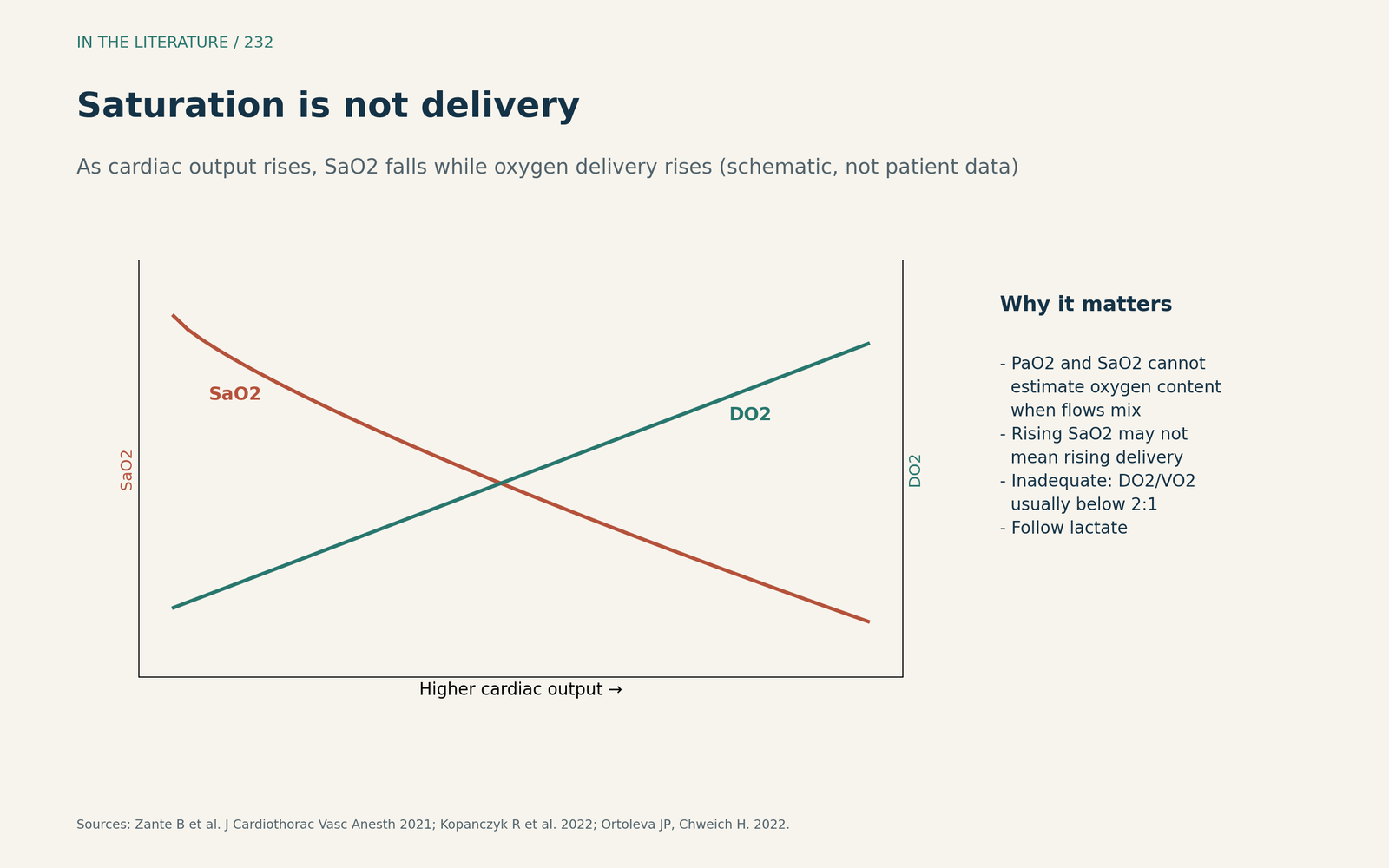
Figure 2. Saturation is not delivery (schematic). Concept redrawn from Zante et al.; interpretation from Kopanczyk et al.[8], [3] View larger.

Step 3: flow, cardiac output and escalation
Adequate oxygenation requires extracorporeal flow of roughly two-thirds of native cardiac output,[6] and if ECMO flow is below 60% of cardiac output, arterial saturation will predictably be below 90%.[4] Oxygen consumption can be lowered with deeper sedation, paralytics and fever reduction.[4] In Masi et al. (Cardiovasc Drugs Ther 2025), cardiac output control was considered only after recirculation, cannula position and membrane function were ruled out, and it decreased DO2, especially with beta-blockers; the study was monocentric and retrospective with a small sample.[5] Lowering cardiac output may decrease oxygen delivery, and hybrid or parallel circuits should be considered only when inadequate oxygenation is certain.[3]
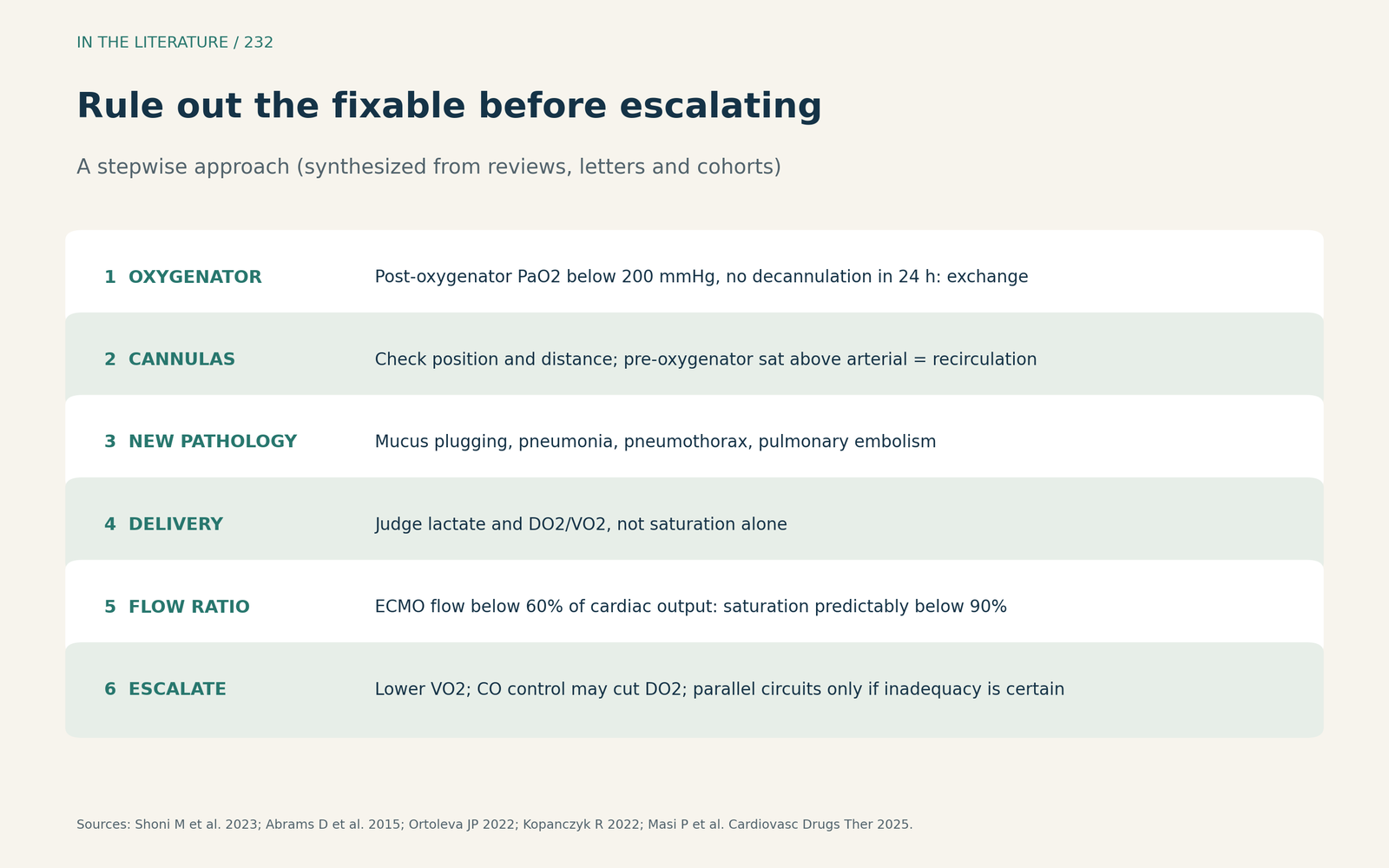
Figure 3. Rule out the fixable before escalating. Synthesized from Abrams et al., Ortoleva and Chweich, Shoni et al., Masi et al. and Kopanczyk et al.[1], [2], [4], [5], [3] View larger.

Three takeaways
- Check the oxygenator, cannula position and recirculation first; pre-oxygenator saturation above arterial saturation points to recirculation.[1], [4]
- Judge oxygen delivery and lactate, not saturation alone, and look for new lung pathology.[3], [2], [8]
- Compare ECMO flow with cardiac output; lowering cardiac output can raise saturation while reducing delivery.[4], [5]
References
- Abrams D, Bacchetta M, Brodie D. Recirculation in venovenous extracorporeal membrane oxygenation. ASAIO J. 2015;61(2):115–121. doi:10.1097/MAT.0000000000000179
- Ortoleva JP, Chweich H. Normalizing the abnormal: hypoxemia in venovenous ECMO. J Cardiothorac Vasc Anesth. 2022;36(8):3433–3434. doi:10.1053/j.jvca.2022.05.002
- Kopanczyk R, Bhatt A, Kumar N, Henson CP. Persistent hypoxemia in COVID-19 patients on ECMO: keep your eyes on the prize. J Cardiothorac Vasc Anesth. 2022;36(9):3710–3711. doi:10.1053/j.jvca.2022.04.011
- Shoni M, Lazar S, Jackson A, et al. Parallel venovenous extracorporeal membrane oxygenation circuits for refractory hypoxemia in a super-super-obese patient. J Cardiothorac Vasc Anesth. 2023;37(7):1304–1314. doi:10.1053/j.jvca.2023.03.008
- Masi P, Tchatat Wangueu L, Bagate F, et al. Control of cardiac output with ivabradine or beta-blockers for refractory hypoxemia under veno-venous ECMO for severe ARDS. Cardiovasc Drugs Ther. 2025;39(5):961–966. doi:10.1007/s10557-024-07650-5
- Gagliardi V, Gagliardi G. Recirculation in veno-venous extracorporeal membrane oxygenation. Medicina (Kaunas). 2024;60(12):1936. doi:10.3390/medicina60121936
- Assouline B, Sieurin C, Looyens C, et al. Impact of cannula configuration on efficiency and mechanical ventilation intensity during venovenous ECMO. BMC Pulm Med. 2026;26:319. doi:10.1186/s12890-026-04363-3
- Zante B, Berger DC, Schefold JC, Bachmann KF. Dissociation of arterial oxygen saturation and oxygen delivery in VV-ECMO: the trend is your friend. J Cardiothorac Vasc Anesth. 2021;35(3):962–963. doi:10.1053/j.jvca.2020.06.084