ITL-0a3e6088b685
Juan Li, MD · Updated November 17, 2026
Watch the video
Key points
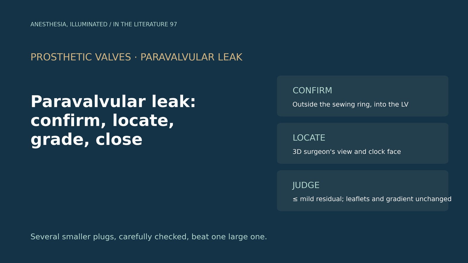
- First confirm that the jet is paravalvular. It must arise outside the sewing ring and enter the ventricle; eccentric transvalvular jets, washing jets and other shunts can mimic a leak.[1], [2], [3]
- Locate the leak with three-dimensional transesophageal echocardiography (3D TEE) in the en face surgeon's view and describe it on a clock face. For mitral prostheses viewed from the left atrium, the aortic valve is placed at 12 o'clock.[1], [2], [4]
- Grade with several parameters. Circumferential extent of more than 30% suggests severe regurgitation, but vena contracta should not be used alone, and the extent bands were developed for the aortic position.[1], [2], [5]
- A successful closure leaves no more than mild residual leak without leaflet impingement or a gradient change. Residual leak greater than mild was associated with higher mortality.[5]
- Transcatheter closure had lower early mortality than surgery but more residual leak; long-term mortality did not differ significantly.[6], [7]
Months after a mitral valve replacement, a patient has new hemolysis and heart failure. TEE shows an eccentric jet near the sewing ring. Is it really paravalvular, where exactly is it, how severe is it, and what should the echocardiographer confirm before and after closure?[1], [2]
Is it really paravalvular?
The 2019 American Society of Echocardiography (ASE) guideline stresses that the jet must enter the left ventricle to be considered true regurgitation, and recommends a coordinated nomenclature for its site using a clock depiction or anatomic landmarks.[1] Smeltz and colleagues list other sources of a left atrial jet that must be distinguished from a mitral leak, including a patent foramen ovale and the normal "washing" jets of mechanical valves.[2]
Eccentric transvalvular jets can mislead. Bykhovsky and colleagues described an apparent paravalvular leak after transcatheter aortic valve replacement in which wire probing failed to find any paravalvular communication, and intracardiac echocardiography showed a restricted cusp as the true source.[3]
Where is it?
The ASE guideline describes 3D TEE with color Doppler as the imaging technique of choice for locating and characterizing prosthetic mitral paravalvular regurgitation and for guiding closure.[1] By convention, the mitral prosthesis is viewed from the left atrium in the surgeon's view, rotated so that the aortic valve is at the top. A leak between 12 and 6 o'clock lies on the septal side, and between 6 and 12 o'clock on the lateral side.[2]
Clock-face reporting is less standardized in the aortic position.[4] With mechanical aortic prostheses, Huang and colleagues used off-axis and deep transgastric views to work around shadowing from the valve.[4]
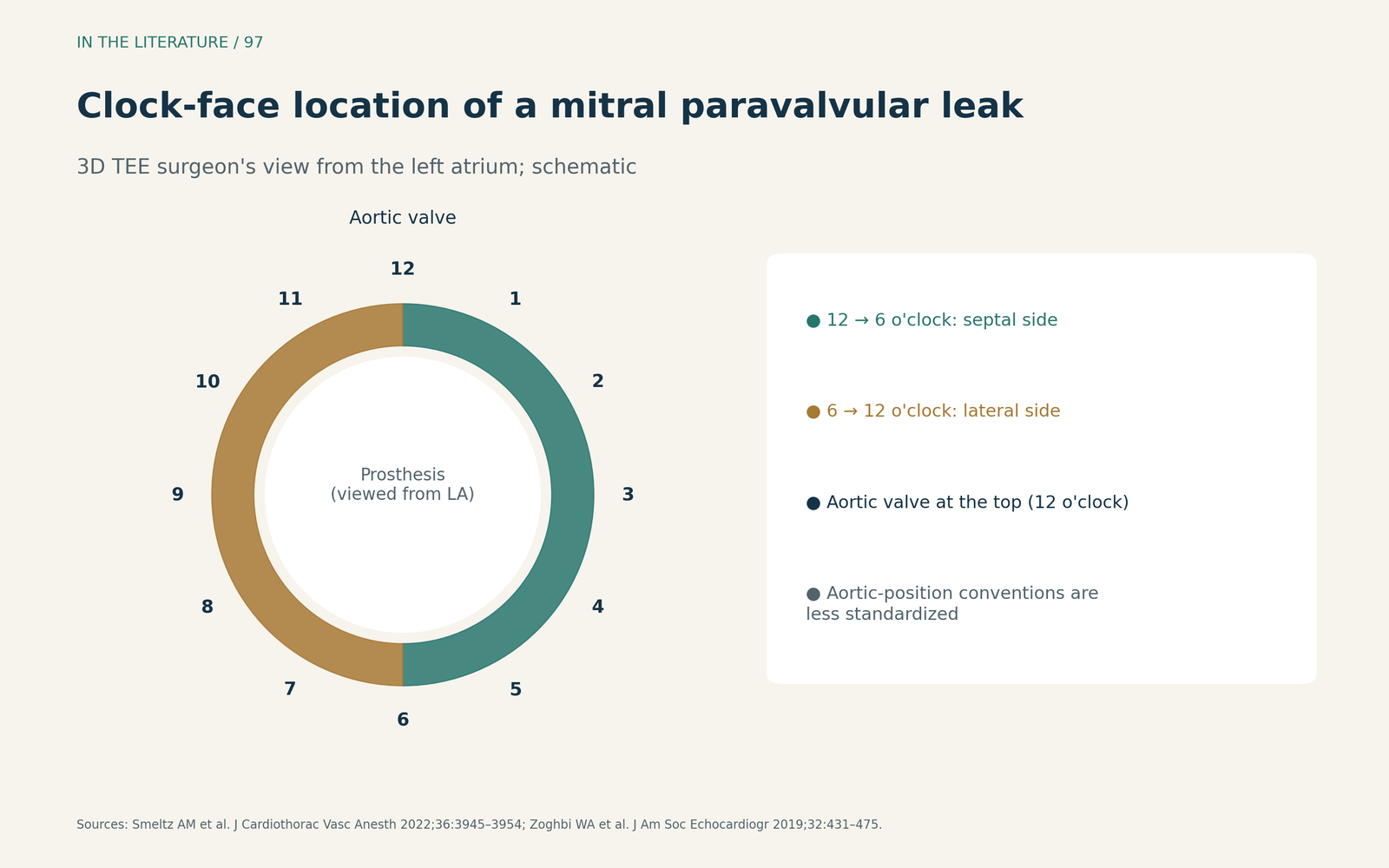
Figure 1. Reporting the location. Schematic based on the convention described by Smeltz et al. and the ASE nomenclature recommendation; not to scale.[1], [2] View larger.

How severe is it?
Severity rests on several measures. The ASE guideline uses the circumferential extent of the jet in the short-axis view, with continuous extent greater than 30% indicating severe regurgitation, together with vena contracta, flow convergence and Doppler signs.[1] Papadopoulos and colleagues add that vena contracta width should not be used as a standalone quantitative measure, and that CT is better for defining the anatomic substrate than for grading severity.[5]
The extent bands come from aortic prostheses. Smeltz and colleagues cite less than 10% as mild, 10% to 30% as moderate and more than 30% as severe, and note uncertainty about applying these criteria to the mitral position.[2] In Huang and colleagues' case, a leak occupying approximately 20% to 25% of the aortic annular circumference was graded moderate.[4]
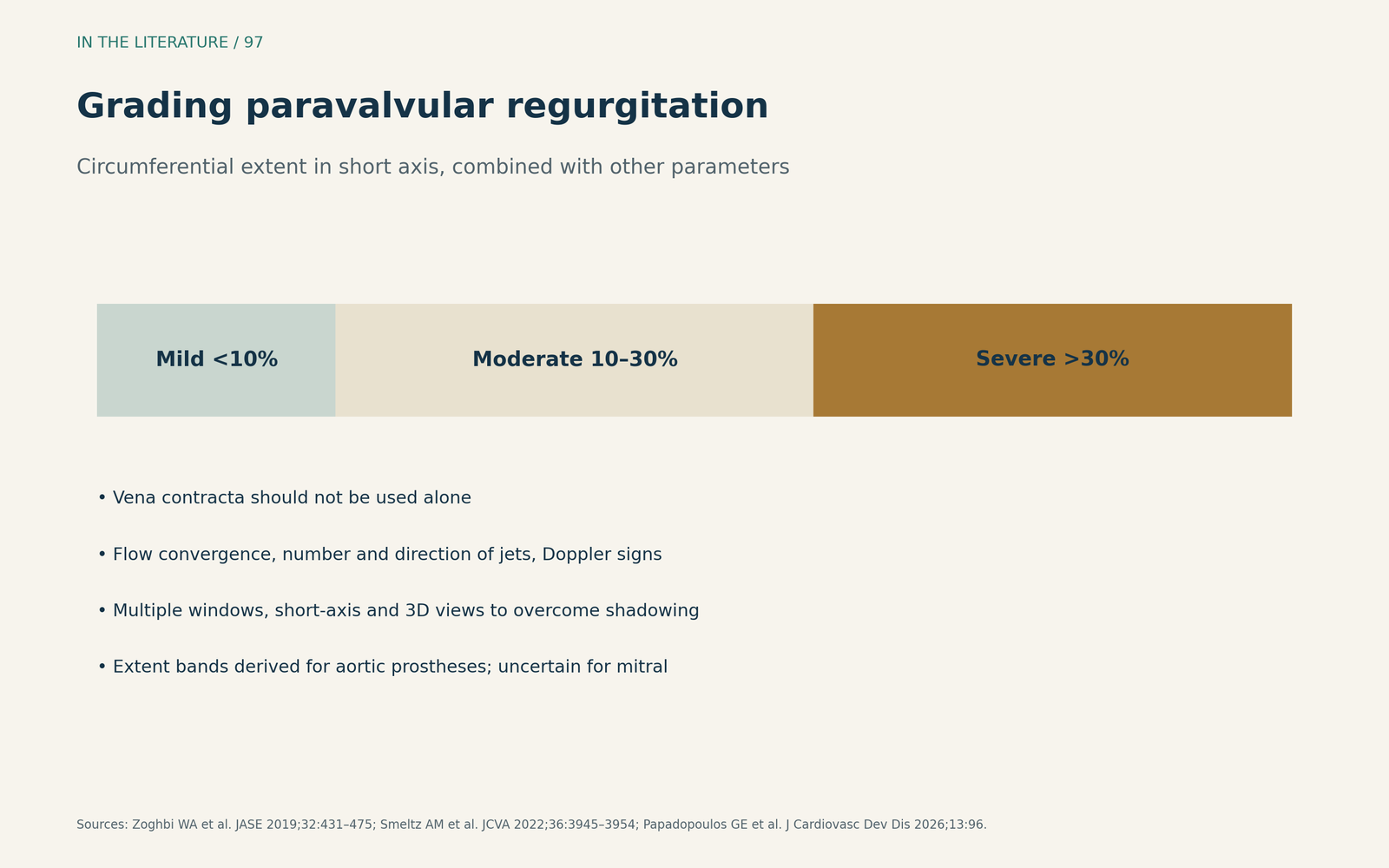
Figure 2. Grade with more than one parameter. Bands as cited from the ASE guideline and by Smeltz et al.; derived for aortic prostheses, uncertain for the mitral position.[1], [2], [5] View larger.

Guiding and judging the closure
Not every leak suits a plug. Huang and colleagues list intracardiac thrombus, valve rocking and a leak larger than one-third of the prosthesis circumference as contraindications to percutaneous closure.[4] Smeltz and colleagues note that surgical leak tests can be falsely negative, and that several smaller plugs are preferable to one large plug.[2]
Papadopoulos and colleagues define the procedural end point as no more than mild residual regurgitation without prosthetic leaflet impairment or device embolization, and call for immediate recapture whenever leaflet motion or transvalvular gradients are affected. In a cohort they cite, patients left with no more than mild residual leak had lower 30-day (1% vs 14%) and 1-year (15% vs 39%) mortality than those with more.[5]
Transcatheter or surgical?
In the multicenter KISS registry, Güner and colleagues found in-hospital death more frequent after surgery than after transcatheter closure (15.9% vs 4.7%), while residual leak after the index procedure was more frequent with transcatheter closure (17% vs 7.3%). Long-term mortality did not differ significantly (adjusted hazard ratio 1.11; 95% CI 0.67–1.81).[6] A meta-analysis by Improta and colleagues, which appears to include the KISS data, similarly found lower 30-day mortality (odds ratio 0.28) but more residual moderate or severe leak (odds ratio 3.56) with transcatheter closure.[7] Both are observational, and patients selected for surgery likely differed.
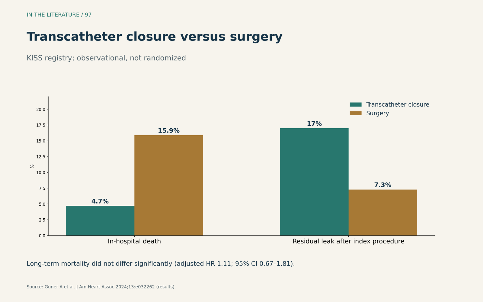
Figure 3. A trade-off between early risk and completeness. Redrawn from the results of the KISS registry (Güner et al.); observational, not randomized.[6] View larger.

Three takeaways
- Prove the jet is paravalvular. Trace its origin outside the sewing ring into the ventricle and exclude mimics.[1], [2], [3]
- Locate and grade consistently. Use the 3D surgeon's view and clock face, and combine circumferential extent with other parameters.[1], [2], [5]
- Define success before deploying. Aim for no more than mild residual leak with unchanged leaflet motion and gradients; weigh early risk against completeness when choosing transcatheter or surgical repair.[5], [6], [7]
References
- Zoghbi WA, Asch FM, Bruce C, et al. Guidelines for the evaluation of valvular regurgitation after percutaneous valve repair or replacement: a report from the American Society of Echocardiography developed in collaboration with the Society for Cardiovascular Angiography and Interventions, Japanese Society of Echocardiography, and Society for Cardiovascular Magnetic Resonance. J Am Soc Echocardiogr. 2019;32(4):431–475. doi:10.1016/j.echo.2019.01.003
- Smeltz AM, Merlo A, Ikonomidis JS, et al. Paravalvular leak immediately following repeat mitral valve replacement: considerations for repair in a high-risk patient. J Cardiothorac Vasc Anesth. 2022;36(10):3945–3954. doi:10.1053/j.jvca.2022.06.019
- Bykhovsky MR, Atianzar K, Agarwal S, et al. Use of intracardiac echocardiography to differentiate post-transcatheter aortic valve replacement valve insufficiency masquerading as paravalvular leak. CASE (Phila). 2020;4(3):111–114. doi:10.1016/j.case.2019.11.004
- Huang L, Mahmood F, Laham R, et al. Percutaneous approach to mechanical aortic valve prosthesis paravalvular leak closure: lessons learned. J Cardiothorac Vasc Anesth. 2024;38(5):1203–1210. doi:10.1053/j.jvca.2024.01.031
- Papadopoulos GE, Ninios I, Evangelou S, et al. Transcatheter paravalvular leak closure: a step-by-step guide. J Cardiovasc Dev Dis. 2026;13(2):96. doi:10.3390/jcdd13020096
- Güner A, Kırma C, Ertürk M, et al. Transcatheter closure or surgery for symptomatic paravalvular leaks: the multicenter KISS registry. J Am Heart Assoc. 2024;13(1):e032262. doi:10.1161/JAHA.123.032262
- Improta R, Di Pietro G, Odeh Y, et al. Transcatheter or surgical treatment of paravalvular leaks: a meta-analysis of 13 studies and 2003 patients. IJC Heart Vasc. 2025;56:101583. doi:10.1016/j.ijcha.2024.101583