ITL-e1b9a09101b9
Juan Li, MD · Updated November 18, 2026
Watch the video
Key points
- A normal prosthesis is mildly obstructive. Pooled data from 7092 aortic and 1684 mitral prostheses show that variation in gradients is mainly due to flow dependence.[1]
- Compare like with like. Doppler values should be compared with reference values for the specific valve type and size, and interpretation is easier with a baseline study.[1]
- A high gradient alone is not dysfunction. Severe mismatch can raise gradients; consensus definitions use EOA, DVI and change from baseline.[2]
- Body size changes the meaning. The same EOA of 1.3 cm2 may give a mean gradient of about 13 mmHg at a BSA of 1.5 m2, but nearly 35 mmHg at 2.5 m2.[3]
- Change over time matters most. A mean gradient rise above 10 mm Hg suggests bioprosthetic thrombosis, and a baseline TTE before discharge is advised.[2], [4]
During cardiac surgery for another lesion, TEE shows a mean gradient across a small aortic bioprosthesis that looks high, and the team wonders whether it is obstructed. How should the number be interpreted before calling it obstruction?[1], [2]
What "normal" looks like
Rosenhek and colleagues pooled normal Doppler values from 7092 aortic and 1684 mitral prostheses. Even normally functioning prostheses are to some degree obstructive, and variation in velocities and gradients is mainly due to flow dependence, so normal values should ideally be given for specific flow rates. Pressure half-time is questionable for normally functioning prostheses, and interpretation is easier when baseline data are available.[1]
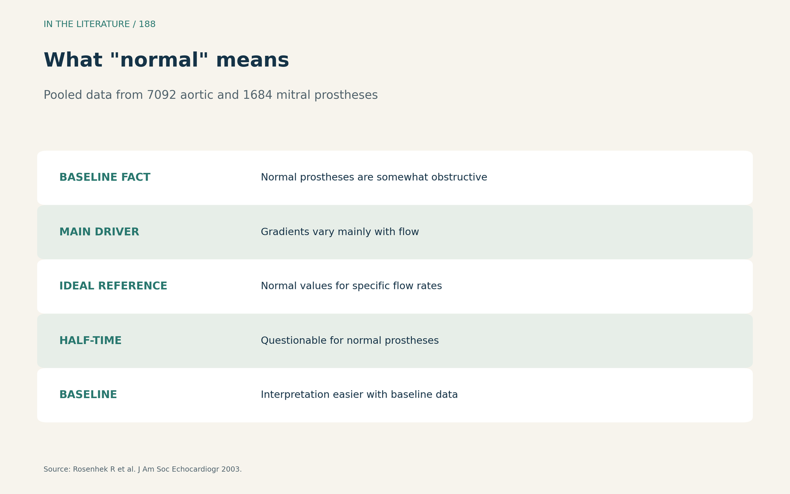
Figure 1. Normal is not zero. Synthesized from Rosenhek et al.[1] View larger.

Mismatch versus acquired obstruction
The Heart Valve Collaboratory treats a small EOA below 1.2 cm2 or a low Doppler velocity index as red flags, but notes that a high gradient may reflect severe mismatch and that dysfunction should not be diagnosed on a high gradient alone. Moderate haemodynamic deterioration is defined as a mean gradient rise of at least 10 mm Hg, and change during follow-up is emphasized.[2] Talapaneni and colleagues define mismatch as an indexed EOA of 0.85 cm2/m2 or less (severe at 0.65 or less), show that, at a normal cardiac index, an EOA of 1.3 cm2 may give a mean gradient of about 13 mmHg at a BSA of 1.5 m2 but nearly 35 mmHg at 2.5 m2, warn that indexed EOA may overestimate mismatch in larger patients, and advise reading it as a continuous variable.[3]
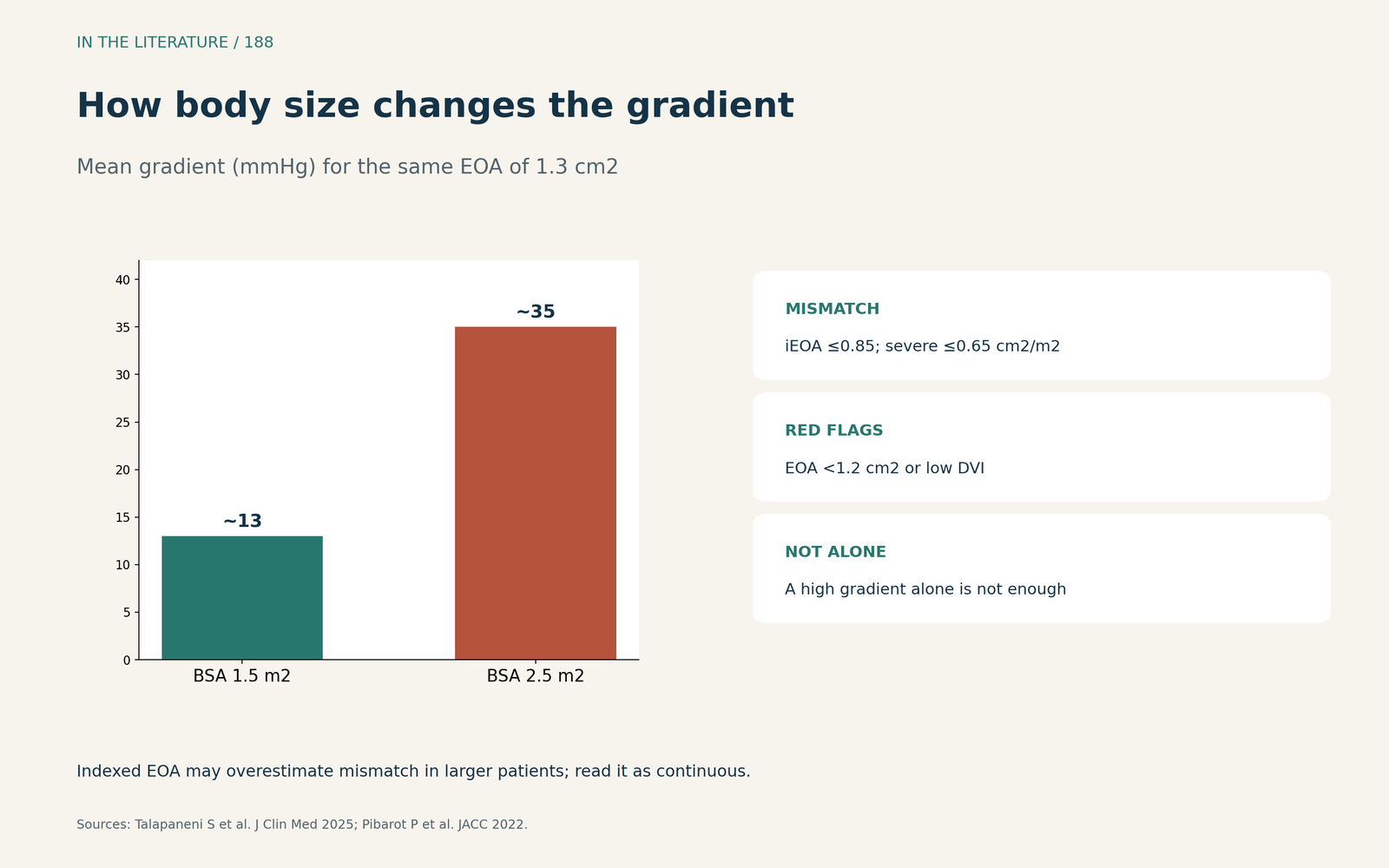
Figure 2. Gradient, orifice, body size. Synthesized from Pibarot et al. and Talapaneni et al.[2], [3] View larger.

Thrombus, pannus and imaging
Dangas and colleagues advise comparing Doppler values with reference values for valve type and size, treat a mean gradient rise above 10 mm Hg as suggesting bioprosthetic thrombosis, and recommend a baseline TTE before discharge after bioprosthetic implantation.[4] In Kim's meta-analysis of seventeen studies with 229 patients, a high gradient on TTE could suggest obstruction but also occurred with high output, mismatch and regurgitation; pannus caused 55.8% of obstruction cases, which is why further imaging is needed.[5]
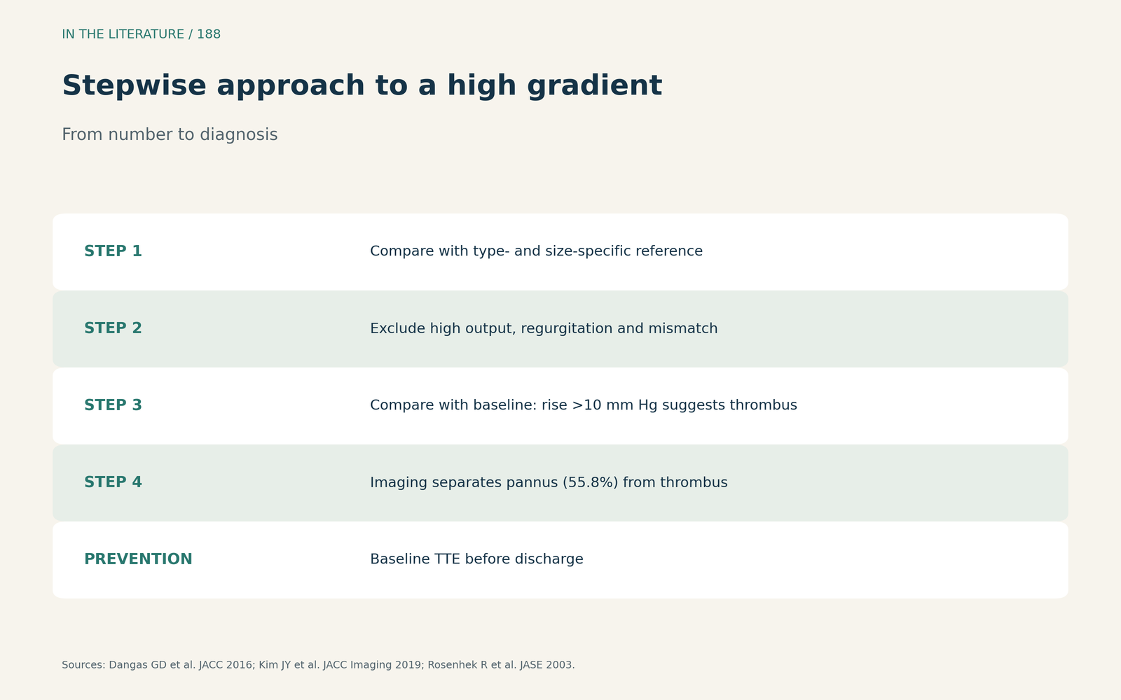
Figure 3. From number to diagnosis. Synthesized from Dangas et al., Kim et al. and Rosenhek et al.[4], [5], [1] View larger.

Three takeaways
- Judge a prosthetic gradient against the valve's own type, size and flow.[1]
- Use flow-independent indices and body size before calling obstruction.[2], [3]
- A baseline study turns a single number into a trend.[1], [4]
References
- Rosenhek R, Binder T, Maurer G, Baumgartner H. Normal values for Doppler echocardiographic assessment of heart valve prostheses. J Am Soc Echocardiogr. 2003;16(11):1116–1127. doi:10.1067/S0894-7317(03)00638-2
- Pibarot P, Herrmann HC, Wu C, et al. Standardized definitions for bioprosthetic valve dysfunction following aortic or mitral valve replacement: JACC state-of-the-art review. J Am Coll Cardiol. 2022;80(5):545–561. doi:10.1016/j.jacc.2022.06.002
- Talapaneni S, Ahmad D, Khandelwal M, et al. Prosthesis–patient mismatch following aortic valve replacement: a comprehensive review. J Clin Med. 2025;14(24):8868. doi:10.3390/jcm14248868
- Dangas GD, Weitz JI, Giustino G, Makkar R, Mehran R. Prosthetic heart valve thrombosis. J Am Coll Cardiol. 2016;68(24):2670–2689. doi:10.1016/j.jacc.2016.09.958
- Kim JY, Suh YJ, Han K, et al. Diagnostic value of advanced imaging modalities for the detection and differentiation of prosthetic valve obstruction: a systematic review and meta-analysis. JACC Cardiovasc Imaging. 2019;12(11):2182–2192. doi:10.1016/j.jcmg.2018.11.033