ITL-4364a15186fc
Juan Li, MD · Updated November 17, 2026
Watch the video
Key points
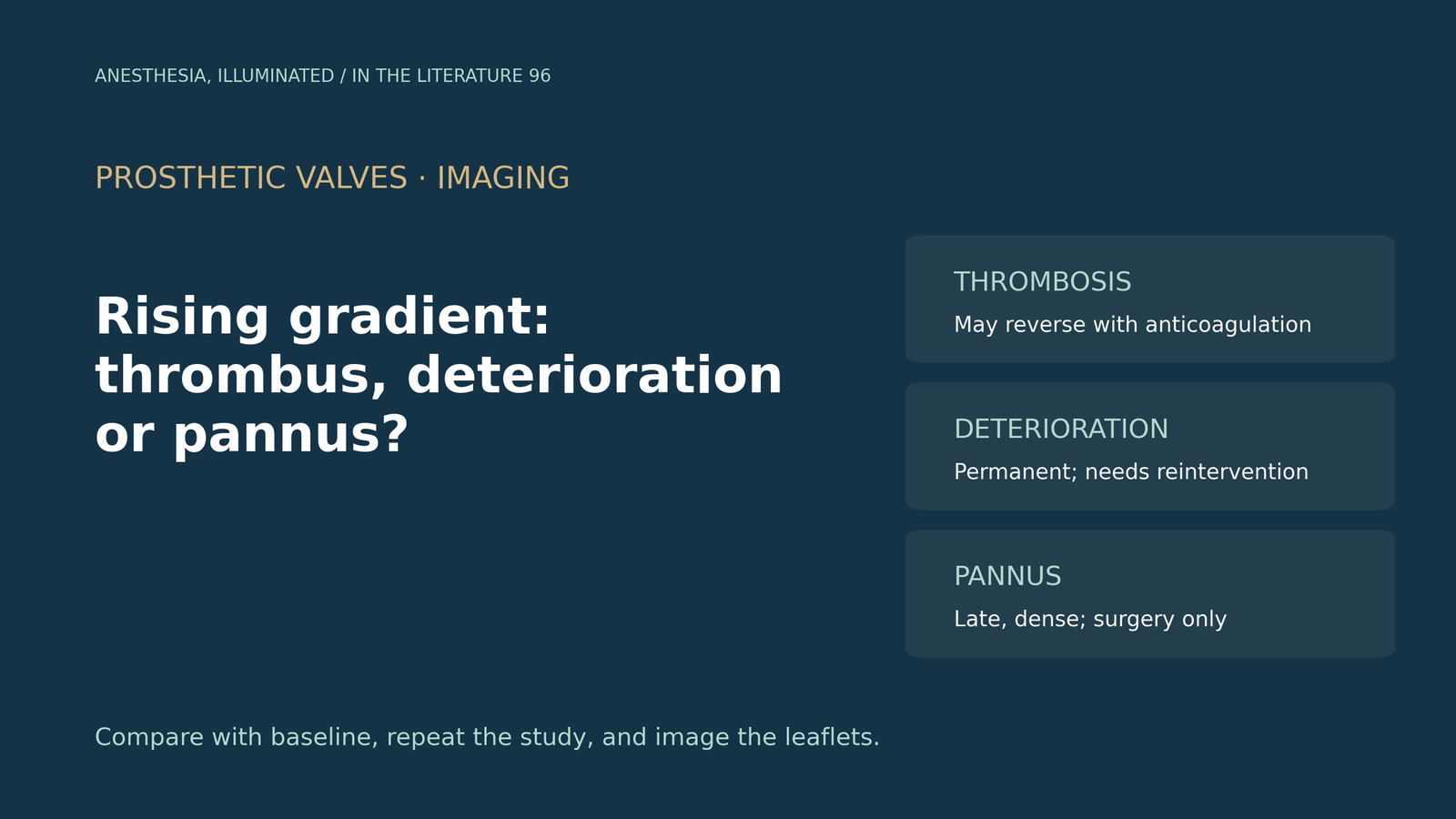
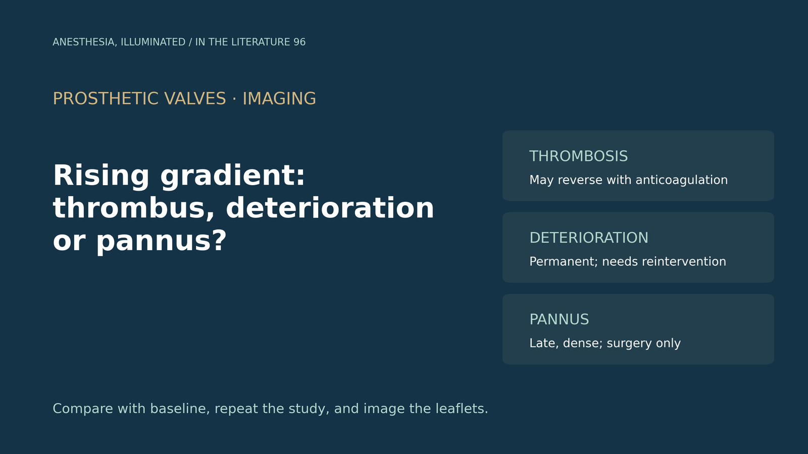
- A rising prosthetic valve gradient has several causes with different treatments. Thrombosis, including subclinical leaflet thrombosis, can respond to anticoagulation, structural valve deterioration (SVD) is permanent and needs reintervention, and pannus needs surgery.[1], [2]
- Timing and morphology help. In a Mayo Clinic series, bioprosthetic valve thrombosis was diagnosed a median of 10–24 months after implantation; on CT, hypoattenuated leaflet thickening points to thrombus.[1], [3], [4]
- Hemodynamic SVD criteria depend on a rise in mean gradient from a baseline study 1–3 months after implantation, but single measurements are unreliable: most patients labelled with SVD at one visit were not labelled at the next.[2], [5]
- An anticoagulation trial is both test and treatment. Median recovery of bioprosthetic thrombosis took 3.8 months, and a gradient fall of more than 25% within 3 months predicted recovery.[3], [4]
- For mechanical valves, thrombus is typically early and soft and pannus late, dense and on the ventricular side. Indeterminate masses are generally managed as pannus.[1], [6]
A patient who had a bioprosthetic aortic valve replacement 3 years ago comes for noncardiac surgery. The mean gradient across the valve has risen from 12 to 30 mmHg, and the leaflets look thickened. Is this thrombosis that anticoagulation can reverse, structural deterioration that needs reintervention, or something else, and how do you tell?[1], [2]
Three causes, three treatments
The 2024 American Society of Echocardiography (ASE) prosthetic valve guideline separates SVD, defined as intrinsic permanent change to the valve such as calcification or leaflet tear, from nonstructural dysfunction, including pannus, and from thrombosis. On echocardiography, thrombus on a bioprosthesis may appear simply as valve thickening, so it can mimic SVD.[1]
This distinction matters because the treatments differ. Patel and colleagues note that contemporary SVD definitions exclude valve thrombosis, and that true SVD is treated by reintervention.[2] Velders and colleagues describe SVD as an irreversible process that does not resolve without reintervention, whereas thrombosis and endocarditis can temporarily produce similar echocardiographic abnormalities.[5]
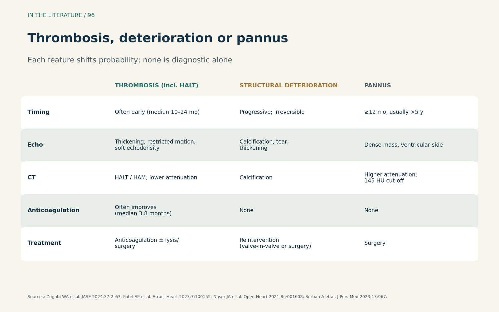
Figure 1. Thrombosis, structural deterioration or pannus. Synthesized from the 2024 ASE guideline and reviews by Patel, Serban and Samadzadeh Tabrizi and colleagues; features shift probability rather than prove a diagnosis.[1], [2], [4], [6] View larger.

What the gradient can and cannot tell you
The Valve Academic Research Consortium 3 (VARC-3) stages hemodynamic deterioration against a reference echocardiogram 1–3 months after the procedure. Moderate deterioration (stage 2) is a rise in mean gradient of 10 mmHg or more to at least 20 mmHg, together with a fall in effective orifice area or Doppler velocity index. Severe deterioration (stage 3) is a rise of 20 mmHg or more to at least 30 mmHg.[2]
These thresholds are not as stable as they look. In 1,118 patients after surgical aortic valve replacement, Velders and colleagues found that after a first SVD label, 59–65% had no SVD at the next visit, depending on the definition used. The 95% prediction interval for within-patient change in gradient ranged from −9.6 to 7.5 mmHg, overlapping the 10 mmHg threshold. VARC-3 itself advises using at least 2 serial echocardiograms rather than a single measurement.[5]
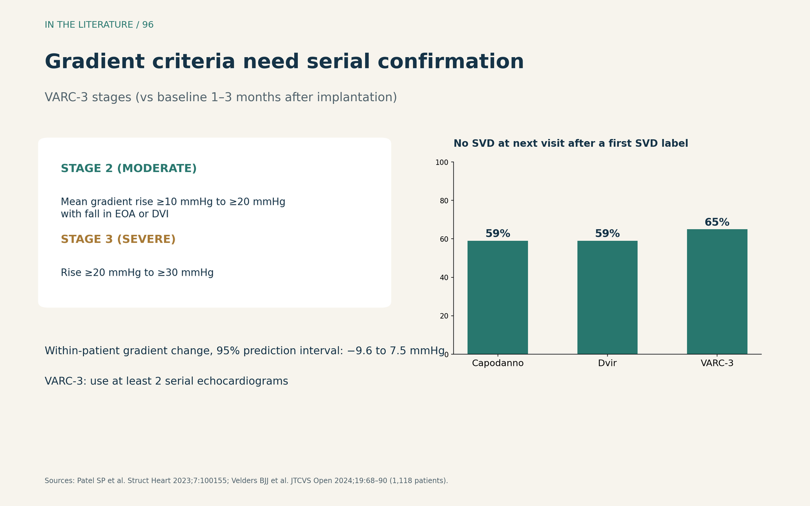
Figure 2. Gradient criteria need serial confirmation. VARC-3 stages as summarized by Patel et al.; consistency data redrawn from Velders et al. (1,118 patients).[2], [5] View larger.

Imaging the leaflets
Timing and imaging add clues. In Naser and colleagues' series of 77 patients with bioprosthetic valve thrombosis, median time to diagnosis was 24, 21 and 10 months for aortic, mitral and tricuspid valves.[3] On CT, the ASE guideline describes hypoattenuated leaflet thickening (HALT), with reduced motion termed hypoattenuation affecting motion, in 3.6% to 40% of bioprostheses.[1] Samadzadeh Tabrizi and colleagues note that HALT is dynamic, with spontaneous resolution reported in up to 56%.[4]
In the ASE guideline's example, the mean gradient fell from 28 to 14 mmHg after anticoagulation, and HALT resolved on repeat CT.[1]
The anticoagulation trial
When thrombosis is suspected, anticoagulation tests the diagnosis. In Naser and colleagues' series, median time to recovery was 3.8 months, faster for mitral than aortic valves (2.5 vs 4.8 months) and for porcine than pericardial aortic valves (2.9 vs 20.3 months). Over half of patients needed more than 3 months, and a gradient reduction of more than 25% within the first 3 months predicted recovery. Five patients responded initially but then had rising gradients from degeneration.[3] In registries cited by Samadzadeh Tabrizi and colleagues, HALT resolved in all 36 patients treated with oral anticoagulants.[4]
A short or incomplete trial can therefore mislabel thrombosis as SVD, and an initial response does not exclude later degeneration.[3]
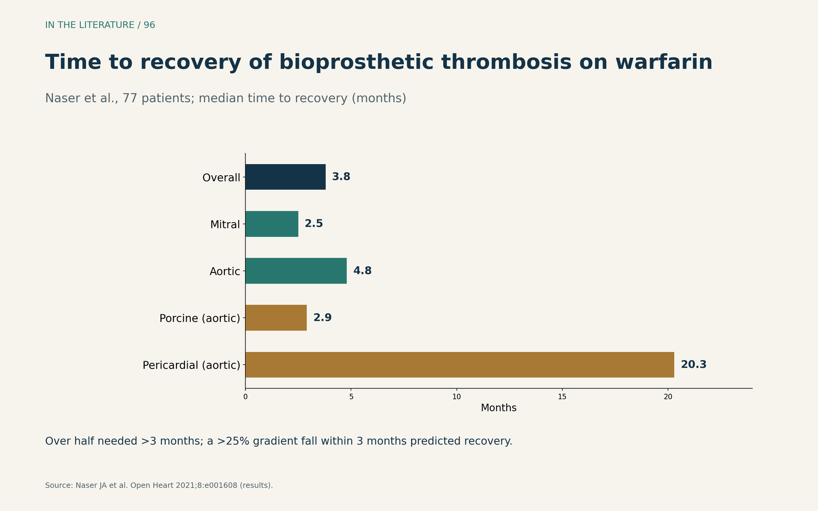
Figure 3. Recovery can take months. Redrawn from the results of Naser et al. (77 patients; median time to recovery).[3] View larger.

Mechanical valves: thrombus or pannus
For mechanical valves, the choice is mainly between thrombus and pannus. The ASE guideline notes that thrombus is seen more commonly early after surgery, whereas pannus is seen late, usually on the ventricular side; on CT, a cut-off of 145 Hounsfield units helps distinguish them.[1] Serban and colleagues add that pannus takes at least 12 months to form (usually more than 5 years), and that leaflet restriction, softer echodensity, a larger mass and rapid gradient development favor thrombus. Thrombus can be treated with anticoagulation, thrombolysis or surgery, but pannus only with surgery, and when imaging is indeterminate, management is generally as for pannus.[6]
Three takeaways
- Name the mechanism, not just the gradient. Thrombosis, SVD and pannus look alike on Doppler but are treated differently.[1], [2]
- Confirm with serial studies and imaging. Compare with the baseline echocardiogram, repeat measurements, and use TEE or CT to look at the leaflets.[1], [5]
- Give anticoagulation enough time. Recovery from bioprosthetic thrombosis often takes more than 3 months; mechanical valve pannus needs surgery.[3], [4], [6]
References
- Zoghbi WA, Jone PN, Chamsi-Pasha MA, et al. Guidelines for the evaluation of prosthetic valve function with cardiovascular imaging: a report from the American Society of Echocardiography developed in collaboration with the Society for Cardiovascular Magnetic Resonance and the Society of Cardiovascular Computed Tomography. J Am Soc Echocardiogr. 2024;37(1):2–63. doi:10.1016/j.echo.2023.10.004
- Patel SP, Garcia S, Sathananthan J, et al. Structural valve deterioration in transcatheter aortic bioprostheses: diagnosis, pathogenesis, and treatment. Struct Heart. 2023;7(3):100155. doi:10.1016/j.shj.2022.100155
- Naser JA, Petrescu I, Ionescu F, et al. Gradient changes in bioprosthetic valve thrombosis: duration of anticoagulation and strategies to improve detection. Open Heart. 2021;8(1):e001608. doi:10.1136/openhrt-2021-001608
- Samadzadeh Tabrizi N, Fishberger G, Musuku SR, Shapeton AD. Hypoattenuated leaflet thickening: a comprehensive review of contemporary data. J Cardiothorac Vasc Anesth. 2024;38(11):2761–2769. doi:10.1053/j.jvca.2024.06.043
- Velders BJJ, Vriesendorp MD, Asch FM, et al. Current definitions of hemodynamic structural valve deterioration after bioprosthetic aortic valve replacement lack consistency. JTCVS Open. 2024;19:68–90. doi:10.1016/j.xjon.2024.02.023
- Serban A, Dadarlat-Pop A, Achim A, et al. Diagnosis of left-sided mechanical prosthetic valve thrombosis: a pictorial review. J Pers Med. 2023;13(6):967. doi:10.3390/jpm13060967