ITL-f7c8a0ee5389
Juan Li, MD · Updated November 17, 2026
Watch the video
Key points
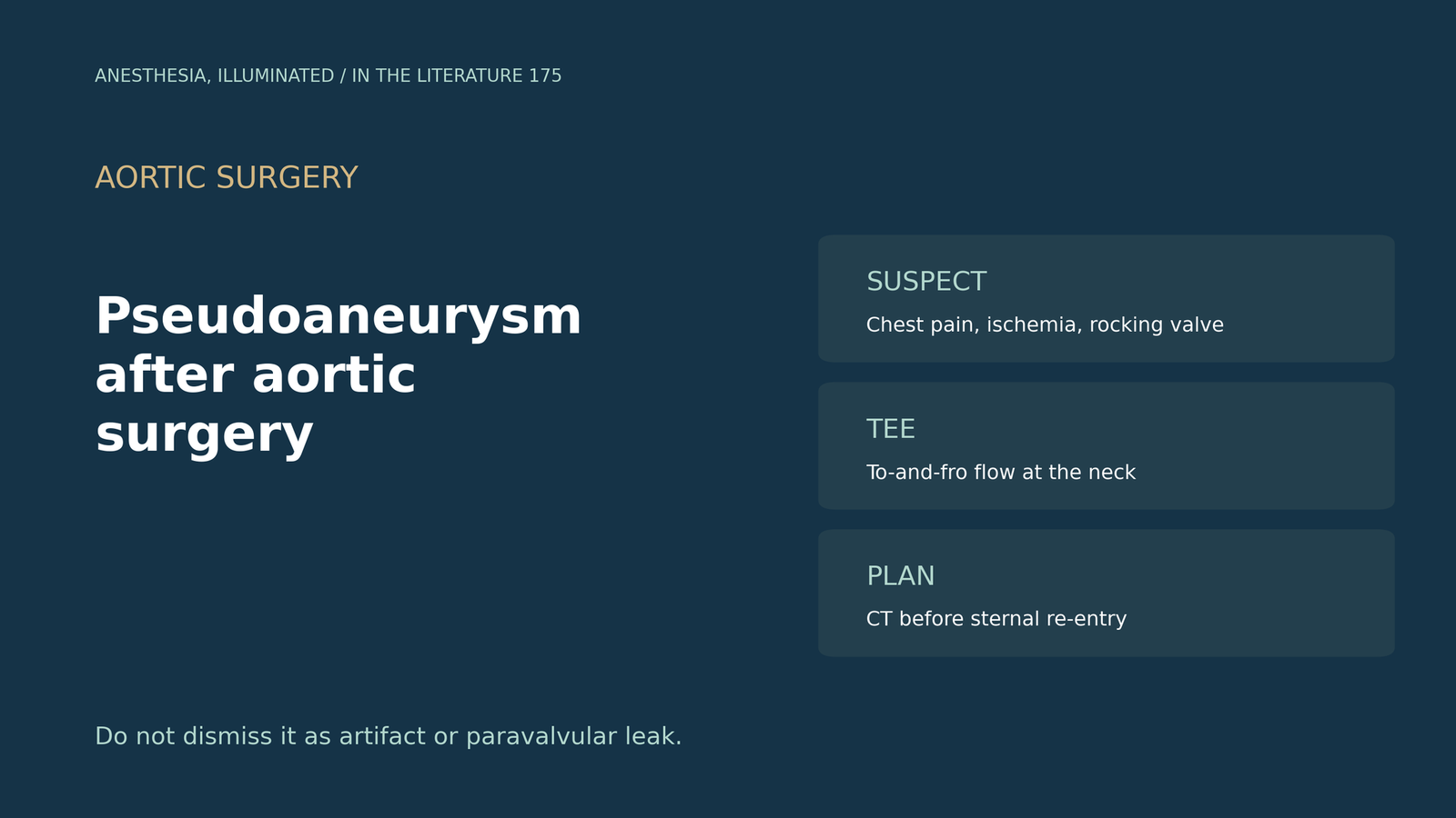
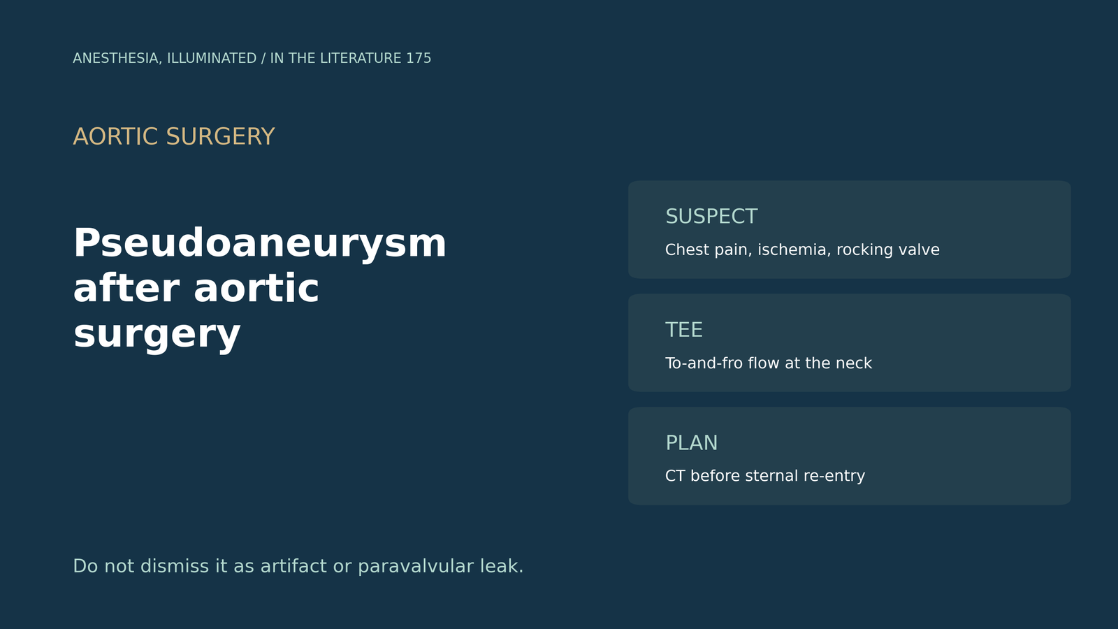
- It hides. Root pseudoaneurysm occurs in about 0.2% to 0.5% of cases, mostly with endocarditis, and in one case it was not suspected until fluoroscopy showed a rocking prosthesis and a left main lesion that worsened in systole.[1]
- Listen to the neck. Pulsed-wave Doppler shows bidirectional to-and-fro flow, and color Doppler a swirling yin-yang pattern.[2]
- Beware of mimics. Diastolic emptying can simulate paravalvular regurgitation, and the graft can collapse in systole while TTE is limited by valve shadowing.[3]
- Look for fistulas and artifacts. A pseudoaneurysm at the cannulation site produced a jet into the right atrium that could have been mistaken for motion artifact.[4]
- Plan the re-entry. Most are asymptomatic, but coronary compression can cause ischemia, and sternal re-entry may breach the sac.[5]
Years after aortic valve replacement, a patient presents with chest pain and dynamic narrowing of the left main coronary artery, and the CT was read only to exclude dissection. Could this be a pseudoaneurysm, and how should it be recognized?[1], [2]
How it presents
Seif and colleagues describe dynamic left main occlusion after aortic valve surgery. Root pseudoaneurysm occurs in about 0.2% to 0.5% of cases, most often with endocarditis; CT aortography had excluded dissection but was not formally reported, and pseudoaneurysm was not suspected until fluoroscopy showed prosthetic valve rocking and the left main lesion worsened markedly in systole, suggesting extrinsic compression. Reoperation can carry mortality above 20% to 30%.[1] Rodriguez-Patarroyo and colleagues note that most thoracic aortic pseudoaneurysms are asymptomatic though chest pain may occur; in their patient the sac compressed the proximal right coronary artery.[5]
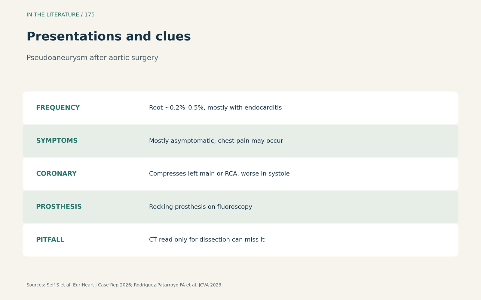
Figure 1. Presentations and clues. Synthesized from Seif et al. and Rodriguez-Patarroyo et al.[1], [5] View larger.

Echo signs and pitfalls
Maraboto Gonzalez and colleagues define a pseudoaneurysm as an outpouching from injury to the vessel wall; pulsed-wave Doppler at the neck showed bidirectional flow, color Doppler shows a swirling yin-yang pattern, a high upper esophageal view was needed to keep the lesion in view, and recognizing these features is important.[2] Li and colleagues found that acoustic shadowing from mechanical valves limited TTE; on TEE the graft collapsed in systole as the pseudoaneurysm expanded, and diastolic emptying into the ventricle simulated paravalvular regurgitation. Signs include a pulsatile echo-free space that fills with blood.[3] Wang and colleagues describe a pseudoaneurysm at the original cannulation site with a high-velocity jet into the right atrium that could have been mistaken for motion artifact.[4]
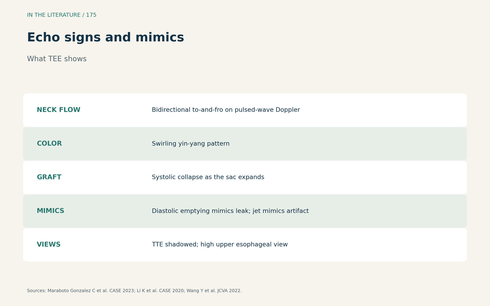
Figure 2. Echo signs and mimics. Synthesized from Maraboto Gonzalez et al., Li et al. and Wang et al.[2], [3], [4] View larger.

Planning before re-entry
Preoperative CT is vital to define the relation to the sternum, and in Li's cited series reoperation carried an operative mortality of 6.9%.[3] Wang and colleagues needed CT angiography for detailed planning,[4] and Rodriguez-Patarroyo and colleagues warn that sternal re-entry may breach the pseudoaneurysm and cause exsanguination.[5]
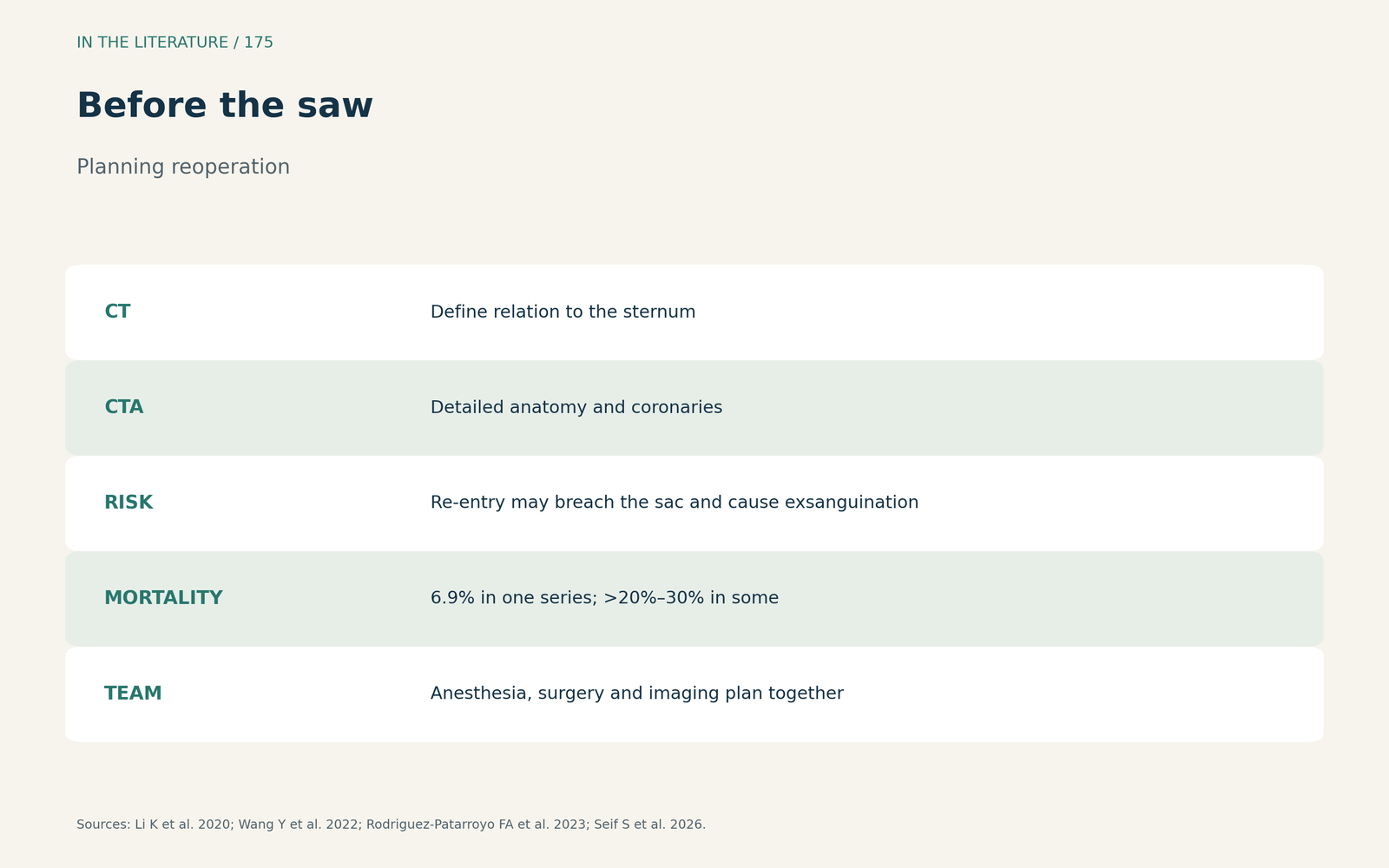
Figure 3. Before the saw. Synthesized from Li et al., Wang et al., Rodriguez-Patarroyo et al. and Seif et al.[3], [4], [5], [1] View larger.

Three takeaways
- Think of it after any aortic or cardiac surgery. Especially with chest pain, coronary compression or a rocking prosthesis.[1], [5]
- Use Doppler at the neck. To-and-fro flow and a yin-yang swirl; do not dismiss it as artifact or paravalvular leak.[2], [3], [4]
- Get CT before re-entry. The sac may lie right under the sternum.[3], [5]
References
- Seif S, Kukoyi B, Mujahid A, Nawaytou O, Ratib K. Dynamic left main occlusion after aortic valve surgery: recognizing extrinsic compression by aortic root pseudoaneurysm—a case report. Eur Heart J Case Rep. 2026;10(10):ytag600. doi:10.1093/ehjcr/ytag600
- Maraboto Gonzalez C, Butala N, Patel NK, et al. Transesophageal echocardiography guidance for percutaneous closure of ascending aortic pseudoaneurysm. CASE (Phila). 2023;7(1):21–26. doi:10.1016/j.case.2022.09.009
- Li K, Ayoub C, Pettersson G, et al. Multimodality imaging in the evaluation of ascending aortic pseudoaneurysms to guide complex surgical management. CASE (Phila). 2020;4(5):433–438. doi:10.1016/j.case.2020.06.003
- Wang Y, Dong L, Shu X. Ascending aortic pseudoaneurysm fistulating into the right atrium: vital diagnosis via intraoperative transesophageal echocardiography. J Cardiothorac Vasc Anesth. 2022;36(11):4226–4227. doi:10.1053/j.jvca.2022.07.010
- Rodriguez-Patarroyo FA, Yalcin EK, Campana MM, et al. Management of a parturient with an aortic pseudoaneurysm. J Cardiothorac Vasc Anesth. 2023;37(1):86–89. doi:10.1053/j.jvca.2022.10.011