ITL-6517cc40ce77
Juan Li, MD · Updated November 17, 2026
Watch the video
Key points
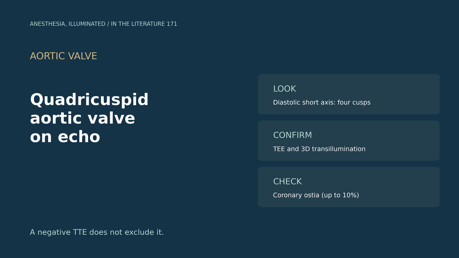
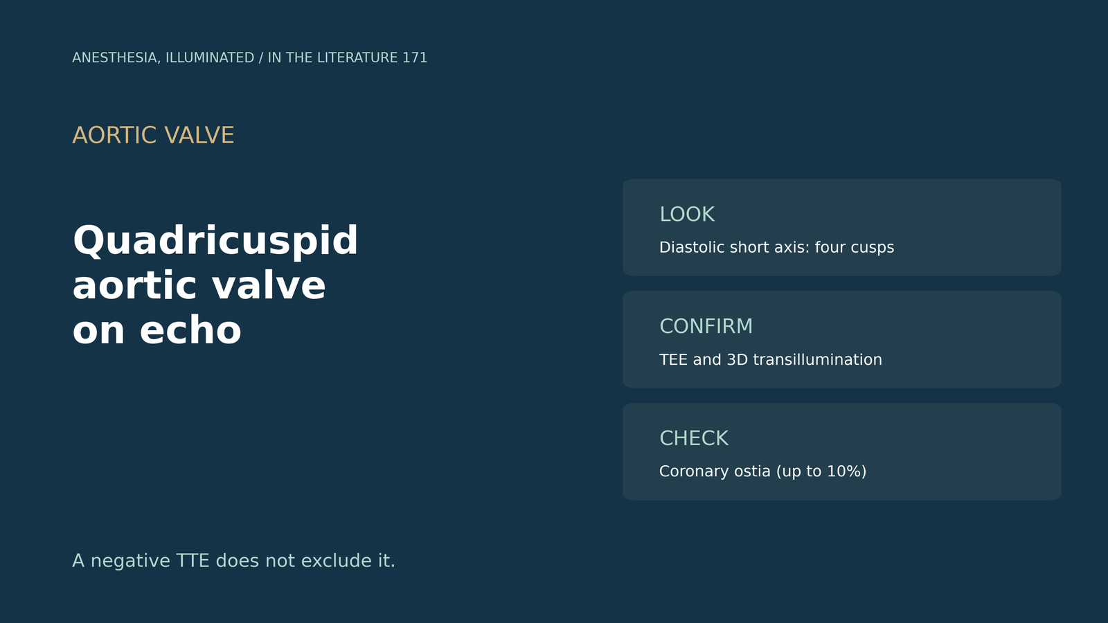
- It is rare and regurgitant. Incidence is 0.013% to 0.043% on echo, type B is most common (30.9%), and about 75% present with aortic regurgitation.[1]
- Look in diastole. The valve is best seen in short axis during diastole; TEE is more sensitive than TTE, and one valve was missed over 12 years and eight TTEs.[2]
- TTE misses it; 3D helps. In the largest cohort, 7 of 50 (14%) were diagnosed by TEE after a negative TTE, and 3D transillumination clarifies the cusps.[3]
- Check the coronaries. Coronary anomalies occur in up to 10%, so the ostia should be examined.[4]
- Expect surgery. Nearly 50% eventually need aortic valve surgery.[2]
During TEE before mitral surgery, the aortic valve in short axis does not show the usual three cusps, and there is a central jet of aortic regurgitation. Is this a quadricuspid aortic valve, and how is it confirmed?[1], [2]
How often and what type
Sohn and Arain pooled 149 reported cases. Incidence is 0.008% to 0.033% at autopsy and 0.013% to 0.043% on echo; the normal valve shows an X-shaped commissural pattern on short axis, whereas quadricuspid valves were most often type B (30.9%), then type A (23.5%) and type C (10.7%). About 75% present with aortic regurgitation. Cases were found by TTE in 39.6% and TEE in 38.9%; the authors suggest TTE for screening and TEE for further investigation.[1]
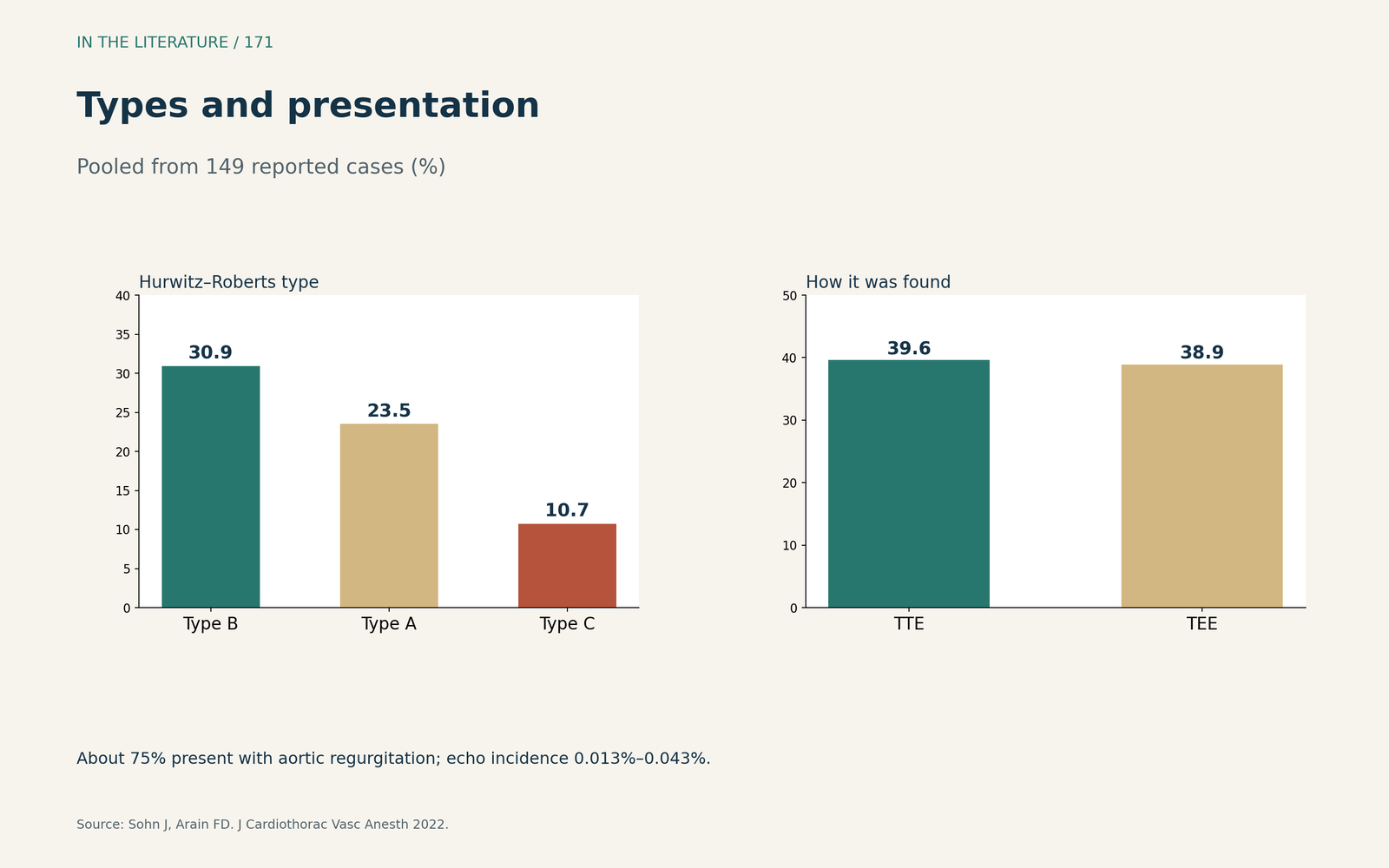
Figure 1. Types and presentation. Redrawn from Sohn and Arain.[1] View larger.

Seeing four cusps
Corsi and colleagues describe TEE showing four distinct cusps with thickening and calcification; the valve is best viewed in short axis during diastole, TEE is more sensitive because of higher image quality and is the current reference standard, one valve went undiagnosed over 12 years and eight TTEs, and nearly 50% eventually need surgery.[2] Kendall and colleagues note that the valve is likely missed on TTE: in the largest cohort, 7 of 50 patients (14%) were diagnosed by TEE after a negative TTE. 3D TEE transillumination revealed their valve, improving depth perception and border delineation; a small accessory cusp can make the valve functionally bicuspid, and TTE clues include central regurgitation and leaflet doming.[3]
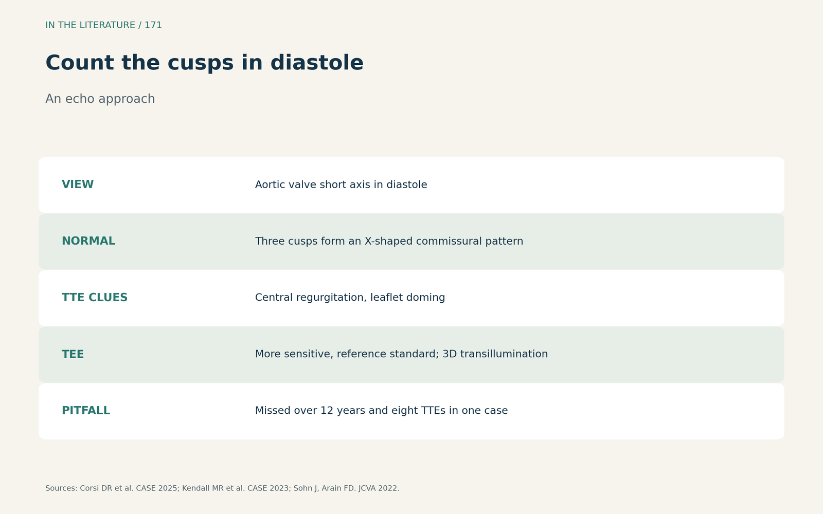
Figure 2. Count the cusps in diastole. Synthesized from Corsi et al., Kendall et al. and Sohn and Arain.[2], [3], [1] View larger.

Check the coronary ostia
Tedga and colleagues describe an infant with a quadricuspid valve and a guarded left coronary ostium of 1.5 mm; coronary anomalies occur in up to 10% of patients and should be ruled out whenever the aortic valve is abnormal.[4]
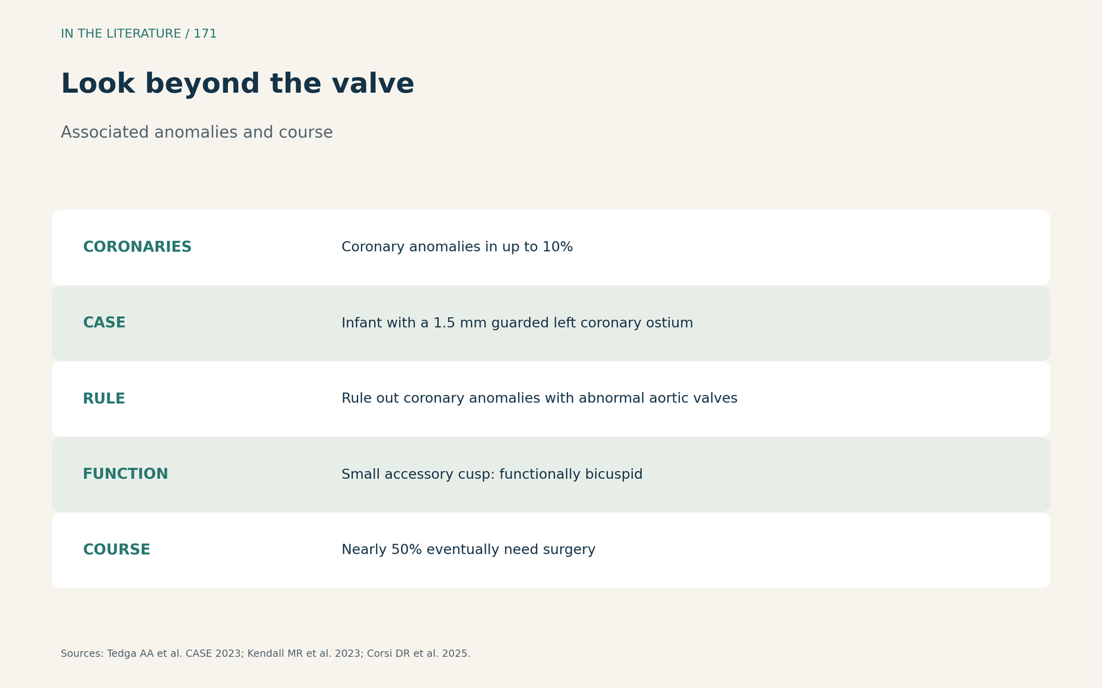
Figure 3. Look beyond the valve. Synthesized from Tedga et al. and Corsi et al.[4], [2] View larger.

Three takeaways
- Count cusps in diastolic short axis. Four cusps, not the X of three.[2], [1]
- Do not trust a negative TTE. Use TEE and 3D transillumination when suspicion remains.[3]
- Check the coronary ostia. Anomalies occur in up to 10%.[4]
References
- Sohn J, Arain FD. Two consecutive cases of quadricuspid aortic valve and a review of 149 cases. J Cardiothorac Vasc Anesth. 2022;36(3):717–723. doi:10.1053/j.jvca.2021.02.033
- Corsi DR, Kelly B, Nair N, et al. Clinical variability in presentation and management of quadricuspid aortic valve: a case series. CASE (Phila). 2025;9(4):130–134. doi:10.1016/j.case.2024.12.008
- Kendall MR, Chacon M, Janardhanan R. Incidental quadricuspid aortic valve demonstrated by transillumination on three-dimensional transesophageal echocardiography. CASE (Phila). 2023;7(3):113–115. doi:10.1016/j.case.2022.11.006
- Tedga AA, Pigula F, Lewis L, Kimball TR. Quadricuspid aortic valve with guarded left coronary artery ostium in an infant with myocardial infarction. CASE (Phila). 2023;7(1):6–9. doi:10.1016/j.case.2022.10.005