ITL-92e32f7d3def
Juan Li, MD · Updated November 17, 2026
Watch the video
Key points
- Compare residual mitral regurgitation (MR) with baseline under similar loading conditions and ultrasound settings, during hemodynamic stability.[1]
- Map every residual jet. When image quality permits, measure each three-dimensional vena contracta area (3D-VCA) in its own perpendicular plane and sum the areas.[1], [2]
- Proximal isovelocity surface area (PISA) calculations for effective regurgitant orifice area are unreliable after TEER; altered valve geometry defeats the usual assumptions.[1]
- Integrate jet measurements with pulmonary venous flow, left atrial pressure and the transmitral gradient. A prognostic cutoff is not automatically a target for another device.[1]–[3]
After leaflet grasp, the original broad jet may become several smaller, eccentric jets. One view looks reassuring; another shows residual color flow. The decision is not simply whether the color display is smaller. How much regurgitation remains, and can it be reduced further without compromising diastolic inflow? That requires two linked assessments: residual leakage during systole and forward passage through the remaining valve orifices during diastole.[1]
Make the before-and-after comparison meaningful
MR changes with loading conditions. Anesthesia, positive-pressure ventilation and altered vascular tone can change the apparent severity and the measured left atrial pressure. A reduction observed under different physiological conditions cannot be attributed entirely to the repair.[1], [3]
The 2026 ASE intraprocedural imaging guideline recommends comparison with baseline using similar loading conditions and instrument settings, with measurements during hemodynamic stability. Establish the blood pressure and rhythm at the time of assessment, then interpret the images and pressures together. This is especially important when the intraoperative appearance differs substantially from the preprocedural study.[1]
Next, locate the residual jets with two-dimensional color Doppler and biplane sweeps through the bicommissural view. Three-dimensional imaging helps establish their relationship to the device and remaining orifices. Identify number, location and eccentricity before trying to assign one severity label. A single favorable imaging plane can miss a jet that lies outside it.[1]
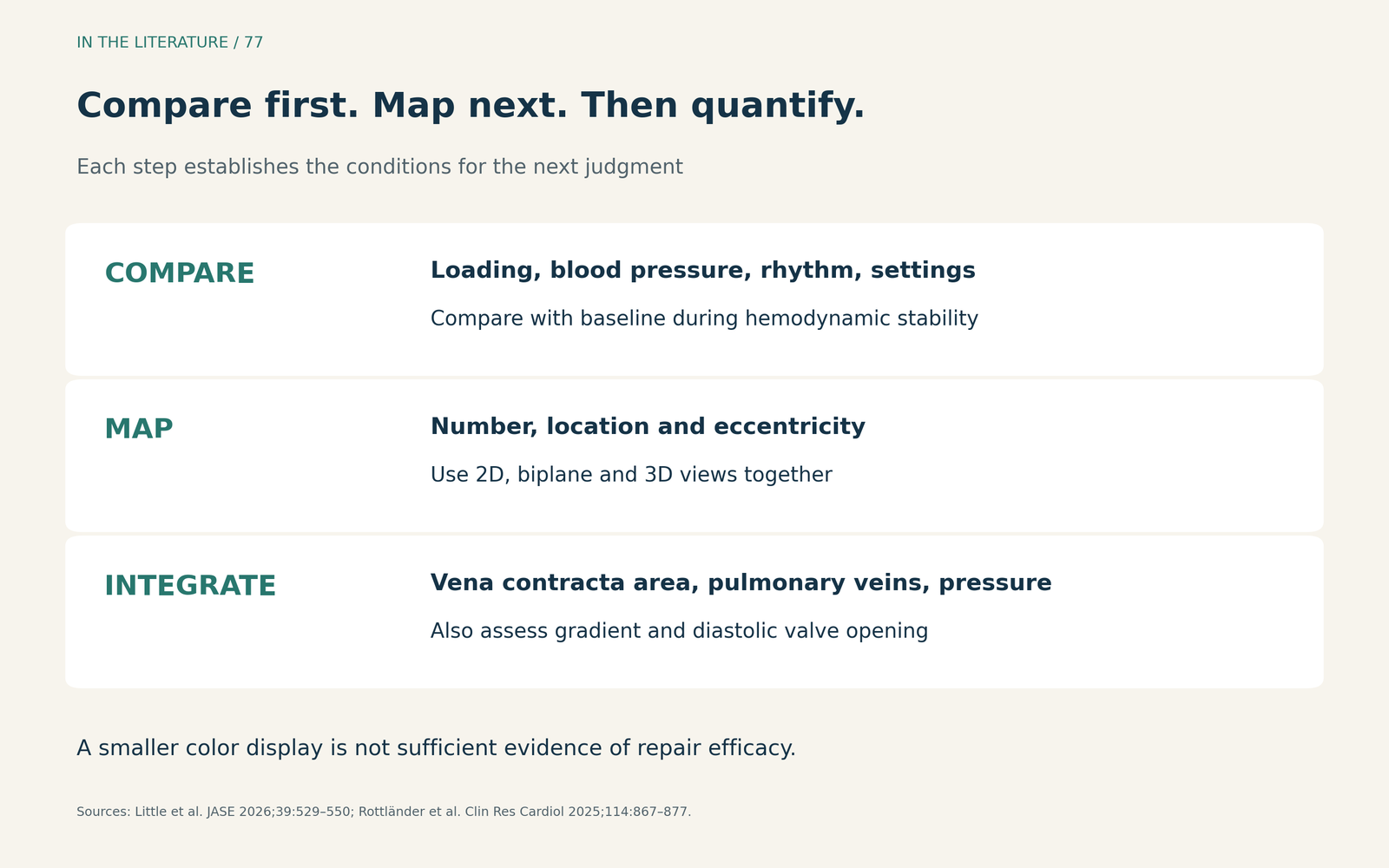
Figure 1. A practical order for assessment. Establish comparability before interpreting a smaller color display as treatment effect; map the jets before quantifying them. Original synthesis of the ASE imaging guidance and the MITRA-PRO measurement protocol.[1], [2] View larger.

Measure each vena contracta, not the whole color plume
The vena contracta is the narrow portion of the regurgitant jet close to its origin. With an adequate three-dimensional color dataset, use multiplanar reconstruction to align with an individual jet, identify its narrowest cross-section and trace it perpendicular to its direction. Repeat this for the other jets and add the measured areas. The jets may travel in different directions, so one common plane can be wrong for several of them.[1], [2]
This is not the same as adding the downstream color-jet areas in the left atrium. Nor is it a reason to calculate a conventional PISA-derived effective regurgitant orifice area after repair. The altered, often multiple orifices make that calculation unreliable. Even 3D-VCA depends on acquisition quality, appropriate cropping and operator experience; an oblique plane or artifacts can invalidate a precise-looking result.[1], [2]
Rottländer and colleagues evaluated 823 patients with available pre- and post-TEER 3D-VCA measurements in the prospective multicenter MITRA-PRO registry. Larger residual 3D-VCA was associated with higher one-year mortality. This supports taking the measurement seriously, but the analysis was observational and involved a selected subset with usable measurements. It did not establish that placing another device to reach a particular area improves survival.[2]
The reported 0.07 cm² prognostic cutoff came from receiver-operating-characteristic analysis in the same cohort. The authors also describe modest prognostic discrimination and caution against using 3D-VCA alone. Treat the cutoff as a research finding, not as a universal procedural endpoint or a complete residual-MR grading system.[2]
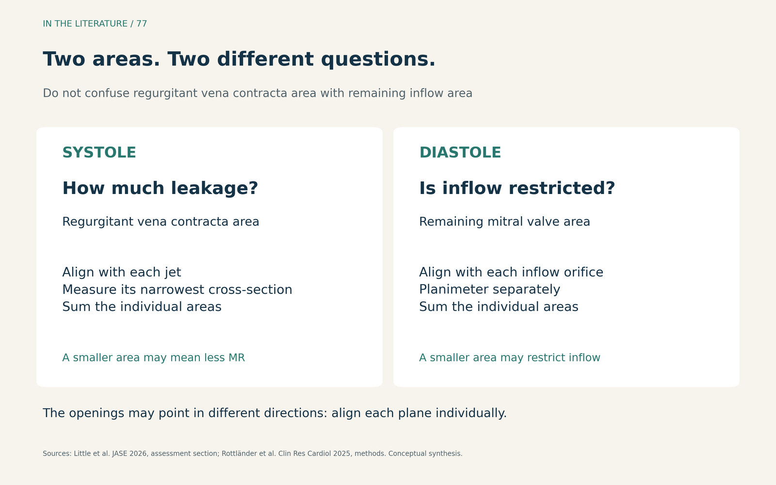
Figure 2. Two areas that must not be confused. Systolic VCA describes residual regurgitant jets; diastolic valve area describes the remaining inflow orifices. Measure each relevant opening in its own plane. These are conceptual categories, not patient measurements or anatomical drawings.[1], [2] View larger.

Use the hemodynamic response to test the interpretation
Improvement in pulmonary venous systolic flow can support MR reduction, particularly when systolic reversal was present at baseline. A fall in left atrial pressure or its v wave, and a change in forward stroke volume, provide additional information. These findings should agree with the anatomical and Doppler assessment; they do not replace it.[1]
Paulus and colleagues prospectively studied 299 patients at one center, measuring left atrial pressure before and after device implantation. Higher residual v-wave pressure was associated with death or heart-failure hospitalization during follow-up of up to two years; median follow-up was 12 months. The study's 25 mmHg cutoff was derived within that cohort. It is a prognostic association, not proof that actively lowering the v wave below that level improves outcomes.[3]
A persistently elevated v wave therefore prompts reassessment rather than an automatic additional device. Residual MR may have been underestimated, the remaining valve opening may be too restrictive, or atrial and ventricular physiology may contribute to the pressure. Conversely, a low v wave should not overrule clearly significant residual MR. General anesthesia and ventilation also influence these pressures, which may differ from those in the awake patient.[3]
Assess what was gained—and what may have been narrowed
Before device release, measure the mean transmitral gradient with continuous-wave Doppler and assess the remaining diastolic orifice area when feasible. Heart rate, flow and residual MR affect the gradient. Three-dimensional planimetry should align with each inflow orifice individually; the orifices are not necessarily parallel. Sum the separately measured areas to describe the remaining opening; pressure half-time is not recommended for this measurement.[1]
Keep this diastolic inflow-area measurement separate from the systolic regurgitant VCA measurement. A smaller VCA may indicate less leakage, while a smaller diastolic valve area may indicate a cost to forward filling. The decision about repositioning or further repair requires both observations, rather than pursuing the lowest possible residual jet measurement in isolation.[1], [3]
Reassess after device release: removal of catheter tension can alter device orientation and the final repair result.[1]
When the measurements disagree, a useful clinical response is to revisit the acquisition and physiological conditions, then discuss the combined findings with the interventional team. This is a synthesis of the available evidence: no single area, pressure or gradient resolves every post-TEER decision.[1]–[3]
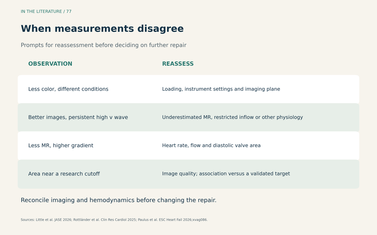
Figure 3. What discordance should make you reconsider. These are prompts for integrated reassessment, not independent diagnostic rules or instructions to deploy another device. Original synthesis.[1]–[3] View larger.

Three takeaways
- Make the comparison fair. Match loading conditions and imaging settings, then locate all residual jets before assigning severity.[1]
- Use 3D-VCA as part of the assessment. Align and measure each jet separately; an observational prognostic cutoff does not define a mandatory repair target.[1], [2]
- Judge leakage and inflow together. Reconcile the images, pulmonary venous flow, left atrial pressure and transmitral gradient before accepting or modifying the repair.[1], [3]
References
- Little SH, Quader N, Brady M, et al. Guidelines for the Intraprocedural Imaging for Mitral Valve Transcatheter Edge-to-Edge Repair (M-TEER): Recommendations from the American Society of Echocardiography. J Am Soc Echocardiogr. 2026;39(6):529–550. doi:10.1016/j.echo.2026.03.003 Published correction: JASE 2026;39:1102; correction text unavailable.
- Rottländer D, Hausleiter J, Schmitz T, et al. Intraprocedural 3D-vena contracta area predicts survival after transcatheter edge-to-edge repair: results from MITRA-PRO registry. Clin Res Cardiol. 2025;114(7):867–877. doi:10.1007/s00392-024-02580-6
- Paulus M, Rösch J, Grewe F, et al. Residual left atrial v wave predicts clinical outcome of transcatheter edge-to-edge mitral valve repair. ESC Heart Fail. 2026;13(2):xvag086. doi:10.1093/eschf/xvag086