ITL-0a6eb1091ac3
Juan Li, MD · Updated November 17, 2026
Watch the video
Key points
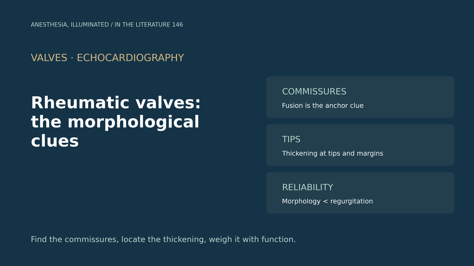
- Commissural fusion is the anchor. It is the main mechanism of mitral obstruction, gives a small central oval orifice, and in short axis helps confirm a rheumatic process.[1]
- Look at the tips. Rheumatic stenosis involves mainly the tips and margins, with a hockey-stick or doming anterior leaflet and chordal thickening, whereas annular calcification rarely causes more than mild stenosis.[1]
- Thickness has age-specific limits. Normal mitral thickness is under 3 mm in children and 3.5 mm in adults, and it should be measured with tissue harmonics turned off.[1]
- Morphology is the less reliable half. Inter-rater agreement was lower for morphological features than for regurgitation, and hand-carried devices were only 66.7% specific for leaflet thickening.[2]
- In the aortic valve, commissural fusion with free-edge thickening points to rheumatic disease, which almost always occurs with rheumatic mitral disease.[1], [3]
Before mitral valve surgery, TEE shows a thickened, stenotic mitral valve and a mildly thickened aortic valve. Is this rheumatic disease or calcific degeneration, and which features settle the question?[1], [3]
Mitral valve clues
The American Society of Echocardiography guideline lists four characteristic features: commissural fusion, leaflet thickening with or without calcification, restricted motion giving a hockey-stick or doming anterior leaflet in diastole, and chordal thickening, shortening and fusion. Commissural fusion is the main mechanism of obstruction and leaves a small central oval orifice; seeing it in the short-axis view helps confirm a rheumatic process.[1]
Rheumatic stenosis involves predominantly the leaflet tips and margins, whereas stenosis from annular calcification is rarely more than mild unless the calcification is extensive and reaches the leaflet bases. Normal mitral thickness is under 3 mm in children and 3.5 mm in adults, and thickness should be measured with tissue harmonics turned off. Under World Heart Federation criteria, rheumatic mitral stenosis requires a mean gradient above 4 mmHg plus rheumatic morphological features.[1]
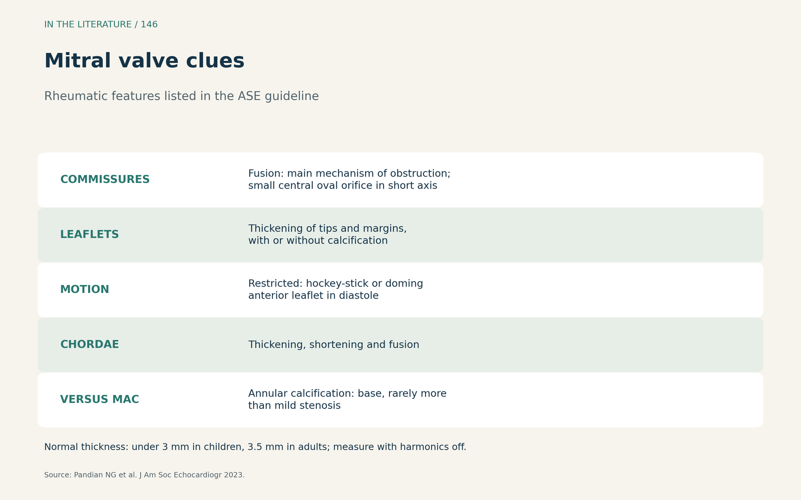
Figure 1. Mitral clues. Synthesized from the ASE guideline.[1] View larger.

Aortic and tricuspid clues
Rheumatic aortic disease is identified by commissural fusion and thickening of the free edges, and almost always occurs with rheumatic mitral disease; regurgitation (47%) is more common than stenosis (14%).[1] Alizadeh and colleagues describe the anatomic basis of rheumatic aortic stenosis as commissural fusion with edge thickening, producing a triangular systolic orifice, whereas commissural fusion is typically absent in nonrheumatic stenosis.[3] Primary rheumatic tricuspid regurgitation is diagnosed when it accompanies diastolic doming or thickening.[1]
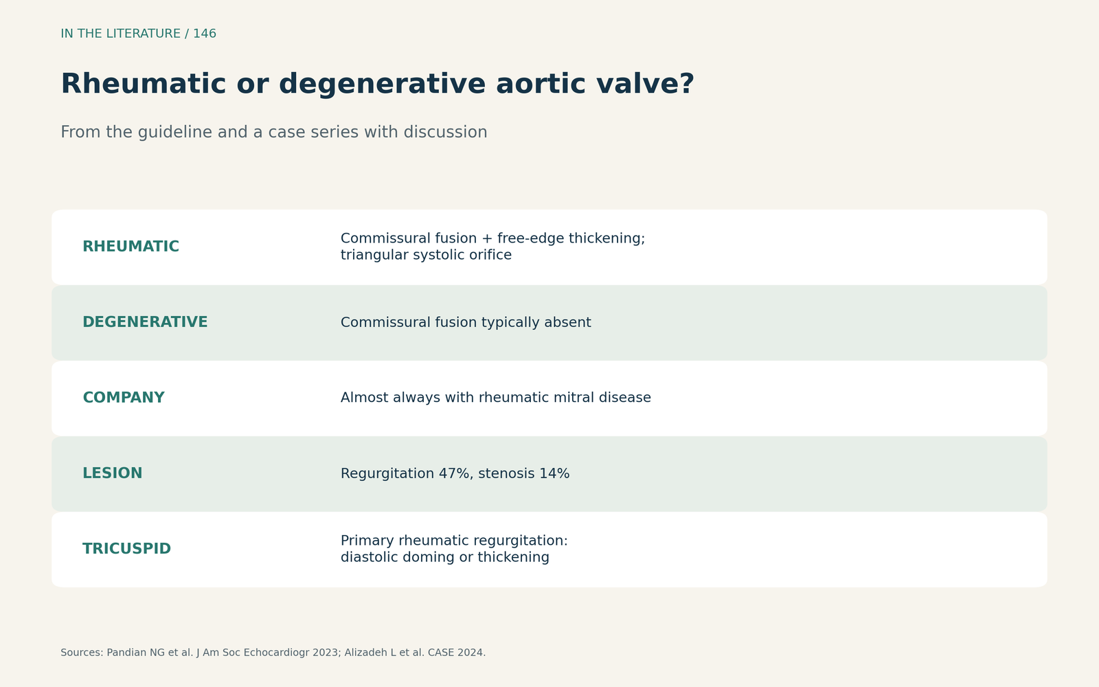
Figure 2. Rheumatic or degenerative aortic valve? Synthesized from the ASE guideline and Alizadeh et al.[1], [3] View larger.

How reliable morphology is
Marangou and colleagues reviewed 34 studies of the 2012 World Heart Federation criteria: inter-rater reliability was lower for morphological features than for regurgitation, and hand-carried devices overestimated morphology, being only 66.7% specific for anterior leaflet thickness above 3 mm.[2] In a screening study by Francis and colleagues, offsite expert review of nonexpert images raised specificity from 61.4% to 77.1% with a sensitivity of 88.4%, and the most commonly missed cases were borderline disease with morphological changes only.[4]
Morphology also mattered less for prognosis. In 165 children and young people with first rheumatic fever, early disease was diagnosed in 64% and resolution occurred in 19%; disease severity, not valve morphology, was the stronger predictor of resolution.[5]
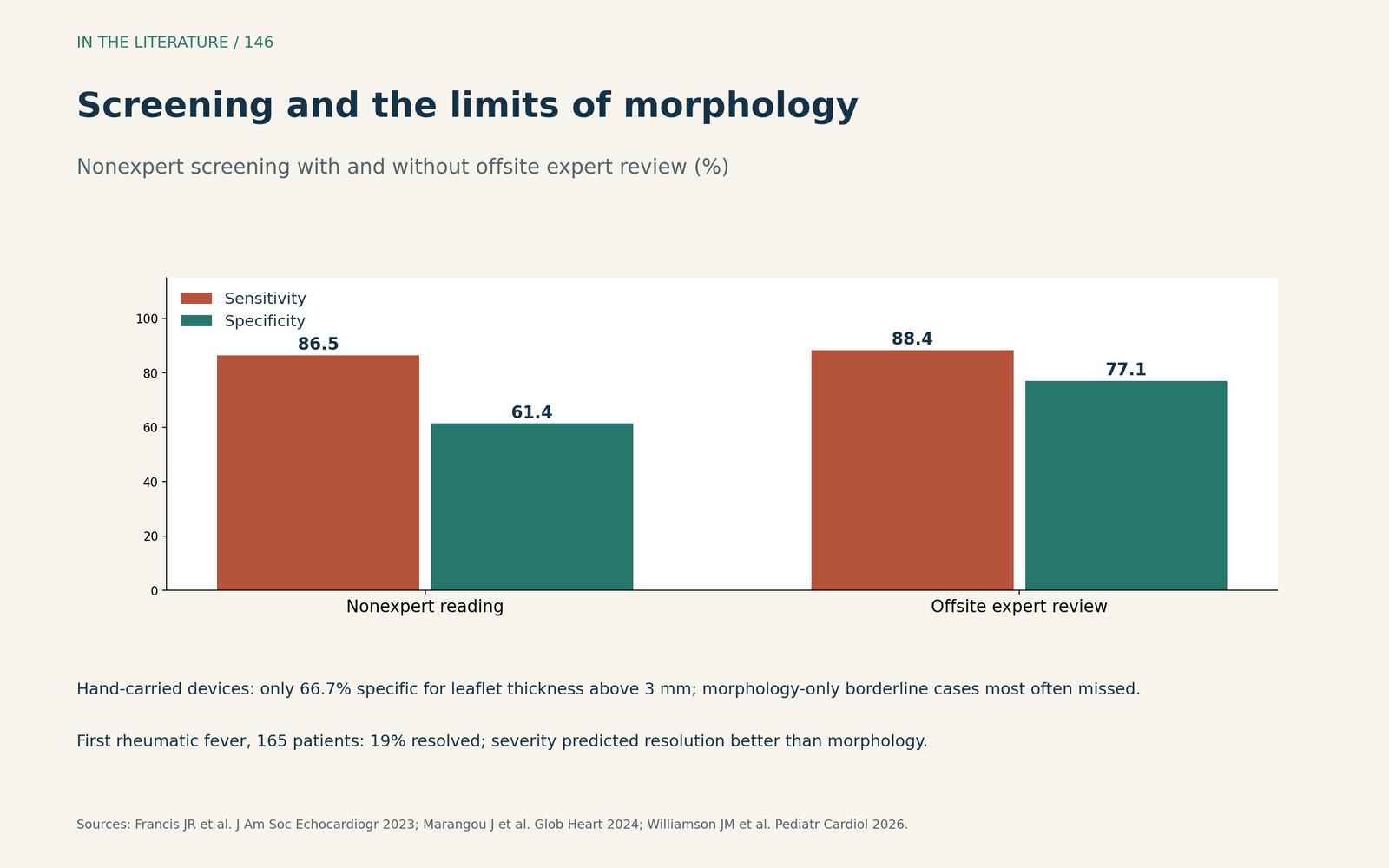
Figure 3. Screening and the limits of morphology. Redrawn from Marangou et al., Francis et al. and Williamson et al.[2], [4], [5] View larger.

Three takeaways
- Find the commissures. Commissural fusion in short axis is the most characteristic clue on both mitral and aortic valves.[1], [3]
- Locate the thickening. Tips and margins suggest rheumatic disease; the annulus and leaflet bases suggest calcific disease.[1]
- Weigh morphology with function. Morphology is less reproducible than regurgitation and less predictive than severity.[2], [5]
References
- Pandian NG, Kim JK, Arias-Godinez JA, et al. Recommendations for the use of echocardiography in the evaluation of rheumatic heart disease: a report from the American Society of Echocardiography. J Am Soc Echocardiogr. 2023;36(1):3–28. doi:10.1016/j.echo.2022.10.009
- Marangou J, Rwebembera J, Mwita J, et al. The echocardiographic diagnosis of rheumatic heart disease: a review of the performance of the World Heart Federation criteria 2012–2023. Glob Heart. 2024;19(1):47. doi:10.5334/gh.1327
- Alizadeh L, Peters F, Vainrib AF, Freedberg RS, Saric M. Rheumatic heart disease: a rare cause of very severe valvular aortic stenosis. CASE (Phila). 2024;8(5):320–324. doi:10.1016/j.case.2024.02.005
- Francis JR, Fairhurst H, Yan J, et al. Abbreviated echocardiographic screening for rheumatic heart disease by nonexperts with and without offsite expert review: a diagnostic accuracy study. J Am Soc Echocardiogr. 2023;36(7):733–745. doi:10.1016/j.echo.2023.02.007
- Williamson JM, Whalley GA, Marangou J, et al. Application of the new 2023 World Heart Federation criteria to rheumatic heart disease in the setting of first episode of acute rheumatic fever: predictors for resolution. Pediatr Cardiol. 2026;47(4):1765–1775. doi:10.1007/s00246-025-03966-7