ITL-fe8a2e052e08
Juan Li, MD · Updated November 17, 2026
Watch the video
Key points
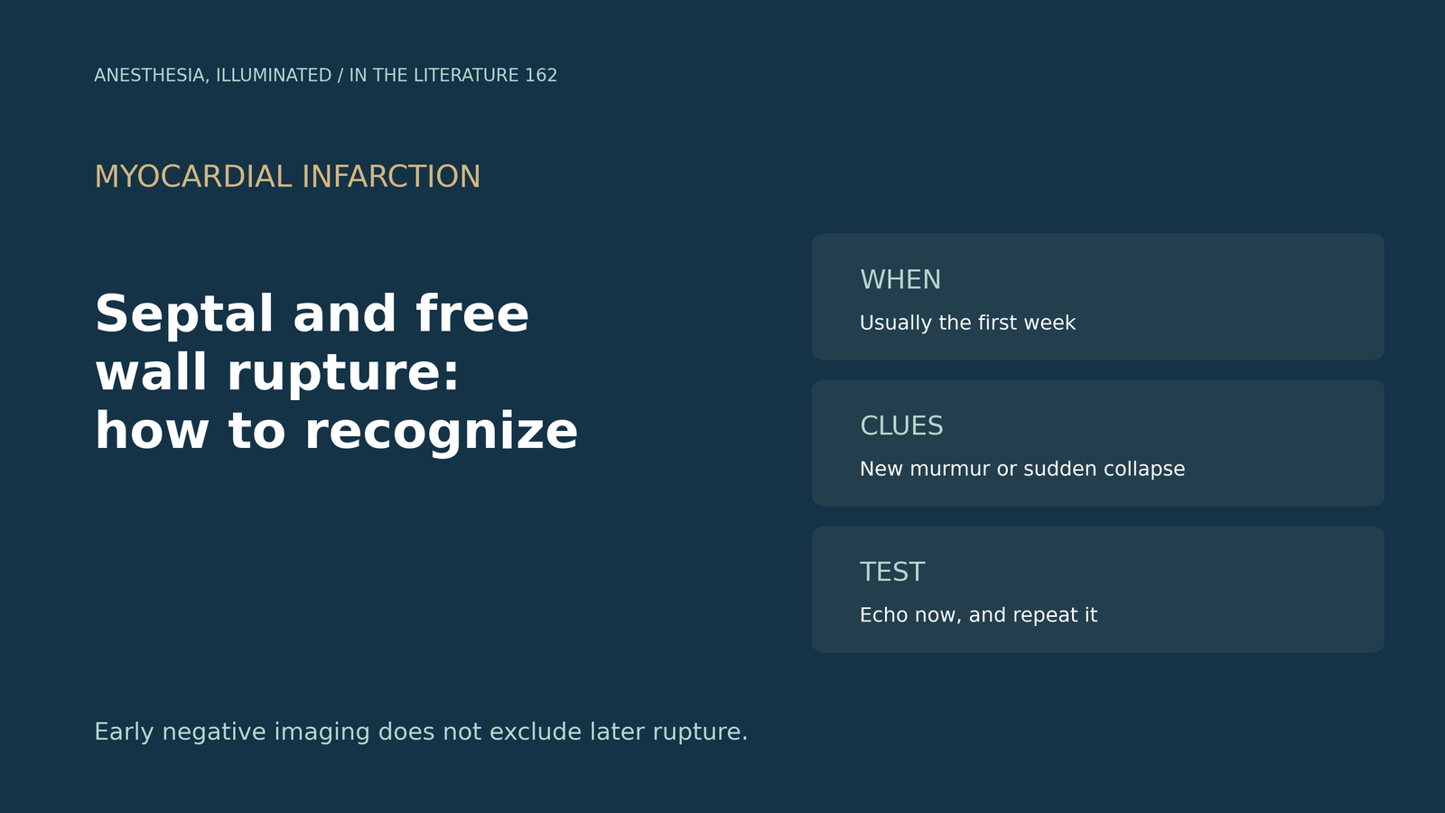
- Think of it early. Rupture typically presents within hours to 1 to 2 days of infarction, and nearly all free wall ruptures appear within 1 week.[1]
- Image at once. Any suspicion warrants emergent imaging; TTE is the first-line test, and echo with color Doppler is nearly 100% sensitive for septal rupture.[1], [2]
- Know the two pictures. Septal rupture brings a new systolic murmur; free wall rupture brings sudden collapse or electromechanical dissociation, with tamponade, shock or dissociation in up to 83%.[1], [3]
- One scan is not enough. Early negative imaging does not exclude later rupture, and in one cohort the median time to rupture was 4.0 days.[2], [3]
- Mind the monitors and the neck. Thermodilution cardiac output should be avoided with a septal defect, and a pseudoaneurysm has a narrow neck, unlike a true aneurysm.[4], [5]
Three days after an inferior myocardial infarction, a patient becomes hypotensive and a new loud systolic murmur is heard. Is this a mechanical complication, and how should it be confirmed?[1], [2]
When and how rupture presents
Yu and colleagues describe presentation within hours to 1 to 2 days of infarction, with nearly all free wall ruptures within 1 week. Up to 83% of free wall ruptures present with tamponade, cardiogenic shock or electromechanical dissociation, and mortality is 40% to 75% for septal and 75% to 90% for free wall rupture.[1] Streian and colleagues note that rupture events classically cluster between days 3 and 7.[2] In Zhang's case-control study, median time from infarction to rupture was 4.0 days and 39.4% ruptured within 3 days; free wall rupture was diagnosed by sudden hemodynamic collapse or electromechanical dissociation, and septal rupture by a new systolic murmur with echo confirmation.[3]
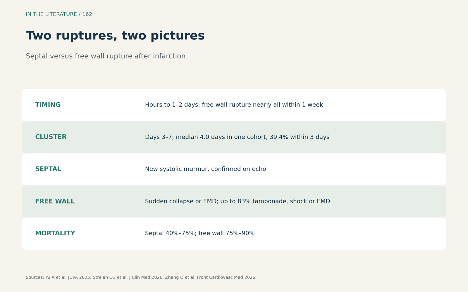
Figure 1. Two ruptures, two pictures. Synthesized from Yu et al., Streian et al. and Zhang et al.[1], [2], [3] View larger.

Confirming the diagnosis
Any suspicion of a mechanical complication warrants emergent imaging. Echo with color Doppler is nearly 100% sensitive for septal rupture and can detect a pericardial effusion suggestive of free wall rupture, and TTE remains a quick, effective and reliable test.[1] Streian and colleagues call TTE the first-line modality and advise evaluating any post-infarction patient with hypotension, a new systolic murmur, unexplained pulmonary oedema or rising lactate; early negative imaging does not exclude subsequent rupture.[2] In Gu's case report, a new loud systolic murmur appeared on hospital day 2, and the authors advise avoiding thermodilution cardiac output in patients with a septal defect.[4] Al-Naabi and colleagues distinguish a pseudoaneurysm, which communicates with the ventricle through a narrow neck, from a true aneurysm, a bulge of infarcted myocardium with a wide neck.[5]
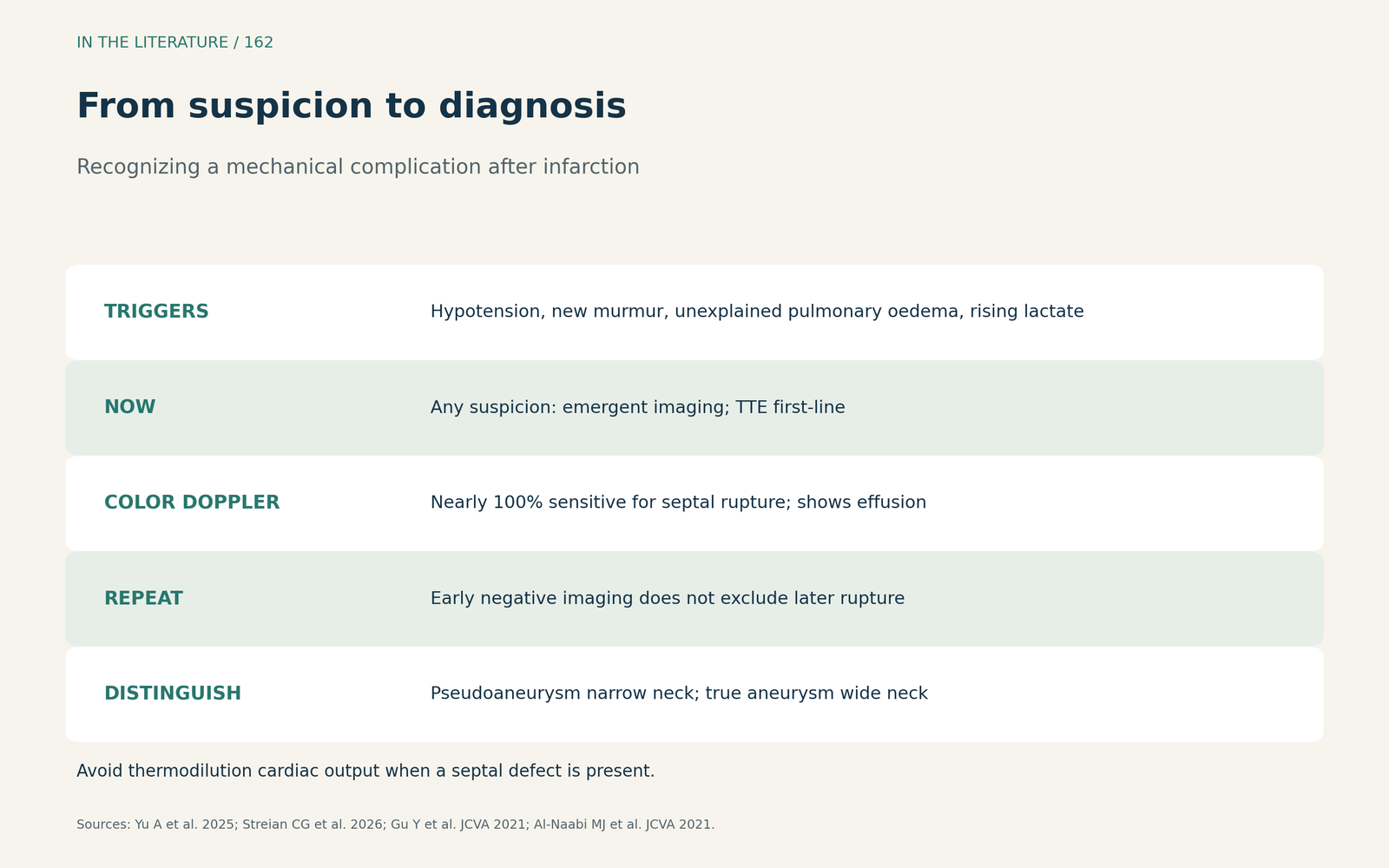
Figure 2. From suspicion to diagnosis. Synthesized from Yu et al., Streian et al., Gu et al. and Al-Naabi et al.[1], [2], [4], [5] View larger.

Why recognition matters
In Zhang's study, rupture cases more often had Killip class III–IV (45.1% vs 8.0%); absence of emergency PCI, Killip class III–IV, female sex and hypoalbuminemia were associated with rupture, and in-hospital mortality was 97.4% for free wall and 57.1% for septal rupture.[3] In data cited by Yu, mortality was 40% to 60% with cardiogenic shock, and operative mortality was 54% for repairs within 7 days.[1]
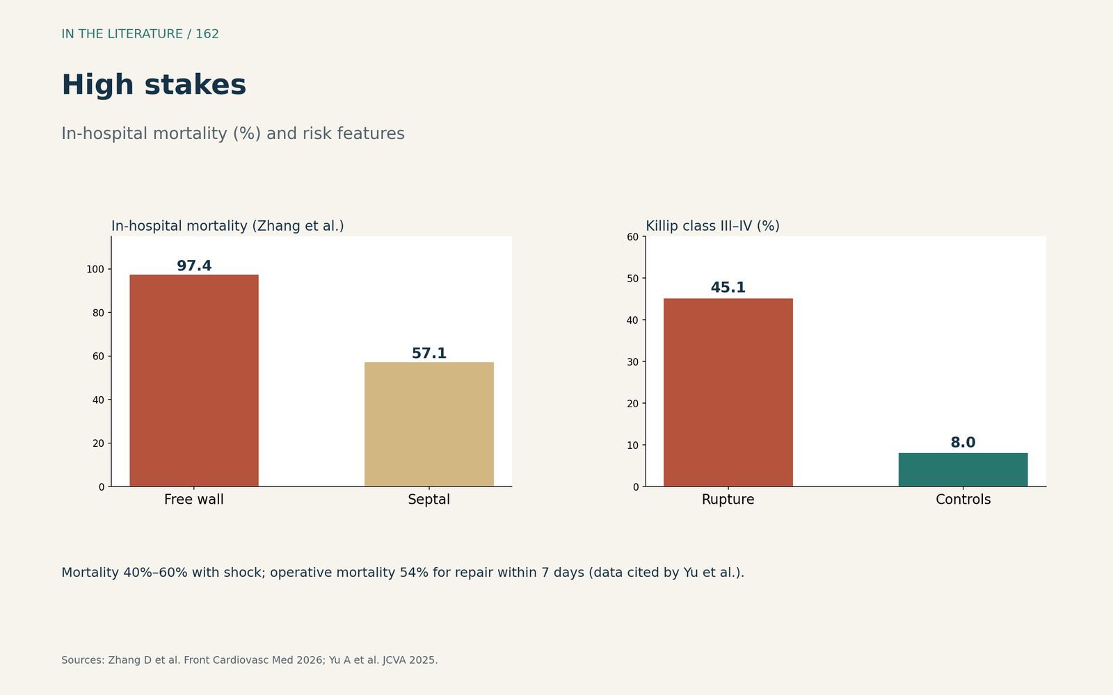
Figure 3. High stakes. Redrawn from Zhang et al. and Yu et al.[3], [1] View larger.

Three takeaways
- A new murmur or sudden collapse in the first week is rupture until proven otherwise.[1], [3]
- Echo first, and repeat it. TTE is first-line, and early negative imaging does not exclude later rupture.[2]
- Interpret monitors carefully. Thermodilution output is unreliable with a septal defect.[4]
References
- Yu A, Maslow A, Sodha N, et al. Evolving management of post-myocardial infarction ventricular septal or free wall rupture. J Cardiothorac Vasc Anesth. 2025;39(12):3589–3608. doi:10.1053/j.jvca.2025.08.045
- Streian CG, Novaconi RC, Munteanu IR, et al. Mechanical complications after acute myocardial infarction: a shock-stage and timing-based management framework. J Clin Med. 2026;15(6):2399. doi:10.3390/jcm15062399
- Zhang D, Huang C, Wang X, et al. Risk factors, subtype profiles, and outcomes of cardiac rupture after acute myocardial infarction: a case-control study. Front Cardiovasc Med. 2026;13:1827832. doi:10.3389/fcvm.2026.1827832
- Gu Y, Panda K, Bensimhon A, Bernstein W. Hybrid approach to post–myocardial infarction ventricular septal rupture repair and its early complications. J Cardiothorac Vasc Anesth. 2021;35(1):274–280. doi:10.1053/j.jvca.2020.06.052
- Al-Naabi MJ, Maddali MM, Venkatachlam R. Mechanical complication after a myocardial infarction. J Cardiothorac Vasc Anesth. 2021;35(2):670–672. doi:10.1053/j.jvca.2020.06.076