
ITL-734ae846e01a
Juan Li, MD · Updated November 18, 2026
Key points
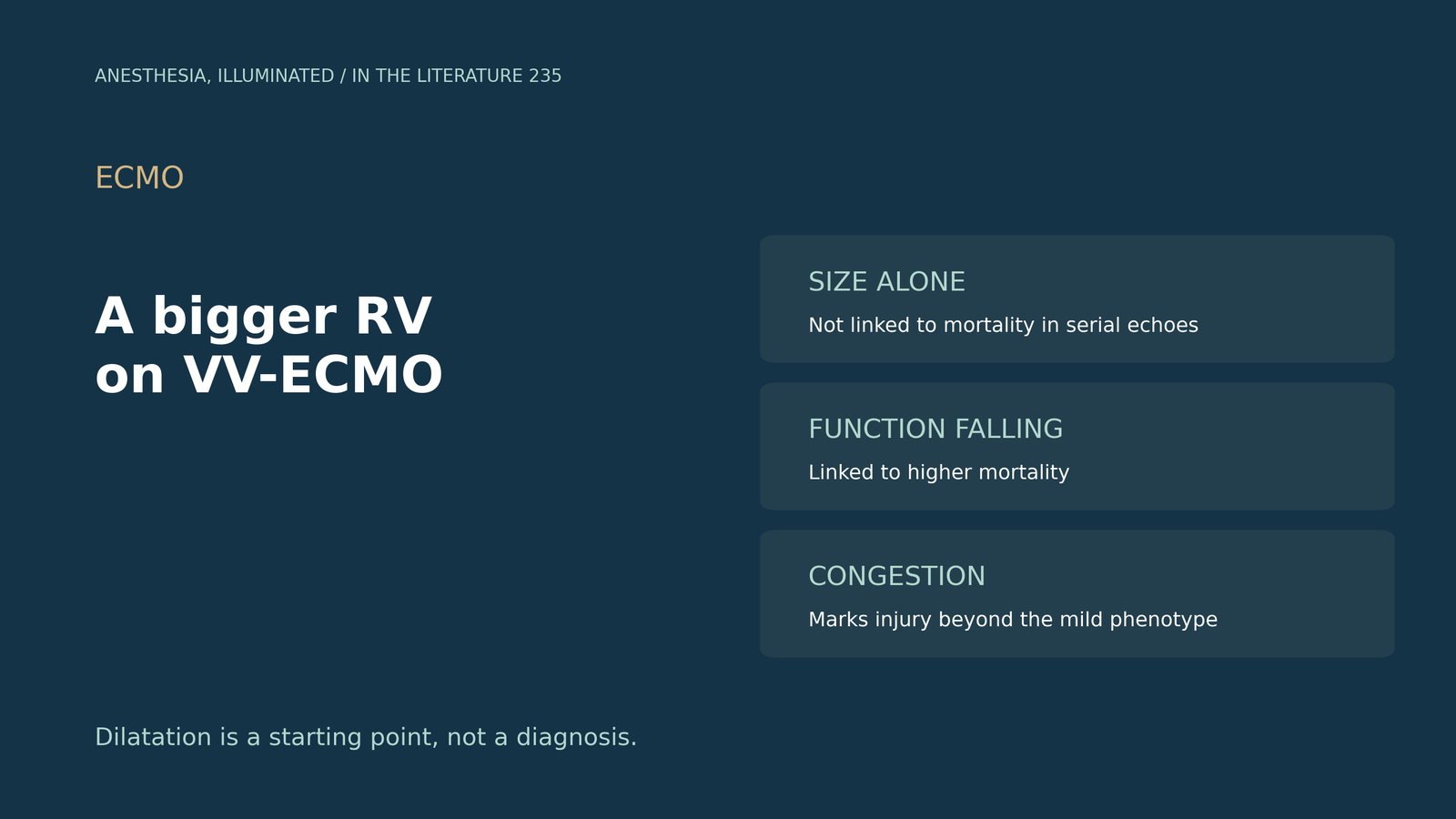
- Size is not function. In serial echoes, an increase in RV size did not correlate with higher mortality, but a progressive decline in RV function did.[1]
- Dilatation can be adaptive. It preserves blood flow, occasionally at the price of systemic venous congestion.[2]
- No agreed definition. Experts consider isolated dilatation without venous congestion a mild injury sub-phenotype.[2]
- Injury predicts worse outcomes. RV injury was linked with higher ICU mortality (48% vs 13%) and, in ARDS, with mortality after adjustment (aOR 2.37).[3], [4]
- It can come late, up to 4 to 6 weeks into VV-ECMO.[2]
A patient has been on venovenous ECMO for ARDS for two weeks. A routine echo shows a larger right ventricle than at cannulation, but TAPSE is unchanged and the patient is not congested. Is this RV failure, and what should change?[1], [2]
Size and function tell different stories
In a retrospective cohort of 70 VV-ECMO patients (Isha et al., ASAIO J 2024), normal RV function fell from 60.6% at ECMO placement to 42% at the second echo, and a severely dilated RV was present in 6.7% on the day of placement.[1] An increase in RV size did not correlate with higher mortality, whereas a progressive decline in RV function was associated with elevated mortality; the study cannot establish causality.[1] Kopanczyk et al. (J Cardiothorac Vasc Anesth 2022) note that initiation of VV-ECMO tends to improve RV function and that RV dilatation and dysfunction may be a marker of COVID disease severity.[5]
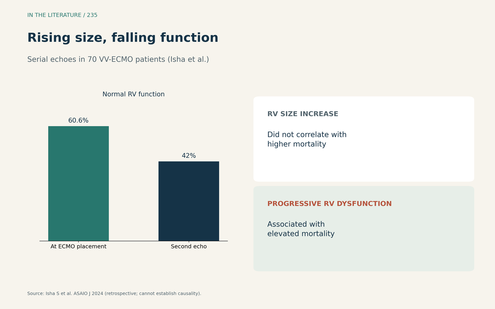
Figure 1. Rising size, falling function: only one tracked with death. Redrawn from Isha et al.[1] View larger.

What experts call RV injury
The PRORVnet Delphi consensus (Zochios et al., Intensive Care Med 2024) brought together 52 experts, who reached consensus on 31 of 35 statements; they note there is no universally accepted definition of RV injury during VV-ECMO, and that injury may persist or even worsen on support.[2] Dilatation preserves blood flow, occasionally at the price of systemic venous congestion; isolated dilatation without venous congestion may be considered a mild sub-phenotype.[2] Proposed markers include RVEDA/LVEDA above 0.6 or at least 1, septal dyskinesia and TAPSE below 17 mm, and acute injury may develop late, up to 4 to 6 weeks into VV-ECMO.[2] RV free-wall longitudinal strain is perhaps a better modality than TAPSE.[5]
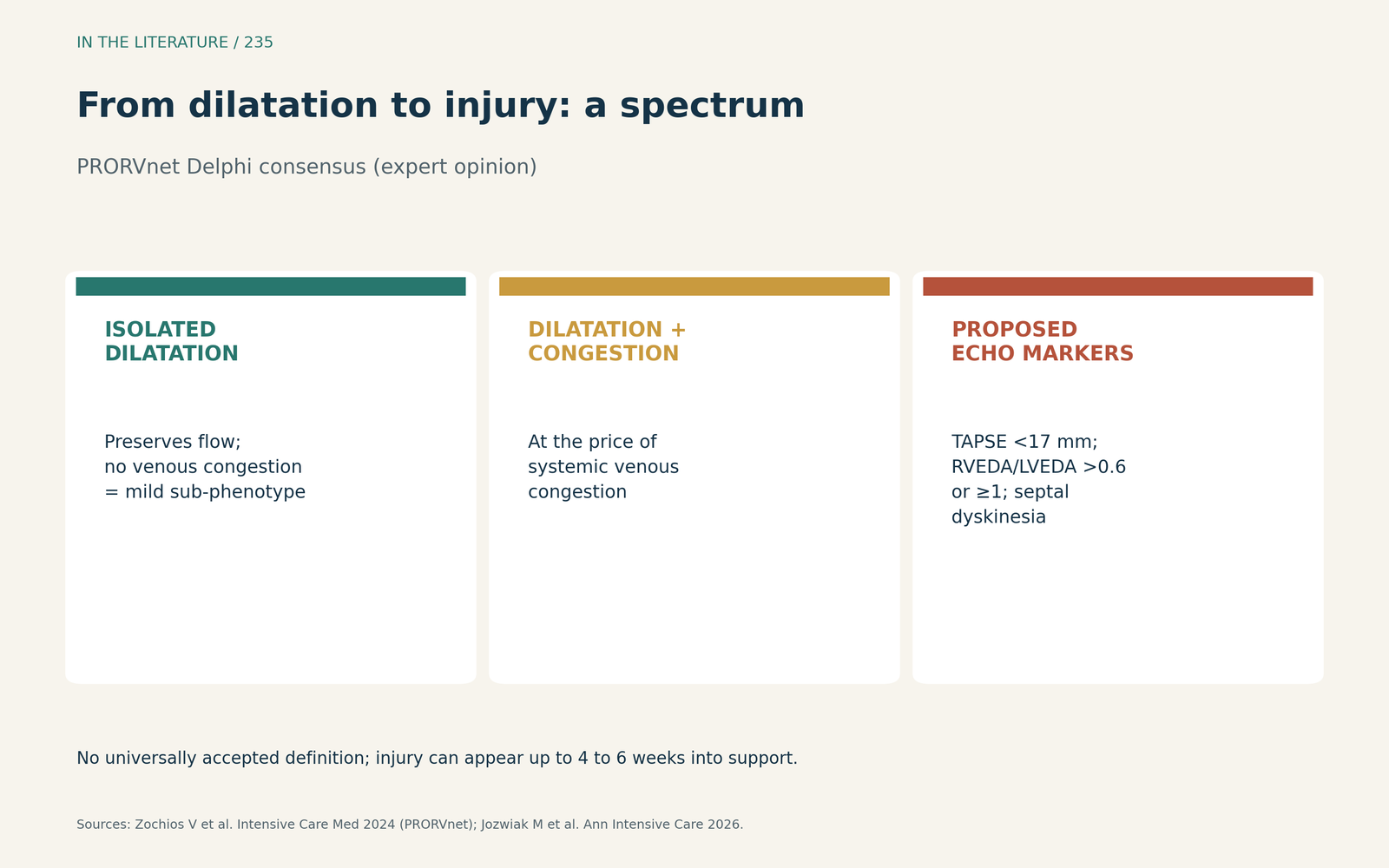
Figure 2. Dilatation is the start of a spectrum, not the diagnosis. Synthesized from the PRORVnet consensus and Jozwiak et al.[2], [4] View larger.

Prognosis
In 40 VV-ECMO patients (McDonald et al., J Intensive Care Soc 2025), RV injury was observed in 63% and was associated with higher ICU mortality (48% vs 13%); late-onset injury could represent a modifiable complication.[3] In ARDS more broadly, Jozwiak et al. (Ann Intensive Care 2026) found a pooled prevalence of 30%, with RV injury still associated with mortality after adjustment (aOR 2.37); definitions ranged from isolated dilation to more comprehensive assessments.[4]
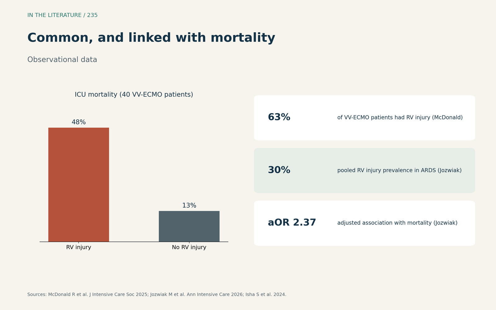
Figure 3. Common, and linked with mortality when it means dysfunction. Data from McDonald et al., Jozwiak et al. and Isha et al.[3], [4], [1] View larger.

What to do
Specific guidelines for regular RV assessment during VV-ECMO are absent.[1] When there is volume overload, diuretics or renal replacement therapy can be used to improve RV dilation and tricuspid regurgitation,[6] and patients with refractory RV failure are generally converted to VA-ECMO or an oxygenated RV assist device.[1]
Three takeaways
- Do not treat isolated RV dilatation as failure; look for falling function and venous congestion.[1], [2]
- Reassess the RV serially; injury can appear weeks into VV-ECMO.[2], [3]
- When dysfunction is established, address congestion and consider escalation for refractory failure.[6], [1]
References
- Isha S, Narra SA, Batool H, et al. Assessing right ventricle over time in patients on veno-venous extracorporeal membrane oxygenation: insights from serial echocardiography. ASAIO J. 2024;70(12):1033–1039. doi:10.1097/MAT.0000000000002235
- Zochios V, Nasa P, Yusuff H, et al. Definition and management of right ventricular injury in adult patients receiving extracorporeal membrane oxygenation for respiratory support using the Delphi method: a PRORVnet study. Expert position statements. Intensive Care Med. 2024;50(9):1411–1425. doi:10.1007/s00134-024-07551-z
- McDonald R, Fowles JA, Gatherer R, Caetano F. Right ventricular injury during VV-ECMO for severe ARDS: does time matter? J Intensive Care Soc. 2025;27(1):82–84. doi:10.1177/17511437251374821
- Jozwiak M, Joseph A, Mourougayen A, et al. Prevalence and prognosis impact of right ventricular injury in patients with acute respiratory distress syndrome: a systematic review and meta-analysis. Ann Intensive Care. 2026;16:100117. doi:10.1016/j.aicoj.2026.100117
- Kopanczyk R, Kumar N, Iyer MH, Bhatt AM. Right ventricular dysfunction in COVID-19 ECMO—a red herring or the real deal? J Cardiothorac Vasc Anesth. 2022;36(3):921–922. doi:10.1053/j.jvca.2021.09.036
- Sutherland LD, Dhawan R. Expert commentary on rescue ECMO for isolated right ventricular dysfunction in a trauma patient. J Cardiothorac Vasc Anesth. 2024;38(4):1037–1040. doi:10.1053/j.jvca.2024.01.013