ITL-75a35b64247d
Juan Li, MD · Updated November 17, 2026
Watch the video
Key points
- Assess systolic anterior motion (SAM), left ventricular outflow tract (LVOT) obstruction and mitral regurgitation (MR) separately; visible SAM alone does not establish its clinical significance.1
- Confirm that the Doppler signal comes from the LVOT: an MR jet can masquerade as a very high outflow gradient.2
- Correct reversible loading and contractile conditions, then repeat TEE; fluid administration and reduction of inotropic support require individual assessment.23
- Persistent important MR deserves attention even without marked LVOT obstruction; the gradient is not the sole criterion for revisiting the repair.13
- Refractory SAM may be amenable to re-repair, although successful intraoperative correction does not guarantee freedom from recurrence.4
During separation from cardiopulmonary bypass (CPB), TEE shows the anterior mitral leaflet moving toward the septum. The immediate task is to determine what that movement is doing to forward flow, valve competence and the patient's circulation. This article concerns SAM after repair for degenerative MR; management experience from hypertrophic cardiomyopathy or after TAVR is not interchangeable with this setting.
Establish the mechanism before treating a gradient
The 2020 ASE/SCA/STS surgical TEE guideline describes the combination of anterior leaflet motion, LVOT flow acceleration, a late-peaking Doppler envelope and often posteriorly directed MR. Examine leaflet coaptation and jet origin in multiple views. A central or anterior MR jet should prompt a search for another repair-related mechanism.1
Torre and colleagues' 2026 narrative review adds a practical warning: use color and pulsed-wave Doppler to localize acceleration before interpreting continuous-wave Doppler. An MR signal may contaminate the tracing. If MR remains after SAM improves, reassess the repair itself.2
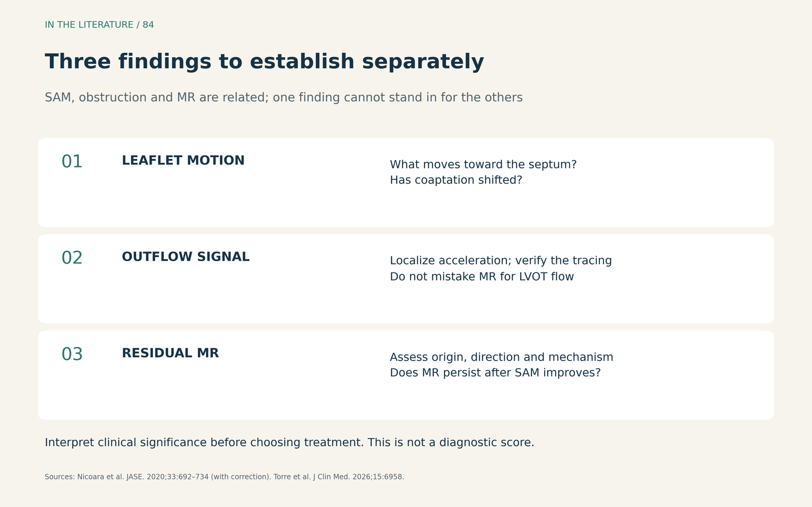
Figure 1. A clinical assessment framework, not a diagnostic score. SAM, obstruction and MR are related findings, but none should be inferred solely from another.1,2 Larger view

Correct the conditions, then look again
In Ashikhmina and colleagues' retrospective series of 761 isolated degenerative mitral repairs, 98 developed SAM after CPB; the definition included chordal motion and did not require obstruction. 89 were managed conservatively with volume augmentation, vasoconstrictors and beta-blockade. This describes selected clinical management, not a randomized comparison or a guaranteed response rate.3
At the bedside, assess filling, vascular tone, heart rate and contractility together. Give volume according to ventricular filling and tolerance; avoid indiscriminate loading. Reconsider catecholamine stimulation, reduce it when clinically feasible, and consider carefully titrated beta-blockade in selected patients. Recheck both MR and outflow after each adjustment. These physiological measures are summarized in the 2026 review; it does not establish one superior drug regimen.2
Persistent MR matters even when the gradient is modest
Of the seven patients who required surgical treatment for SAM in the Ashikhmina cohort, all had at least moderate MR, whereas only one had LVOT obstruction. Two other patients returned to CPB for intracoronary air; they should not be counted as SAM re-repairs.3
That distinction is clinically useful: a low gradient cannot certify an adequate repair. The surgical TEE guideline calls for investigation of greater-than-mild residual MR and assessment under appropriate loading conditions. Persistent SAM-related obstruction and MR despite optimization may warrant re-repair, with the decision incorporating the patient, imaging and operative findings.1
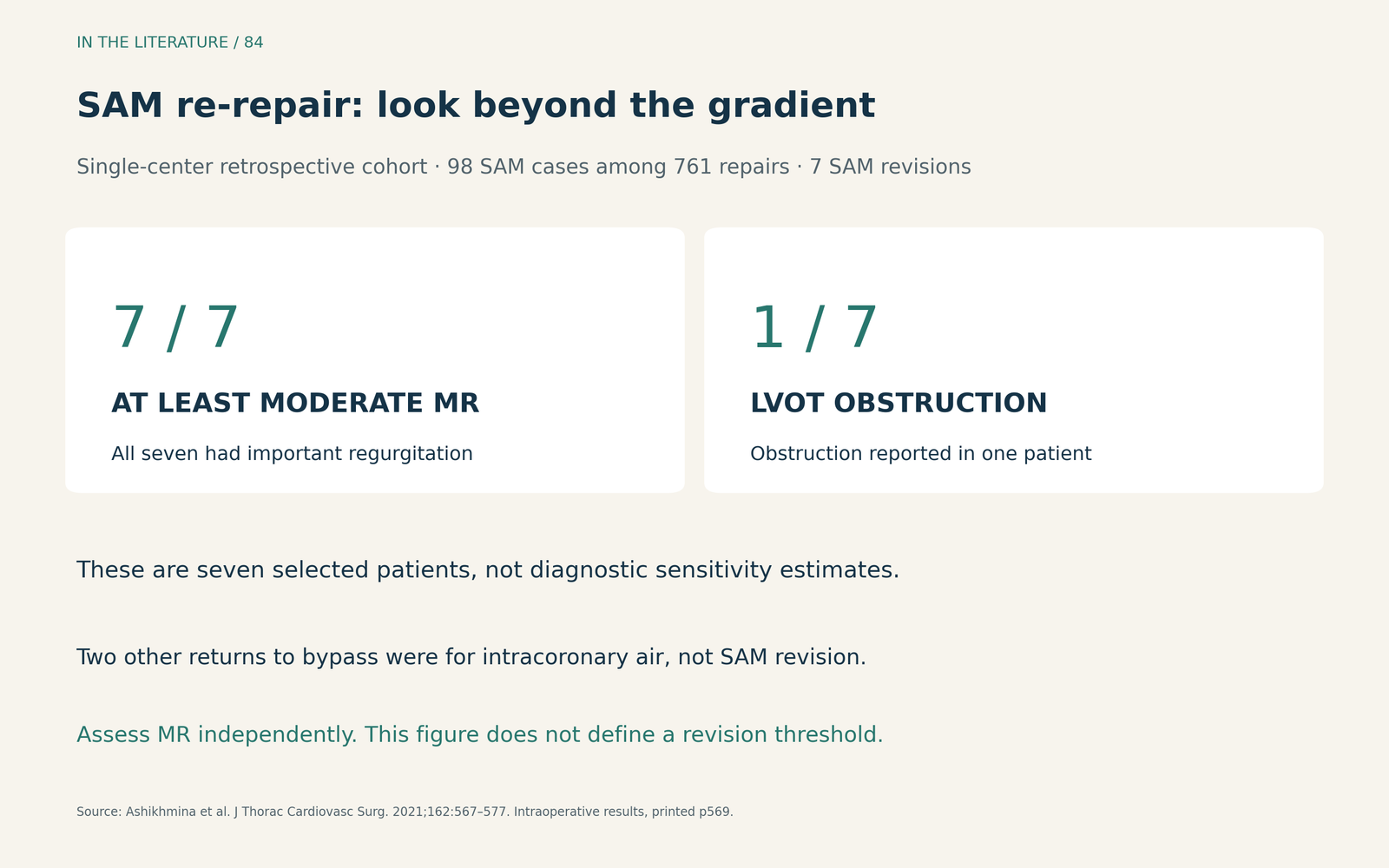
Figure 2. Descriptive findings among seven selected SAM re-repairs, not estimates of test sensitivity or an operative threshold. The cohort used posterior annuloplasty and excluded several concomitant procedures.3 Larger view

Make the return-to-bypass discussion specific
Report what persists after optimization: MR mechanism and severity, verified obstruction, valve geometry and hemodynamic tolerance. Persistent instability or inability to separate safely from CPB needs prompt team reassessment. An isolated 50 mmHg threshold is not a universally validated instruction to reoperate.2
Re-repair is a meaningful option. Fu and colleagues reported 17 SAM re-repairs among 2217 degenerative mitral operations (0.7%) at one expert center. All 17 were repaired without intraoperative replacement for SAM, but two required another repair for recurrent SAM within three months. The series supports feasibility, not a promise of universal success or superiority over replacement.4
When reviewing geometric measurements, use the formal correction to the 2020 guideline: leaflet lengths are measured in mid-diastole, while leaflet-height ratio and perpendicular coaptation–septal distance use early systole. These anatomical risk measurements are not stand-alone indications for repeat CPB.1
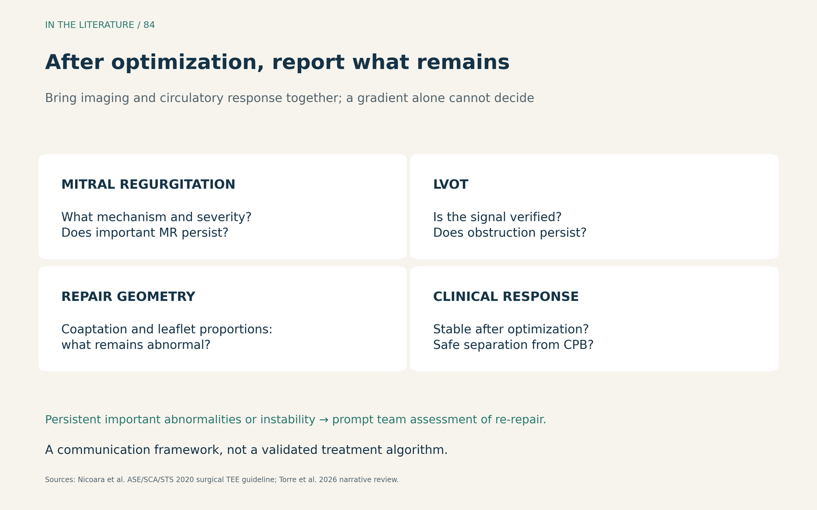
Figure 3. Information to communicate before accepting or revising the repair. Reassess under appropriate loading conditions and document the associated hemodynamics.1,2 Larger view

Three takeaways
- Verify the signal and the mechanism. A moving leaflet or a high-velocity tracing is not the whole diagnosis.
- Judge the response in both directions. Reassess forward outflow and MR after individualized hemodynamic optimization.
- Escalate on the whole clinical picture. Persistent important MR, obstruction or instability warrants a specific discussion of repair adequacy and possible re-repair.
References
- Nicoara A, Skubas N, Ad N, et al. Guidelines for the Use of Transesophageal Echocardiography to Assist with Surgical Decision-Making in the Operating Room: A Surgery-Based Approach. J Am Soc Echocardiogr. 2020;33(6):692-734. Correction: 2020;33(11):1426. doi:10.1016/j.echo.2020.03.002 Correction
- Torre DE, Mangino D, Zoffoli G, et al. Systolic Anterior Motion After Mitral Valve Repair: Echocardiographic Prediction, Surgical Prevention and Perioperative Management. J Clin Med. 2026;15(18):6958. doi:10.3390/jcm15186958
- Ashikhmina E, Schaff HV, Daly RC, et al. Risk factors and progression of systolic anterior motion after mitral valve repair. J Thorac Cardiovasc Surg. 2021;162(2):567–577. doi:10.1016/j.jtcvs.2019.12.106
- Fu W, Wagner C, Pawar G, et al. A surgical armamentarium for correcting systolic anterior motion with re-repair rather than replacement. JTCVS Open. 2024;19:91–93. doi:10.1016/j.xjon.2024.03.010