
ITL-0d7c2c383c08
Juan Li, MD · Updated November 17, 2026
Watch the video

Key points
- There is no consensus echocardiographic definition of septic cardiomyopathy. Studies have used different thresholds for left ventricular (LV) ejection fraction, strain, diastolic indices and right ventricular (RV) function.[1], [2]
- Ejection fraction is strongly load-dependent; a normal value under low afterload can hide impaired contractility that appears once vasopressors raise blood pressure.[1], [2]
- Global longitudinal strain detects dysfunction more often than ejection fraction, but the diagnosis depends on the criterion and day of examination.[2], [3]
- The link with mortality varies by phenotype and timing: RV dysfunction showed a clearer association than LV systolic dysfunction in one meta-analysis.[4]
- Echocardiography is how cardiac dysfunction is recognized, but treatment evidence is weak: guidelines suggest dobutamine or epinephrine only with persistent hypoperfusion, and VA-ECMO data come from selected severe cases.[5], [6], [7]
A patient in septic shock remains hypotensive and hypoperfused despite fluids and norepinephrine. The ejection fraction was "normal" on admission, but a repeat study now shows a dilated, globally hypokinetic ventricle. Is this septic cardiomyopathy, an effect of the vasopressor load, or a different cause of shock, and what does each answer mean for treatment?[1], [5]
No single definition
Plack and colleagues note that there is no consensus echocardiographic definition of septic cardiomyopathy. They describe three commonly accepted features: LV dilatation with normal or low filling pressure, reduced contractility, and RV systolic or diastolic dysfunction with a reduced response to volume.[1] Zakynthinos and colleagues report that the working definition most often used in studies is an LV ejection fraction below 40% in sepsis without acute coronary syndrome that improves within a short period, usually 7 to 10 days.[2]
The lack of a standard matters because prevalence and prognosis depend on the definition. In the meta-analysis by Lin and colleagues, most studies defined LV systolic dysfunction as an ejection fraction below 50%, but RV and diastolic criteria varied.[4]
Loading conditions can hide or create the finding
Ejection fraction depends on preload and afterload. Plack and colleagues describe how correcting preload with early fluids has been associated with unmasking systolic dysfunction.[1] Zakynthinos and colleagues give an example in which ejection fraction fell from 70% after initial resuscitation to 40% a few hours into norepinephrine infusion, revealing impaired contractility.[2]
De Backer and colleagues warn about the opposite errors. LV dysfunction may follow an afterload increase as hypotension is corrected. Dynamic outflow tract obstruction may be caused by inotropes or vasopressors, and RV dysfunction may reflect mechanical ventilation.[5] Each echocardiogram should therefore be interpreted with the blood pressure, vasopressor dose, volume status and ventilator settings at the time of the study.
Timing also changes what is seen. Zakynthinos and colleagues cite LV dysfunction in 22% of patients examined within 24 hours of sepsis onset, rising to 31.8% after 72 hours, and in another cohort from 18% within 6 hours to 60% after 72 hours.[2]
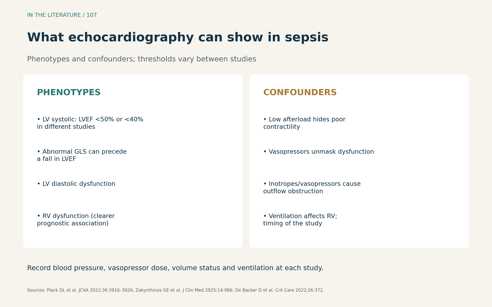
Figure 1. What echocardiography can show in sepsis. Phenotypes and confounders summarized from the reviews by Plack, Zakynthinos and De Backer and colleagues; thresholds vary between studies.[1], [2], [5] View larger.

Strain versus ejection fraction
Global longitudinal strain (GLS) can be abnormal when ejection fraction is preserved.[1] Gonzalez and colleagues prospectively examined 98 patients with septic shock on days 1, 7 and 30 using both conventional and strain criteria. On day 1, myocardial dysfunction was identified in 70 of 98 (71.4%) with strain criteria and 57 of 98 (58.2%) with conventional criteria (P = 0.072). LV systolic dysfunction was found in 60.2% by GLS (worse than −15%) and 48.0% by ejection fraction below 50%. Recovery by day 7 appeared more often with conventional criteria (35.3%) than with strain criteria (17.5%). Among patients with normal function on day 1, 17 of 27 had developed dysfunction by day 7 on strain criteria.[3]
These results show that the label depends on the method and the day. They come from one center, and the strain cut-offs used were chosen by the investigators rather than validated for sepsis.[3]
Does recognizing it predict outcome?
Lin and colleagues pooled 20 observational studies including 4,410 septic patients. Septic cardiomyopathy was not significantly associated with in-hospital mortality (risk ratio 1.28; 95% CI 0.96–1.71). It was associated with 1-month mortality (risk ratio 1.47; 95% CI 1.17–1.86). RV dysfunction was associated with higher mortality (risk ratio 1.72; 95% CI 1.27–2.34), whereas LV dysfunction was not significant (1.33; 95% CI 0.87–2.02). Heterogeneity was moderate, definitions varied, and the association was strongest when echocardiography was done on day 1.[4]
The prognostic message is therefore nuanced. RV involvement and early, severe dysfunction carry more weight than a single mildly reduced ejection fraction.[2], [4]
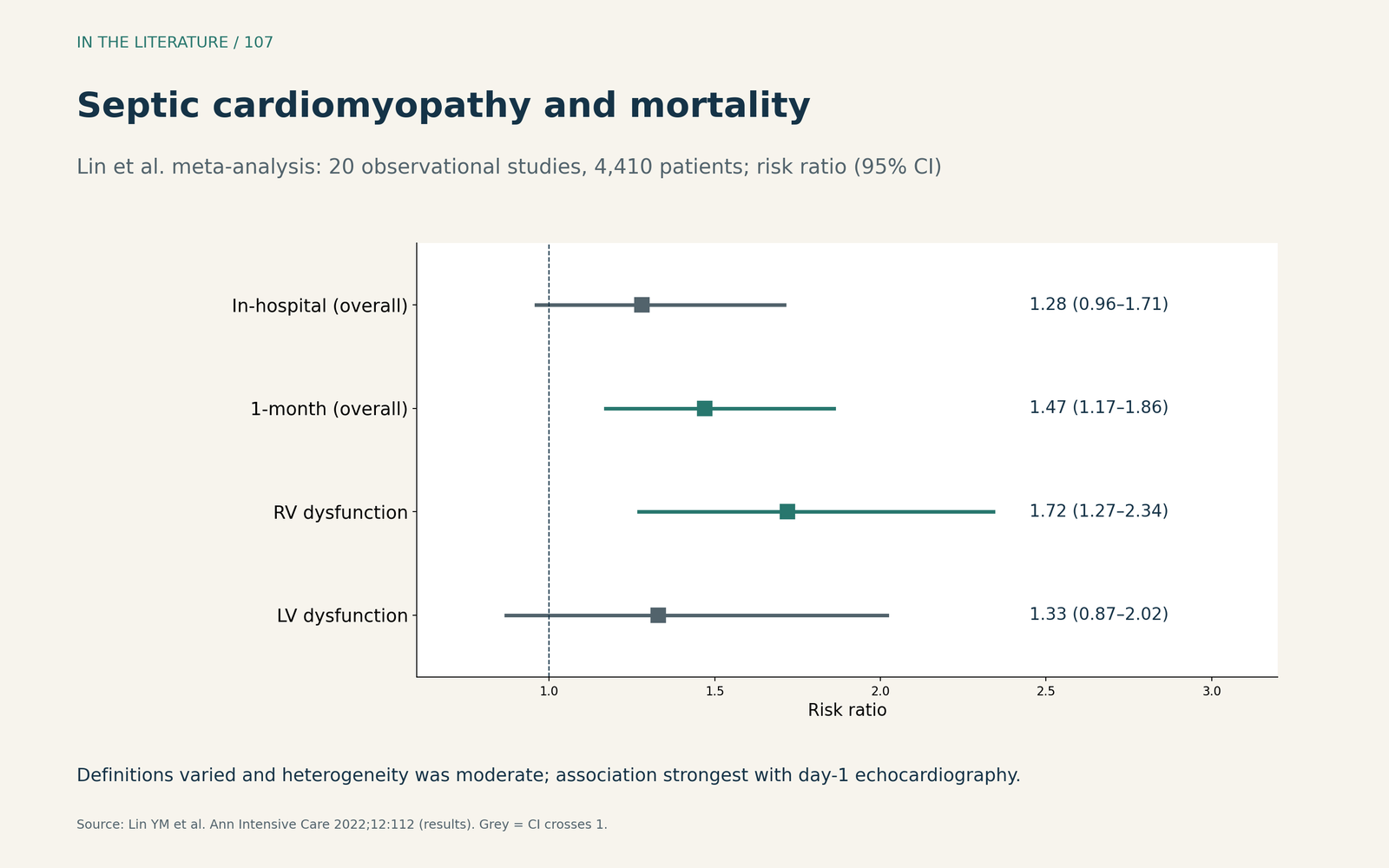
Figure 2. Mortality associations depend on outcome and phenotype. Redrawn from the results of Lin et al. (20 studies, 4,410 patients); risk ratios with 95% confidence intervals.[4] View larger.

What recognition changes in management
The 2026 Surviving Sepsis Campaign guideline states that cardiac dysfunction should be suspected after bedside echocardiography or cardiac output monitoring. For adults with septic shock and cardiac dysfunction with persistent hypoperfusion despite adequate volume status and blood pressure, it suggests inotropes over none, either adding dobutamine to norepinephrine or using epinephrine alone. Both are conditional recommendations with very low certainty evidence. It suggests against levosimendan (conditional, low certainty).[6]
De Backer and colleagues emphasize that inotropes are indicated only when impaired function is associated with low or inadequate stroke volume and poor tissue perfusion. Patients with high cardiac output or dynamic outflow obstruction should not receive inotropes, even with impaired function or high biomarkers.[5]
For the most severe cases, Bréchot and colleagues compared 82 patients with sepsis-induced cardiogenic shock treated with VA-ECMO against 130 controls without it. Inclusion required severe myocardial dysfunction (LV ejection fraction of 35% or less or cardiac index of 3 L/min/m² or less) and severe hemodynamic compromise (high inotrope score or lactate of at least 4 mmol/L). Ninety-day survival was 60% versus 25%, and 51% versus 14% after propensity weighting. The study was retrospective, imbalances remained after weighting, and the results apply to a narrow group with severe myocardial dysfunction rather than to vasoplegic shock.[7]
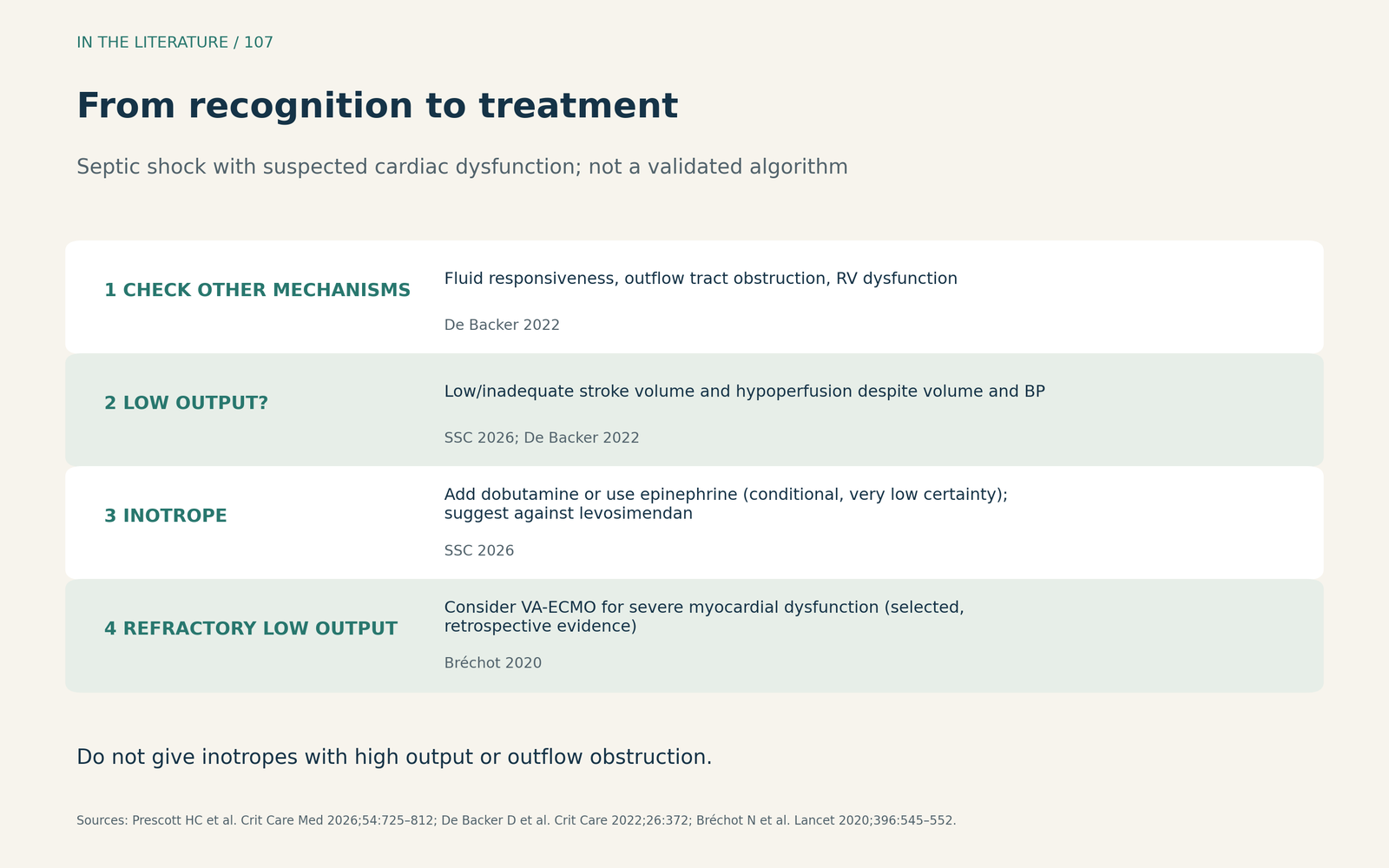
Figure 3. From recognition to treatment. Synthesized from the 2026 Surviving Sepsis Campaign guideline, the personalization review by De Backer and colleagues and the VA-ECMO cohort by Bréchot and colleagues; not a validated algorithm.[5]–[7] View larger.

Three takeaways
- Name the phenotype, not just the ejection fraction. Describe LV systolic, diastolic and RV function, and add strain when image quality allows.[1]–[3]
- Interpret every study in its loading context. Record vasopressor dose, blood pressure, volume status and ventilation; repeat the examination as conditions change.[2], [5]
- Treat the physiology, not the label. Consider inotropes only with low output and persistent hypoperfusion, and reserve VA-ECMO for selected severe low-output cases.[5]–[7]
References
- Plack DL, Royer O, Couture EJ, et al. Sepsis-induced cardiomyopathy reviewed: the case for early consideration of mechanical support. J Cardiothorac Vasc Anesth. 2022;36(10):3916–3926. doi:10.1053/j.jvca.2022.04.025
- Zakynthinos GE, Giamouzis G, Xanthopoulos A, et al. Septic cardiomyopathy: difficult definition, challenging diagnosis, unclear treatment. J Clin Med. 2025;14(3):986. doi:10.3390/jcm14030986
- Gonzalez FA, Bacariza J, Varudo AR, et al. Sepsis-induced myocardial dysfunction diagnosed with strain versus non-strain echocardiography parameters: incidence, evolution and prognosis. Ann Intensive Care. 2025;15(1):141. doi:10.1186/s13613-025-01561-w
- Lin YM, Lee MC, Toh HS, et al. Association of sepsis-induced cardiomyopathy and mortality: a systematic review and meta-analysis. Ann Intensive Care. 2022;12(1):112. doi:10.1186/s13613-022-01089-3
- De Backer D, Cecconi M, Chew MS, et al. A plea for personalization of the hemodynamic management of septic shock. Crit Care. 2022;26(1):372. doi:10.1186/s13054-022-04255-y
- Prescott HC, Antonelli M, Alhazzani W, et al. Surviving Sepsis Campaign: international guidelines for management of sepsis and septic shock 2026. Crit Care Med. 2026;54(4):725–812. doi:10.1097/CCM.0000000000007075
- Bréchot N, Hajage D, Kimmoun A, et al. Venoarterial extracorporeal membrane oxygenation to rescue sepsis-induced cardiogenic shock: a retrospective, multicentre, international cohort study. Lancet. 2020;396(10250):545–552. doi:10.1016/S0140-6736(20)30733-9