ITL-580d151f60c8
Juan Li, MD · Updated November 17, 2026
Watch the video
Key points
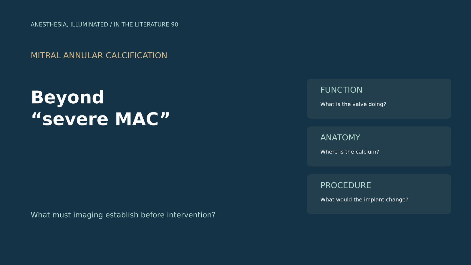
- Extensive mitral annular calcification (MAC) does not, by itself, establish severe mitral valve dysfunction or an indication for intervention.1
- Echocardiography establishes the functional problem; ECG-gated CT maps calcium and the anatomy needed for procedural planning.1,2
- A useful CT report describes calcium distribution and adjacent structures, rather than stopping at a single MAC score.2
- Three-dimensional TEE and CT calcium scores are not interchangeable, and contrast-enhanced CT measurements require attention to the acquisition and scoring method.3,4
- A simulated neo-LVOT must be interpreted with the proposed device, implantation position and cardiac phase; no single number guarantees procedural safety.2
The referral says “severe MAC,” but the team still needs to decide whether a mitral intervention could help and whether the proposed procedure is anatomically feasible. The next imaging request should make those two questions explicit: What is the valve doing to the patient, and what anatomy will the procedure encounter?1,2
Establish the functional problem before planning a device
MAC is an anatomical finding. It may coexist with stenosis, regurgitation or mixed dysfunction, and substantial calcification can also occur without significant valve dysfunction. A calcium burden cannot substitute for a functional assessment.1
Start with transthoracic echocardiography to define the valve disorder and its cardiac consequences. TEE, including three-dimensional imaging, can clarify leaflet morphology and motion when further definition is needed. In degenerative mitral stenosis, however, acoustic shadowing from calcium can obscure the very structures being assessed. A structure hidden behind shadow should be reported as incompletely assessed, rather than assumed to be normal.1,2
The practical output is a description of stenosis and/or regurgitation, the mechanism and leaflet restriction, the relevant cardiac response, and any unresolved imaging uncertainty. CT then answers the anatomical planning questions. In patients with MAC being considered for intervention, the 2025 ESC/EACTS guideline emphasizes ECG-gated CT to establish the extent and location of calcification.1
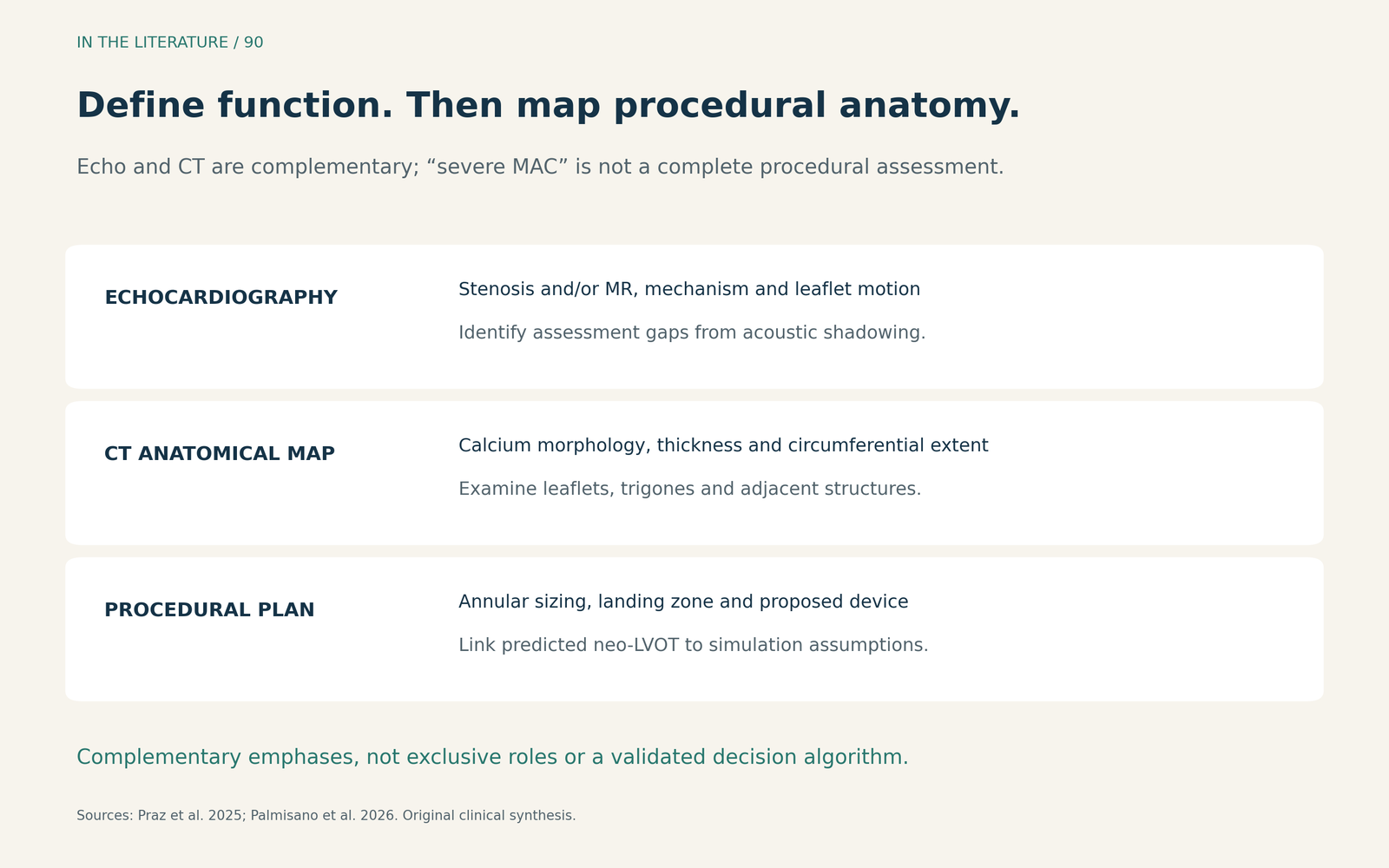
Figure 1. An original clinical synthesis of the imaging roles. These are complementary emphases, not exclusive capabilities or a validated decision algorithm.1,2 Enlarge

Ask for an anatomical map, not only a severity label
The November 2026 ESCR/NASCI expert consensus provides a useful framework for transcatheter mitral valve replacement (TMVR) planning. Describe the morphology, thickness and circumferential extent of calcium, together with involvement of the leaflets and fibrous trigones. Also examine extension toward the ventricular myocardium and outflow tract. Two patients carrying the same “severe MAC” label may present very different landing zones and procedural hazards.2
The report should relate annular measurements to the contemplated device and identify nearby structures that matter to that procedure. The left circumflex coronary artery deserves explicit attention: its patency, course and proximity to the annulus are relevant, but the implication depends on the intervention being considered. A distance cited for one annuloplasty technique should not become a contraindication to every mitral procedure.2
These recommendations represent expert consensus about imaging practice. They help structure the assessment; they do not establish that a particular calcium score identifies patients who will benefit from treatment.2
What recent scoring studies actually add
Quelal Analuisa and colleagues compared a three-dimensional TEE MAC score with a CT-based score in 164 patients from 13 centres undergoing TMVR screening. Overall correlation was modest (Spearman rₛ=0.34) and agreement was limited (ICC=0.35). The two methods did not provide interchangeable descriptions of MAC.3
The useful implication is to combine the functional and anatomical information, not to replace CT planning with an echo score. This was a cross-sectional imaging comparison without procedural-outcome validation. It did not show that selecting a treatment by either score improves outcomes.3
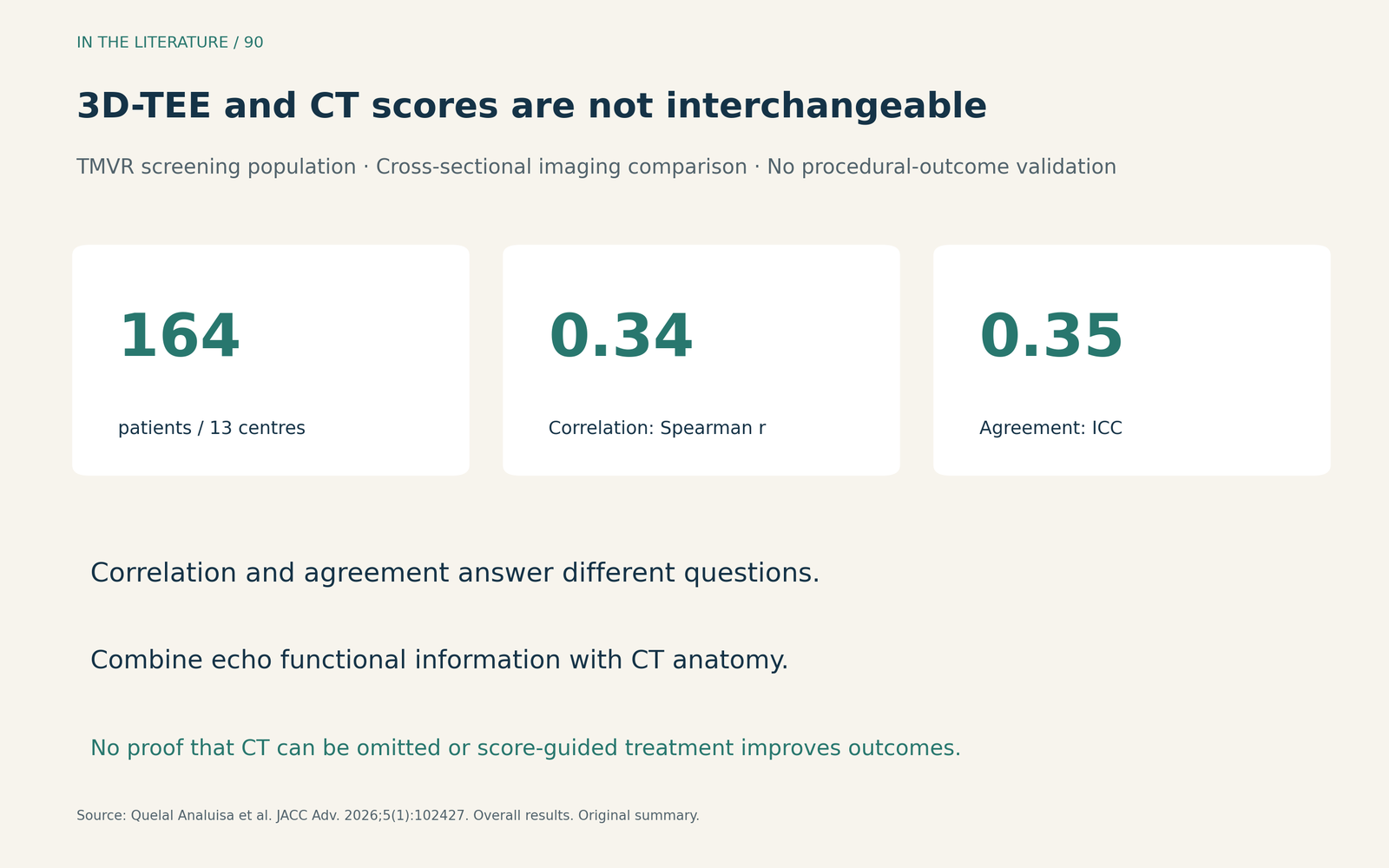
Figure 2. Original summary of the Quelal Analuisa study. Correlation and agreement are different properties; neither establishes that the modalities can replace one another for procedural planning.3 Enlarge

A second, November 2026 study by Al Zein and colleagues examined 303 surgical patients with preoperative contrast-enhanced CT. Its calcium-score calibration used 50 patients within that cohort who also had noncontrast scans. The contrast method identified calcium using a threshold at least 4 standard deviations above mean ascending-aortic blood-pool attenuation, then derived contrast-specific score cutoffs.4
For the clinical reader, the important point is methodological: ask whether the reported score came from contrast or noncontrast imaging and how it was calculated. Those cutoffs were not independently externally validated and may depend on acquisition and analysis protocols. They should not be transferred unchanged between methods or used as universal criteria for procedural eligibility.4
Make the virtual implantation assumptions visible
If TMVR is being considered, the remaining outflow tract after a simulated implant—the neo-LVOT—must be assessed in relation to the proposed device. Device geometry, implantation depth and orientation, leaflet anatomy and cardiac phase influence the predicted result. The consensus recommends assessment across systolic phases because the outflow tract is dynamic.2
A clinically useful handover therefore pairs the predicted area with the simulated valve and position, the phase assessed, and the anatomical features driving concern. This is a practical synthesis of the consensus, not a new eligibility rule. A low-risk-looking value under one simulation does not settle the question for a different device or position.2
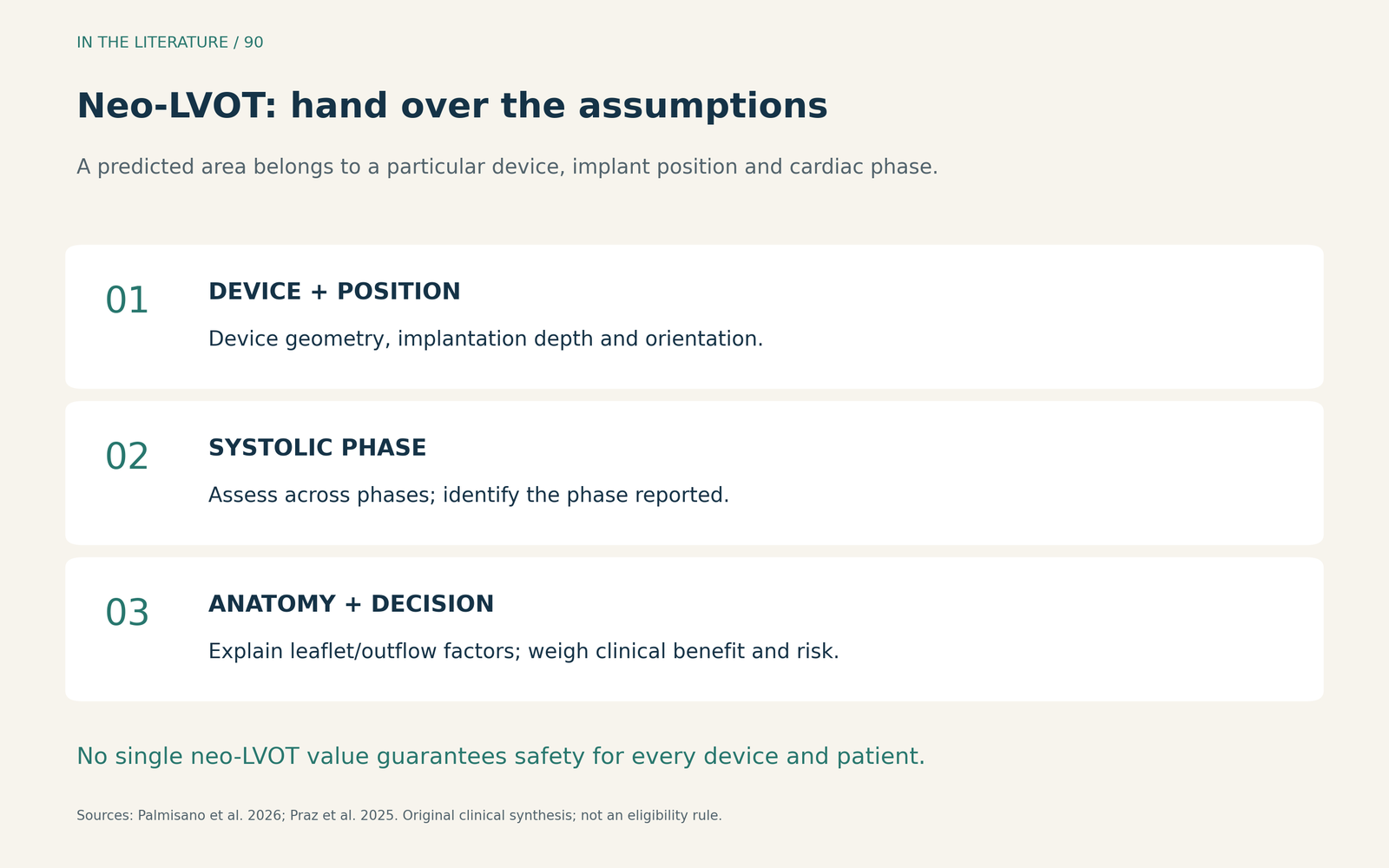
Figure 3. Conditions to retain when communicating a neo-LVOT prediction. Annular sizing and predicted outflow obstruction answer different planning questions; a single calcium score or area cannot replace the complete assessment.2 Enlarge

Finally, anatomical feasibility is only part of the decision. For severe MAC with important mitral dysfunction, the Heart Team must weigh expected clinical benefit, procedural risk and the possibility of futility. Completing a CT plan does not itself establish that intervention is warranted.1
Three takeaways
- State the functional problem first. Describe the mitral dysfunction and what remains uncertain before using “severe MAC” to frame treatment.
- Request a map with a method. Record calcium distribution, relevant adjacent structures, and how any calcium score was obtained.
- Attach the assumptions to the prediction. Communicate device, position and cardiac phase with neo-LVOT assessment, then place feasibility within the Heart Team's benefit–risk decision.
References
- Praz F, Borger MA, Lanz J, et al. 2025 ESC/EACTS Guidelines for the management of valvular heart disease. Eur J Cardiothorac Surg. 2025;67(8):ezaf276. doi:10.1093/ejcts/ezaf276
- Palmisano A, Francone M, Rajiah P, et al. CT and MRI in periprocedural transcatheter mitral valve replacement: expert recommendations by the European Society of Cardiovascular Radiology (ESCR) and the North American Society for Cardiovascular Imaging (NASCI). Eur Radiol. Published online November 6, 2026. doi:10.1007/s00330-026-12955-0
- Quelal Analuisa K, Challa A, Lane C, et al. Mitral Annular Calcification Score Determined by 3-D Transesophageal Echocardiography vs Cardiac Computed Tomography. JACC Adv. 2026;5(1):102427. doi:10.1016/j.jacadv.2025.102427
- Al Zein M, Okushi Y, Unai S, et al. Contrast-enhanced computed tomography for quantifying mitral annular calcification in mitral valve surgery and association with outcomes. Eur Heart J Imaging Methods Pract. 2026;4(4):qyag184. doi:10.1093/ehjimp/qyag184