ITL-064b37132008
Juan Li, MD · Updated November 18, 2026
Key points
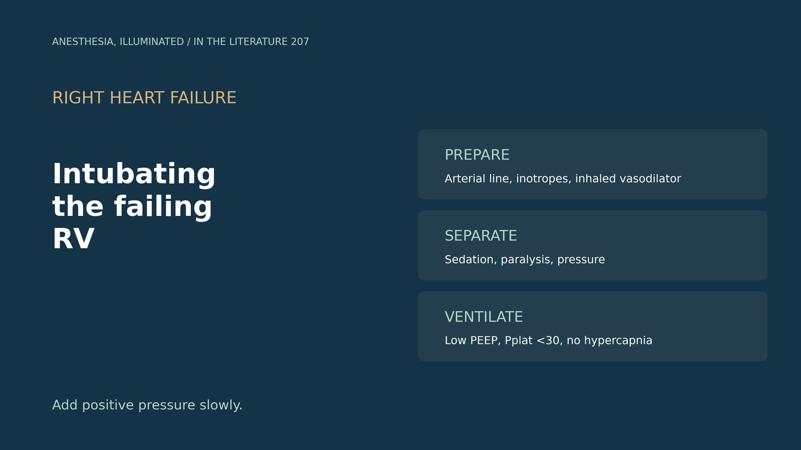
- Positive pressure hurts a failing RV. It rapidly increases RV afterload, and the principal goal is to maximize RV output.[1]
- Instability is common. Cardiovascular instability complicated 42.6% of ICU intubations, and NIV also imposes positive intrathoracic pressure.[2]
- Screen the RV first. Lower TAPSE independently predicted post-intubation hypotension, which occurred in 41.2% of emergency intubations.[3]
- Take the steps apart. SAVIOR separates sedation, paralysis and positive pressure; awake intubation, ketamine or etomidate over propofol, and an arterial line before induction are suggested.[1], [2]
- Ventilate gently. Keep plateau pressure below 30 cmH2O, avoid driving pressure of 18 cmH2O or more and PaCO2 of 48 mmHg or more.[4]
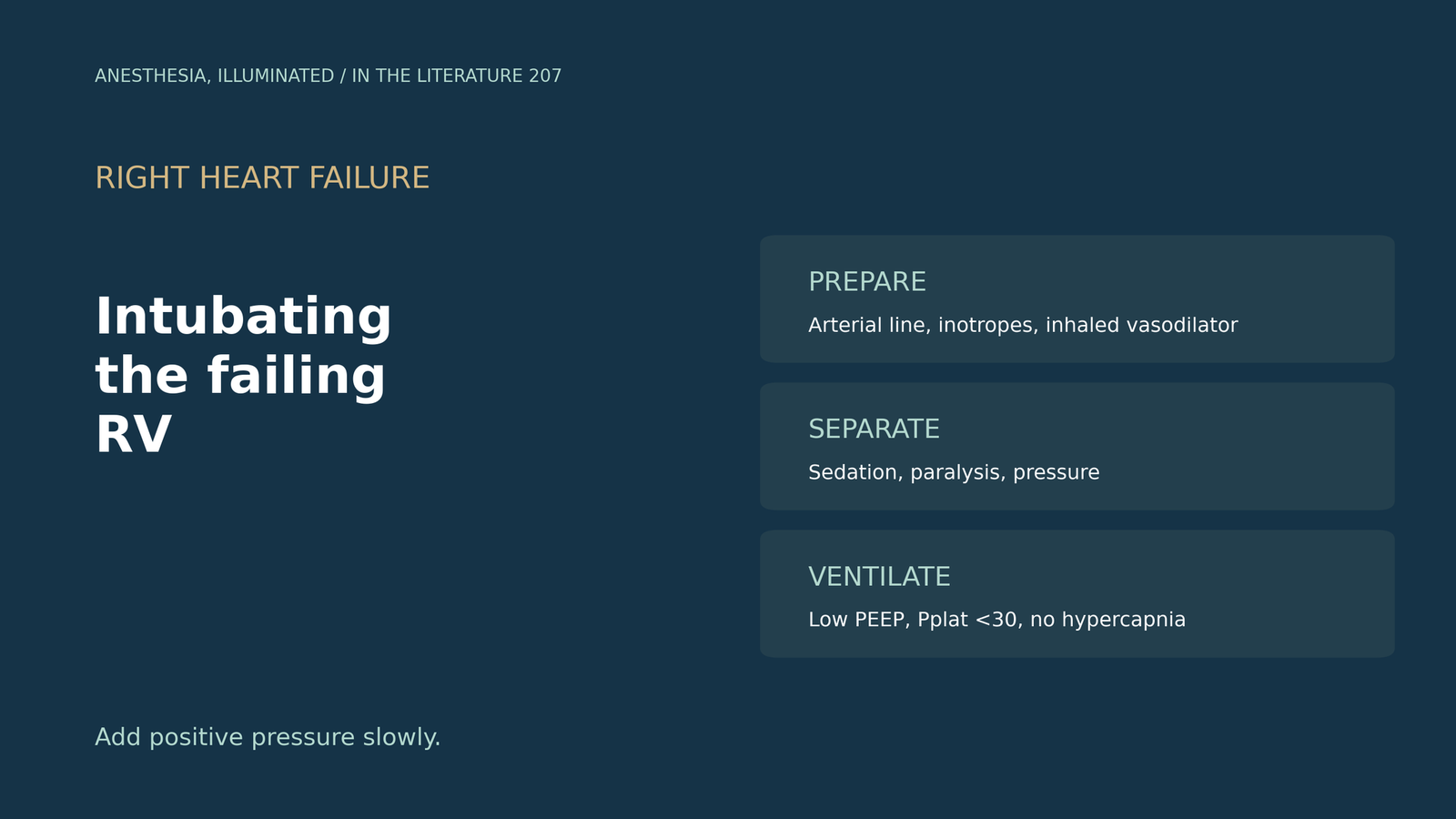
A patient with severe pulmonary hypertension and a failing right ventricle is tiring on high-flow oxygen and needs intubation in the ICU. How should induction and the first breaths of positive pressure be planned to avoid cardiovascular collapse?[1], [2]
Why intubation is dangerous
Srour and colleagues explain that positive pressure rapidly increases afterload on a failing RV and that the principal goal is to maximize RV output.[1] Kotani and colleagues report that cardiovascular instability complicated 42.6% of ICU intubations, that rising RV afterload reduces LV filling and coronary flow, and that NIV also imposes positive intrathoracic pressure.[2] In Prakash and colleagues' emergency department cohort, post-intubation hypotension occurred in 71 of 172 patients (41.2%), and lower TAPSE independently predicted it (OR 0.81), although TAPSE has low specificity and should be combined with other parameters.[3]
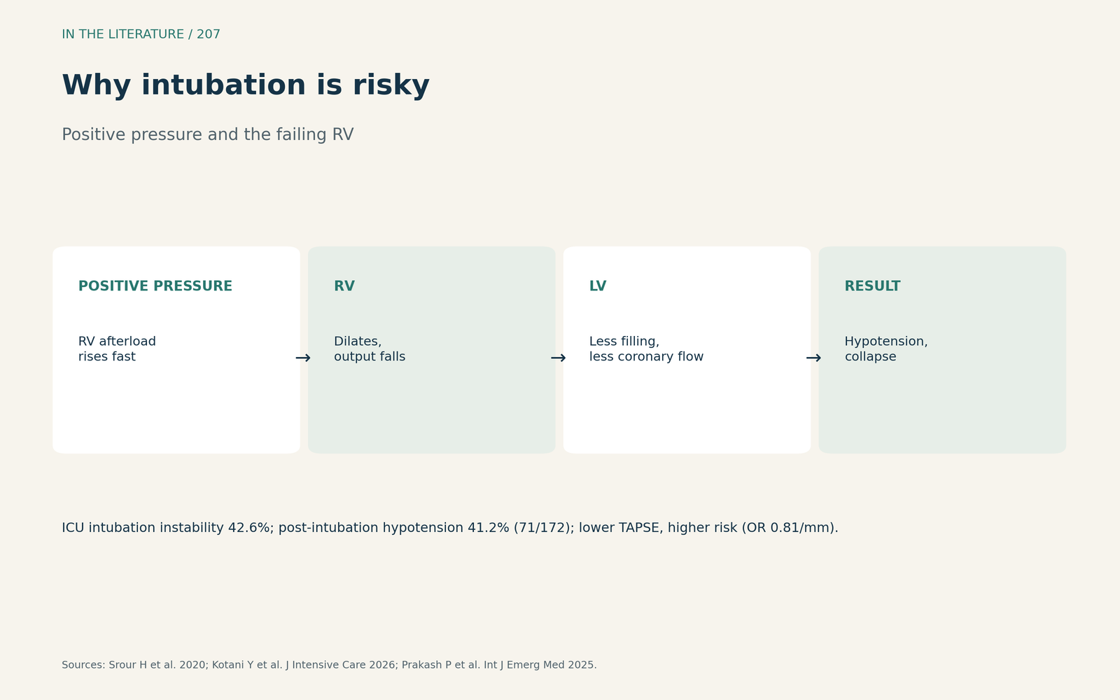
Figure 1. Afterload up, filling down, pressure falls. Synthesized from Srour et al., Kotani et al. and Prakash et al.[1], [2], [3] View larger.

How to intubate
The SAVIOR algorithm emphasizes spontaneous awake ventilation, inotropic support and optimized respiratory physiology, and separates sedation, paralysis and positive pressure. Pressure support is titrated up over 20 to 30 minutes, with an inhaled vasodilator in the circuit to reduce RV workload; the support for this approach is the authors' own case series.[1] Kotani and colleagues add that awake bronchoscopic intubation under topical anesthesia has been suggested, that ketamine or etomidate is preferred over propofol in at-risk patients, that a routine fluid bolus is not supported by evidence, and that an arterial line should be considered before induction.[2] Zochios and colleagues note that the injured RV is best supported by optimizing myocardial perfusion.[5]
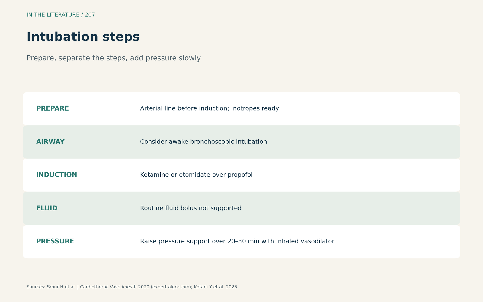
Figure 2. Prepare, separate the steps, add pressure slowly. Synthesized from Srour et al., Kotani et al. and Zochios et al.[1], [2], [5] View larger.

How to ventilate
Peng and colleagues note that ventilation can worsen RV stress through overdistension and raised intrathoracic pressure. Plateau pressure should stay below 30 cmH2O; a driving pressure of 18 cmH2O or more and a PaCO2 of 48 mmHg or more are risk factors for cor pulmonale; and stepwise PEEP increases reduced biventricular stroke work.[4] Kotani and colleagues suggest that initial PEEP may be safer set low.[2] Impaired RV physiology is a major determinant of mortality in ARDS.[5]
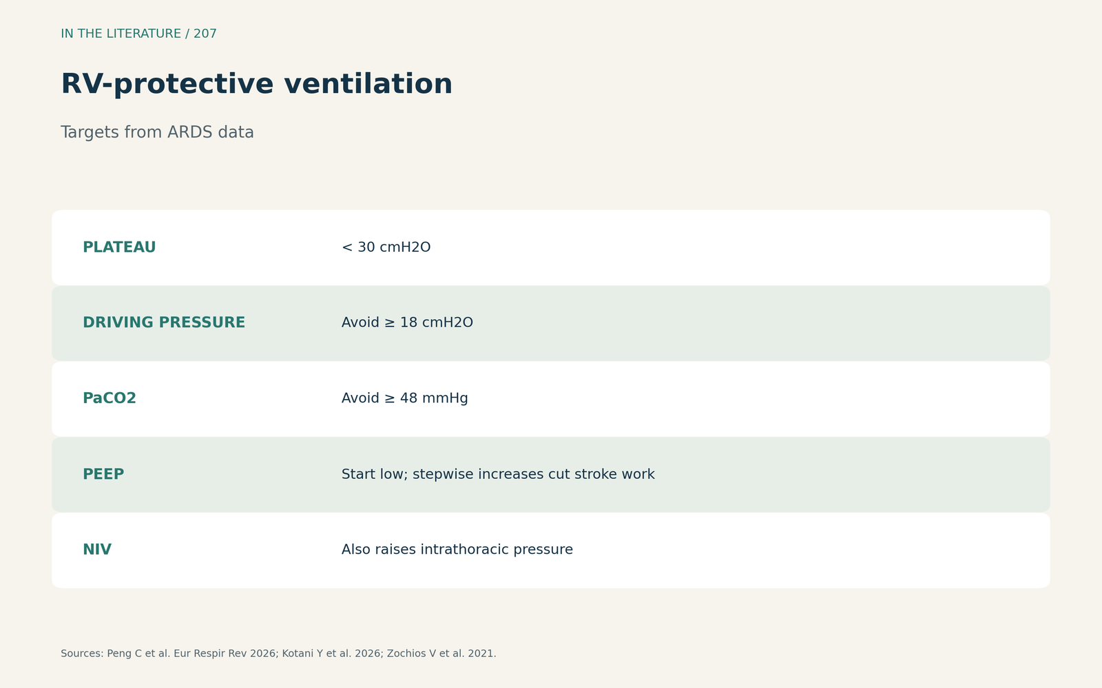
Figure 3. Limit pressure, avoid hypercapnia, start PEEP low. Redrawn from Peng et al., Kotani et al. and Zochios et al.[4], [2], [5] View larger.

Three takeaways
- Before induction, screen the RV, place an arterial line and have inotropes and an inhaled vasodilator ready.[1], [2], [3]
- Separate sedation, paralysis and positive pressure; consider awake intubation and avoid propofol in at-risk patients.[1], [2]
- Ventilate with low PEEP, limited plateau and driving pressures, and no hypercapnia; these targets come from ARDS data.[4]
References
- Srour H, Shy J, Klinger Z, Kolodziej A, Hatton KW. Airway management and positive pressure ventilation in severe right ventricular failure: SAVIOR algorithm. J Cardiothorac Vasc Anesth. 2020;34(1):305–306. doi:10.1053/j.jvca.2019.05.046
- Kotani Y, Koroki T, Hayashi Y, Russotto V. The hemodynamics of tracheal intubation in critically ill patients: a narrative review. J Intensive Care. 2026;14(1):42. doi:10.1186/s40560-026-00877-4
- Prakash P, Nisarg S, Balakrishnan JM, et al. Right ventricular dysfunction: a key predictor of post-intubation hypotension in the emergency department. Int J Emerg Med. 2025;18(1):183. doi:10.1186/s12245-025-00987-0
- Peng C, Yang L, Wu Y, et al. Right ventricular-protective ventilation in acute respiratory distress syndrome: phenotypes, monitoring and ventilatory management. Eur Respir Rev. 2026;35(181):260038. doi:10.1183/16000617.0038-2026
- Zochios V, Lau G, Conway H, Yusuff HO. Protecting the injured right ventricle in COVID-19 acute respiratory distress syndrome: can clinicians personalize interventions and reduce mortality? J Cardiothorac Vasc Anesth. 2021;35(11):3325–3330. doi:10.1053/j.jvca.2021.05.059