
ITL-3b3394cdb180
Watch the video

Figure 1. Closing the communication must preserve both systemic and pulmonary venous drainage. Conceptual illustration, not a deployment diagram.
Key points
- Covered stenting is an option for selected adults with a superior sinus venosus atrial septal defect (SVASD); the 2025 ACHD guideline gives it a Class IIb, B-NR recommendation when technically feasible.[1]
- The critical question is whether the right pulmonary venous pathway to the left atrium remains unobstructed when the communication is excluded.
- Cross-sectional imaging and balloon interrogation answer complementary questions; a smaller color-flow jet alone is insufficient evidence of a satisfactory repair.[1, 2]
- In the international registry, technical success was 97.4%, but this was a selected, retrospective cohort without a randomized surgical comparison.[2]
- Assess residual shunting, venous patency and stent stability separately, and arrange surveillance beyond the procedure.
Start with the pathway, not the hole
An adult has a superior SVASD with right-heart volume loading and anomalous right pulmonary venous drainage. The team is considering a catheter approach. The important imaging task is to establish where blood will flow after the covered stent is expanded.
Unlike a typical secundum ASD, this lesion involves the relationship between the superior vena cava (SVC), the cavoatrial junction and the right pulmonary veins. A covered stent is intended to separate the abnormal communication while preserving pulmonary venous return toward the left atrium. It is therefore a reconstruction of flow pathways, not simply placement of a plug across a septal opening.[1, 2]
Two decisions precede implantation. First, is closure physiologically appropriate? Pulmonary vascular disease, left-heart disease and associated lesions can change that answer. Second, can this particular anatomy be corrected with a stent? The guideline's conditional recommendation is not a declaration that every superior SVASD should undergo catheter treatment.[1]
Use the scan to plan, then test the proposed result
CT or cardiovascular magnetic resonance should delineate the pulmonary veins, including additional veins entering high in the SVC, and the anticipated route to the left atrium. The registry's selection process excluded patients with another reason for cardiac surgery and used cross-sectional imaging before catheter assessment. Patients unsuitable on imaging or balloon testing were not included in its outcome denominator.[2]
Balloon interrogation then simulates exclusion of the communication at the cavoatrial junction. In the registry, TEE, angiography and pressure measurements were used to determine whether the pulmonary venous pathway remained open. Failure to eliminate the shunt or obstruction of that pathway was a reason not to proceed with covered stenting.[2]
For the imaging clinician, the practical question is not just “has the shunt disappeared?” It is “has it disappeared while the intended venous drainage remains satisfactory?” If Doppler, angiography or pressure findings disagree, the discrepancy needs resolution before the apparent success of closure becomes an irreversible obstruction.
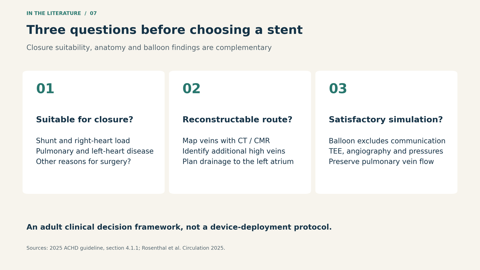
Figure 2. A decision framework based on the guideline and registry selection process. Balloon interrogation of the pulmonary venous pathway does not replace assessment of overall closure suitability.[1, 2]

Read the success rate with its definition attached
The international registry included 381 treated patients from 54 centers; median age was 44 years, and 37 patients were aged 18 or younger. Technical implantation succeeded in 371/381 patients (97.4%). Ten stents embolized and required surgical retrieval and defect repair. The more demanding reported outcome—successful correction without major complications or additional catheter intervention—was achieved in 359/381 (94.2%).[2]
Neither percentage means that every patient had immediate complete elimination of shunting. Immediately after the procedure, no leak was reported in 194 patients, a trivial leak in 121, a small leak in 36 and a moderate leak in seven; leak status was unreported in 13. Some leaks resolved during follow-up, while seven patients required a further catheter procedure for residual shunting within the first year.[2]
These results support feasibility in selected anatomy. They do not establish superiority over surgery or the proportion of all referred patients who can receive a stent. The registry did not capture the full population screened out, follow-up was heterogeneous, and formal follow-up MRI shunt quantification was available in only 51 patients.[2]
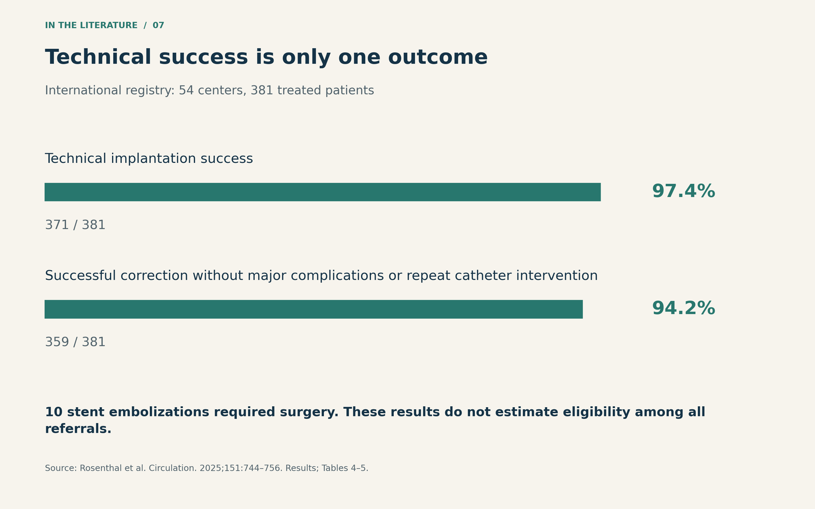
Figure 3. These are different outcomes within one selected cohort, not competing treatment arms. Technical success did not require immediate absence of every residual leak.[2]

Finish with three separate assessments
After deployment, document the residual communication, venous drainage and device position independently. A reassuring result in one domain cannot compensate for a problem in another.
Pulmonary venous obstruction deserves particular attention. In the registry, eight patients required intraprocedural balloon dilation to relieve pulmonary venous pathway narrowing. Another patient developed clinically consequential pulmonary venous obstruction requiring later surgery. Stent embolization was not exclusively an immediate event: four of the ten cases were recognized after leaving the catheter laboratory or by the next day.[2] These observations make a clear rescue plan and an explicit postprocedure handoff clinically relevant.
Surveillance also matters. A 2026 study, available here as an abstract only, reported thrombus in 11/153 patients (7.2%) after SVASD closure, at a median follow-up of one month; all detected thrombi were asymptomatic and nonobstructive, without systemic or pulmonary thromboembolism. Imaging was predominantly TEE. This is not a 7.2% rate of symptomatic embolic events, and the observational comparison of antithrombotic regimens does not establish which regimen is best.[3]
The follow-up plan should therefore state who will reassess the repair, which imaging question remains open and who owns antithrombotic management. The available studies do not justify inventing one universal surveillance schedule or drug regimen.
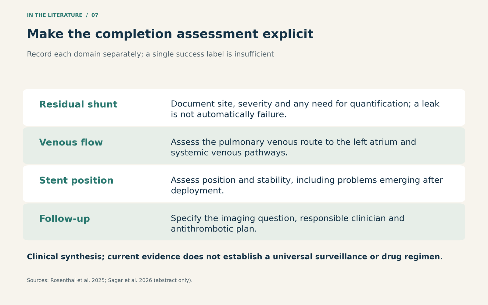
Figure 4. A practical imaging handoff derived from the reported complications and follow-up gaps; not a validated scoring system.[2, 3]

Three takeaways
- Establish the route before excluding the communication. A patent pulmonary venous pathway to the left atrium is central to selecting and assessing the repair.
- Define success precisely. Technical implantation, residual shunt and freedom from complications are different outcomes.
- Carry the assessment beyond deployment. Document venous patency and stent position, with a named follow-up and antithrombotic plan.
References
- Gurvitz M, Krieger EV, Fuller S, et al. 2025 ACC/AHA/HRS/ISACHD/SCAI Guideline for the Management of Adults With Congenital Heart Disease: a report of the American College of Cardiology/American Heart Association Joint Committee on Clinical Practice Guidelines. Circulation. 2026;153:e115–e251. doi:10.1161/CIR.0000000000001402.
- Rosenthal E, Qureshi SA, Sivakumar K, et al. Covered stent correction for sinus venosus atrial septal defects, an emerging alternative to surgical repair: results of an international registry. Circulation. 2025;151(11):744–756. doi:10.1161/CIRCULATIONAHA.124.070271.
- Sagar P, Thejaswi P, Athira M, Sivakumar K. Incidence and predictors of stent thrombosis following transcatheter sinus venosus defect closure. Pediatr Cardiol. Published online May 12, 2026. doi:10.1007/s00246-026-04275-3. Abstract only.