ITL-b324cdf147a6
Juan Li · In the Literature · Updated November 16, 2026
Watch the video
Key points
- CT remains the preferred imaging tool for TAVR planning; a reliable TEE annular measurement answers only part of the planning question.
- Measure the virtual basal ring in systole using aligned multiplanar reconstruction, rather than the leaflet opening or one convenient two-dimensional diameter.
- Automated analysis still requires inspection of the acquisition, annular plane and blood–tissue contour.
- Good correlation or repeatability does not establish that TEE and CT will select the same prosthesis size.
- When CT is difficult to interpret or relatively contraindicated, use TEE within a plan that also addresses coronary, aortic and access-vessel anatomy.
Start with what the measurement needs to decide
A TAVR candidate has renal impairment, and the team is considering a contrast-sparing evaluation. The 3D TEE software returns a clean annular contour and an area close to the boundary between two valve sizes. The useful question is whether that number is sufficiently trustworthy to guide the implant—and what information is still missing.
The 2025 ESC/EACTS guideline identifies cardiac CT as the preferred tool for TAVR planning because it assesses the annulus together with the root, calcium distribution, coronary relationships and vascular access. TEE or cardiac magnetic resonance may be considered when CT is difficult to interpret or relatively contraindicated, including selected patients with renal failure. This is an alternative imaging strategy, not permission to replace the whole assessment with one TEE number. [1]
Make the plane correct before making the contour precise
For sizing, the aortic annulus is the virtual basal ring defined by the lowest cusp attachments. It is different from the open valve orifice at the leaflet tips. Tracing the latter can produce an attractive image of the wrong structure. [2]
A practical measurement sequence follows the ASE screening standard and the workflow used in contemporary validation research:
- Acquire the entire annulus with adequate detail. Limit unnecessary 3D volume, and inspect for dropout, reverberation and stitching. A multibeat dataset can distort measurements even when the rendered image looks smooth.
- Choose the systolic annular frame. Mid-systole was used in the Massie validation study. Keep this distinct from the diastolic convention used for some aortic-root dimensions.
- Align the orthogonal planes at the basal attachments. Use multiplanar reconstruction to confirm the same annular level in both long-axis views before tracing the short-axis plane.
- Inspect the inner contour and report the dimensions. Review area, perimeter and orthogonal diameters together. If calcium shadowing obscures the boundary, the software's closed contour does not make that boundary visible. [2, 3]
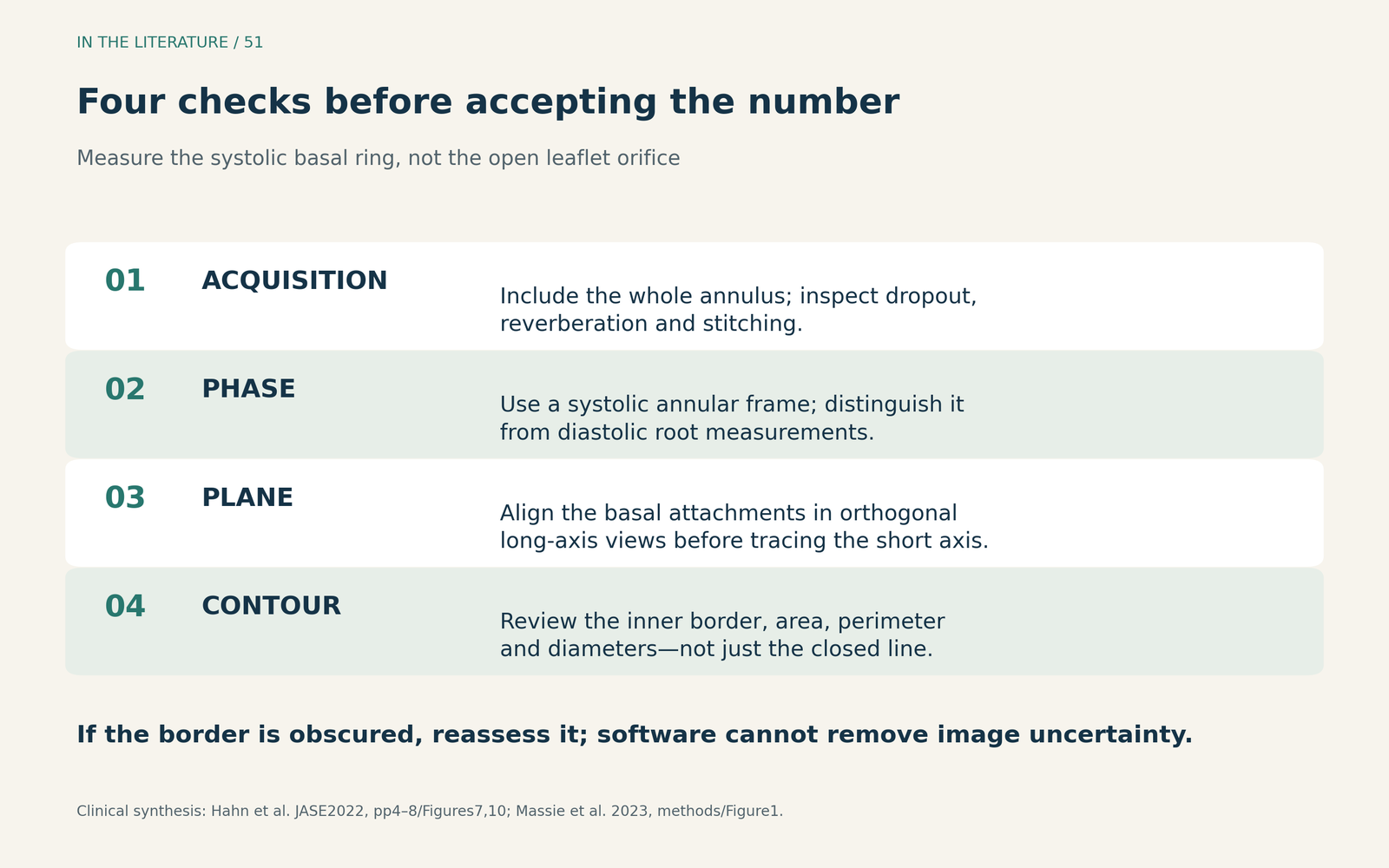
Figure 1. A measurement workflow synthesized from the ASE standard and the Massie study. It does not provide a universal image-quality cutoff or a device-sizing chart. [2, 3] View figure

Ask whether the number would change the valve size
Massie and colleagues prospectively studied 89 patients with severe native aortic stenosis using CT and three TEE approaches: automated, semi-automated and manual. Their GE analysis workflow still required selecting the loop and adjusting the annular planes; semi-automated analysis additionally allowed contour editing. All three approaches numerically underestimated annular area and perimeter compared with CT, with the automated method showing the greatest underestimation. Better agreement between readers did not remove this measurement bias. [3]
The clinically useful comparison involved the 70 patients who underwent implantation. CT-based hypothetical sizing matched the implanted size in 64/70 patients (91%). Across the six TEE analyses—three methods evaluated by expert and novice readers—the corresponding counts ranged from 46/70 to 52/70, approximately 66% to 74%. [3]
These are size-agreement results, not procedural success rates. The actual device was selected primarily using CT, so agreement with that device is not an independent test proving CT-guided treatment superior. Nevertheless, it shows why a high correlation coefficient cannot settle a borderline size decision. [3]
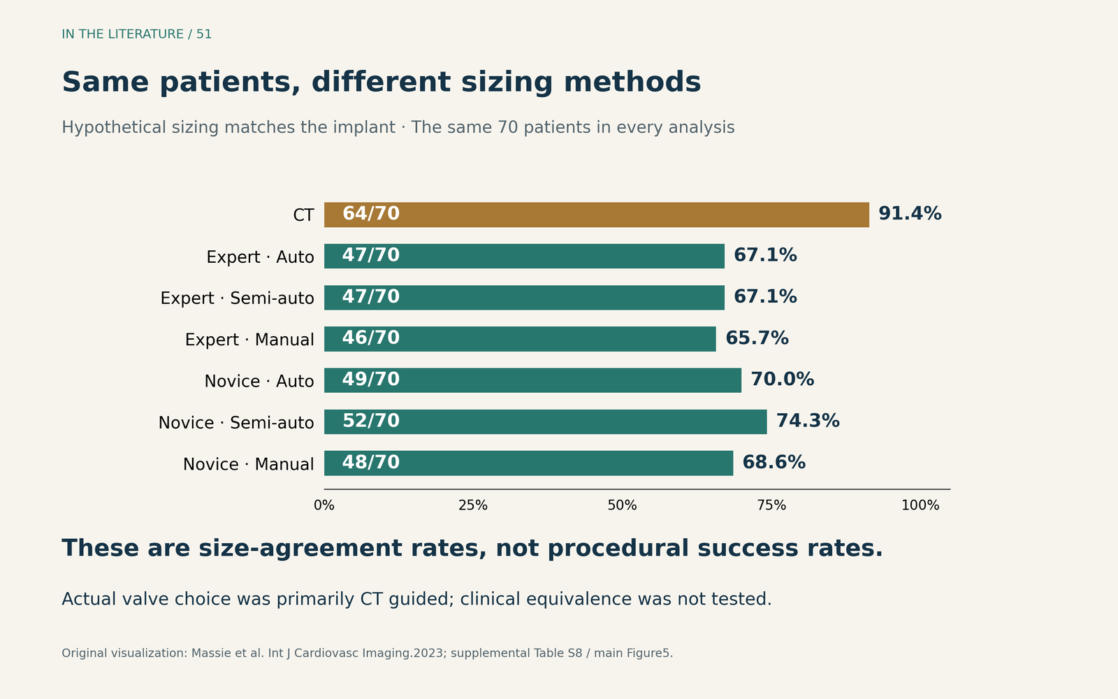
Figure 2. Original visualization of the counts in Massie et al., supplemental Table S8, corresponding to main Figure 5. Each analysis concerns the same 70 implanted patients; the bars are not seven independent patient groups. Actual valve selection was primarily CT guided. [3] View figure

Bias is also software specific. In Biaggi and colleagues' earlier fusion-imaging study, automated annular measurements were generally larger than CT measurements, and actual valves were selected by CT. That study evaluated an experimental segmentation system, not the safety of using its automatic measurements alone. It provides a useful warning against applying a universal correction factor—or automatically choosing a larger valve whenever TEE and CT disagree. [4]
When contrast is a concern, complete the missing anatomy
There is clinical experience with combined alternatives. A 2025 retrospective SAPIEN 3 study compared 83 patients evaluated with TEE plus noncontrast CT with 445 evaluated by CT angiography. It did not detect significant differences in paravalvular leak or permanent pacemaker implantation through one year. The groups differed substantially at baseline, and follow-up was incomplete. This supports feasibility in a selected practice; it does not establish equivalent outcomes, kidney protection, or TEE-only planning. [5]
The ESC/EACTS guideline notes that TEE does not provide the complete coronary and peripheral vascular assessment needed for planning. Thus, before accepting a contrast-sparing plan, the team still needs an adequate assessment of calcium distribution, root and coronary relationships, and the intended access route. The appropriate combination of modalities depends on what remains uncertain in this patient. [1]
For a borderline or discordant annular result, a practical response is to revisit the systolic frame, plane alignment and contour, then resolve the remaining anatomical uncertainty with the imaging team. Averaging incompatible measurements or applying a fixed TEE-to-CT correction is not a validated solution from these studies. [2–4]
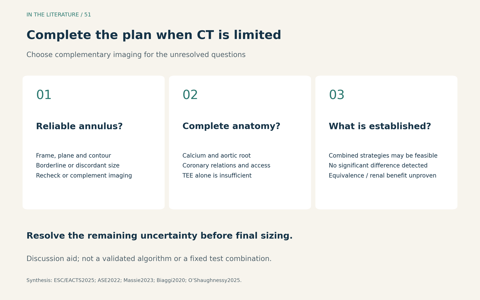
Figure 3. Questions to resolve before final prosthesis sizing. This is a clinical discussion aid, not a validated algorithm or a claim that every patient needs the same combination of tests. [1–5] View figure

Three takeaways
- Verify the structure and plane first. Use a systolic basal-ring measurement with a visible, reviewed contour; a precise trace of the wrong plane is still wrong.
- Evaluate the decision, not just the correlation. Repeatability and correlation with CT do not guarantee the same valve-size selection, especially near a device boundary.
- Make TEE part of a complete plan. When CT is unsuitable, resolve the remaining coronary, aortic and access questions before committing to the prosthesis.
References
- Praz F, Borger MA, Lanz J, et al. 2025 ESC/EACTS Guidelines for the management of valvular heart disease. Eur J Cardiothorac Surg. 2025;67(8):ezaf276. doi:10.1093/ejcts/ezaf276.
- Hahn RT, Saric M, Faletra FF, et al. Recommended Standards for the Performance of Transesophageal Echocardiographic Screening for Structural Heart Intervention: From the American Society of Echocardiography. J Am Soc Echocardiogr. 2022;35(1):1–76. doi:10.1016/j.echo.2021.07.006. Correction, 2022.
- Massie C, Parent M, Berthelot-Richer M, et al. Automated and semi-automated 3D echocardiographic software for aortic annulus sizing in transcatheter aortic valve implantation helps bridge the gap between expert and novice operators. Int J Cardiovasc Imaging. 2023;39(9):1707–1717. doi:10.1007/s10554-023-02885-z.
- Biaggi P, Sager DF, Külling J, et al. Potential Value of Fusion Imaging and Automated Three-Dimensional Heart Segmentation During Transcatheter Aortic Valve Replacement. J Am Soc Echocardiogr. 2020;33(4):516–517.e1. doi:10.1016/j.echo.2019.12.012.
- O'Shaughnessy M, Tabrizi R, Pham D, et al. A Retrospective Analysis Assessing Paravalvular Leak and Pacemaker Implantation Using TEE and Non-Contrast CT for CKD Patients Compared With CT Angiography for Annular Sizing Pre-TAVR. Health Sci Rep. 2025;8(5):e70847. doi:10.1002/hsr2.70847.