ITL-234381a46211
Juan Li, MD · Updated November 17, 2026
Watch the video
Key points
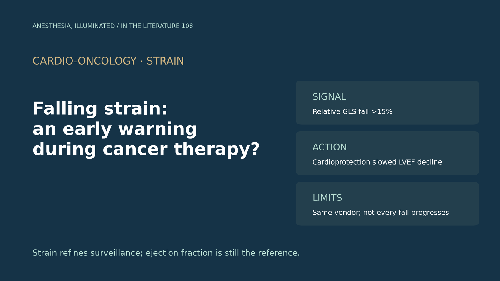
- A relative fall in global longitudinal strain (GLS) of more than 15% from baseline is considered meaningful. GLS varies about 5% to 10% between measurements, less than 2D ejection fraction, but serial values must come from the same vendor and software.[1]
- In adults, GLS often falls before the ejection fraction. In the SUCCOUR trial, starting cardioprotection when GLS fell did not significantly change the primary 1-year ejection fraction outcome, but fewer patients met criteria for cardiac dysfunction (5.8% vs 13.7%).[1], [2]
- At 3 years, SUCCOUR found no difference in ejection fraction change between GLS-guided and ejection fraction-guided care, although twice as many GLS-guided patients had received cardioprotection.[3]
- In SUCCOUR-MRI, about one-third of at-risk patients developed an isolated GLS fall. Cardioprotection reduced the 12-month decline in MRI ejection fraction, but the difference in cancer therapy-related cardiac dysfunction was not significant.[4]
- Not every fall progresses. In low-risk patients, a GLS fall did not always precede dysfunction, and in children the predictive value of an early fall has not been studied.[5], [6], [7]
A woman who received anthracycline chemotherapy followed by trastuzumab for breast cancer comes for elective surgery. Her ejection fraction is normal, but the report notes that GLS has fallen from −20% to −16% since baseline. Does this fall mean early cardiotoxicity, how reliable is the measurement, and what does the evidence say about acting on it?[1], [4]
What counts as a meaningful fall
The 2023 American Society of Echocardiography (ASE) recommendations for children summarize the adult thresholds: a relative fall in GLS of more than 15% from baseline is important, for example from −20% to −17%. GLS is highly reproducible, with variability of 5% to 10%, compared with 15% to 20% for 2D ejection fraction. Values vary slightly between vendors and software packages, so serial studies should use the same platform.[1]
The basis for the threshold is modest. Marwick and colleagues note that the 15% cutoff in guidelines is based on expert opinion, and they stress that sequential GLS must be assessed with the same software.[4] The ASE document also cautions that a fall below normal during treatment may be temporary.[1]
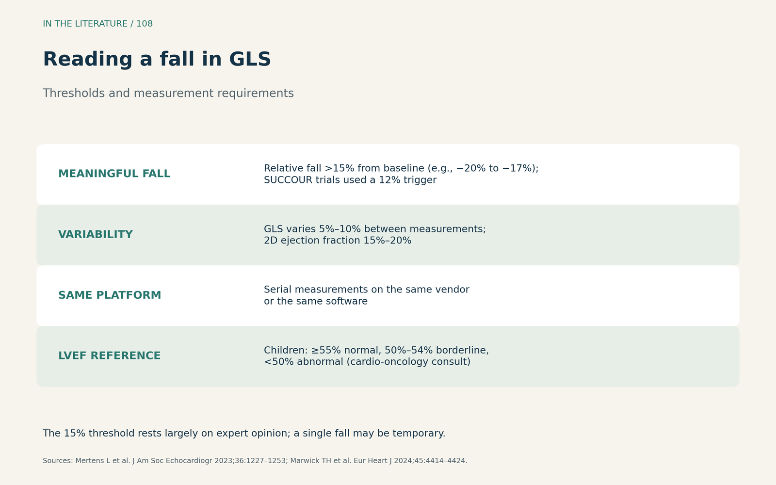
Figure 1. Reading a fall in GLS. Synthesized from the ASE pediatric recommendations and the SUCCOUR-MRI report; thresholds are consensus-based.[1], [4] View larger.

Does GLS fall before the ejection fraction?
The ASE document states that several adult studies found a fall in GLS preceded a fall in ejection fraction.[1] Marwick and colleagues add that most patients who develop dysfunction do not show a significant fall in ejection fraction during chemotherapy.[4]
A meta-analysis by Siddiqui and colleagues looked at baseline measures before chemotherapy. Baseline GLS predicted symptomatic cardiotoxicity (hazard ratio 1.46; 95% CI 1.34–1.58), whereas baseline ejection fraction did not show a significant association. Only 4 studies were included, and the certainty of evidence was low.[6]
Does acting on a GLS fall help?
The SUCCOUR trial randomized anthracycline-treated patients with another heart failure risk factor to cardioprotection triggered by a relative GLS fall of 12% or more, or by an absolute ejection fraction fall of more than 10%. The primary outcome, change in ejection fraction at 1 year, was not significantly different, but fewer patients in the GLS-guided arm met criteria for cancer therapy-related cardiac dysfunction (5.8% vs 13.7%).[2] At 3 years, cardioprotection had been given to 31% of the GLS-guided group and 14.6% of the ejection fraction-guided group, and the change in ejection fraction did not differ.[3] We had access only to the abstracts of these two reports.
SUCCOUR-MRI tested the question more directly. Of the patients followed, about one-third developed a GLS fall of more than 12% without a change in ejection fraction, and 105 were randomized. Ejection fraction on MRI fell by 2.5% with cardioprotection and by 5.6% with usual care; the adjusted between-group difference was 3.6% (95% CI −5.5% to −1.8%). Cardiac dysfunction at 12 months occurred in 1 of 49 versus 6 of 56 patients, a difference that was not significant (P = .075). Median doses reached 75% and 50% of target for renin–angiotensin blockade and beta-blockade.[4]
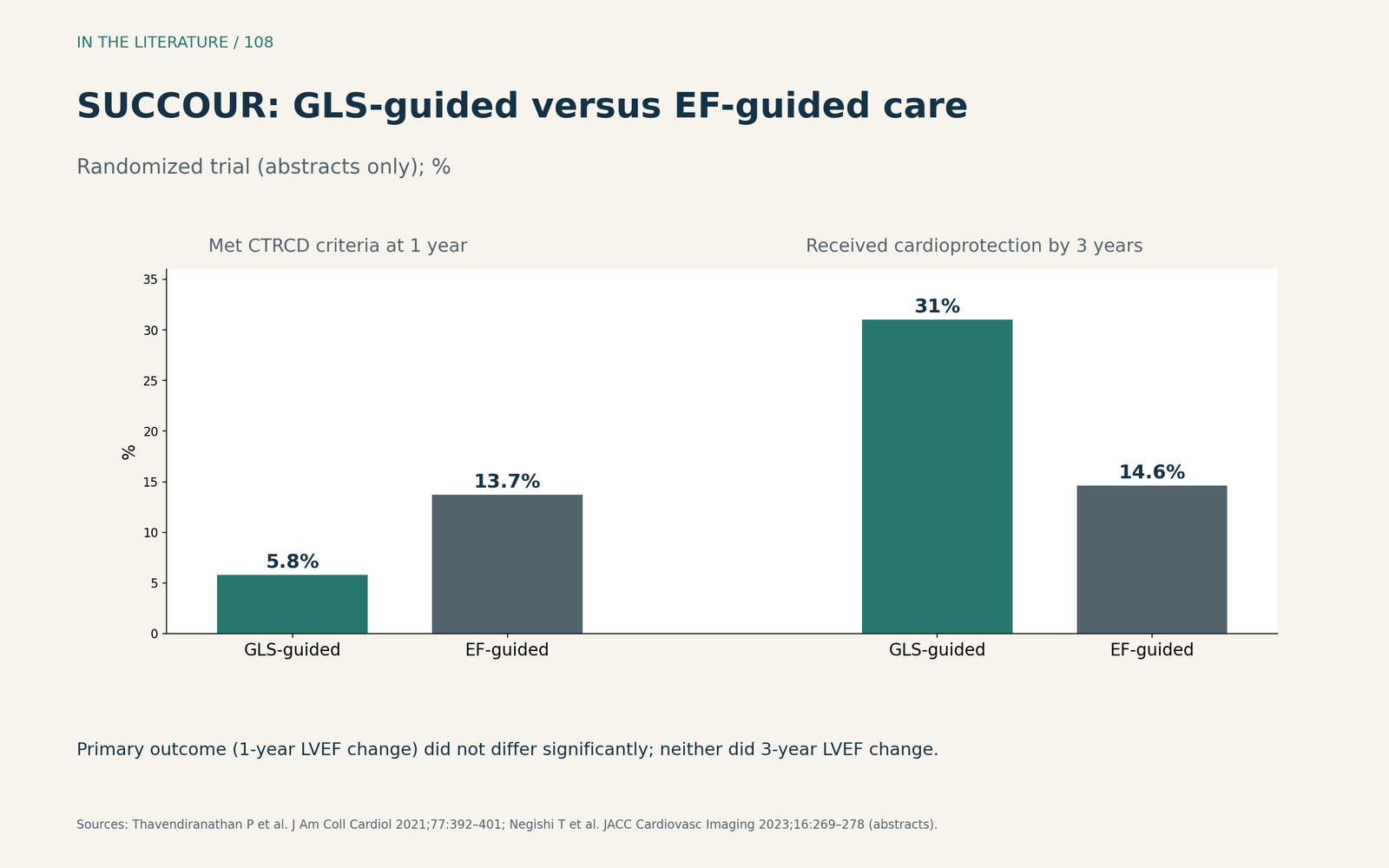
Figure 2. SUCCOUR at 1 and 3 years. Redrawn from the abstracts of Thavendiranathan et al. and Negishi et al.[2], [3] View larger.

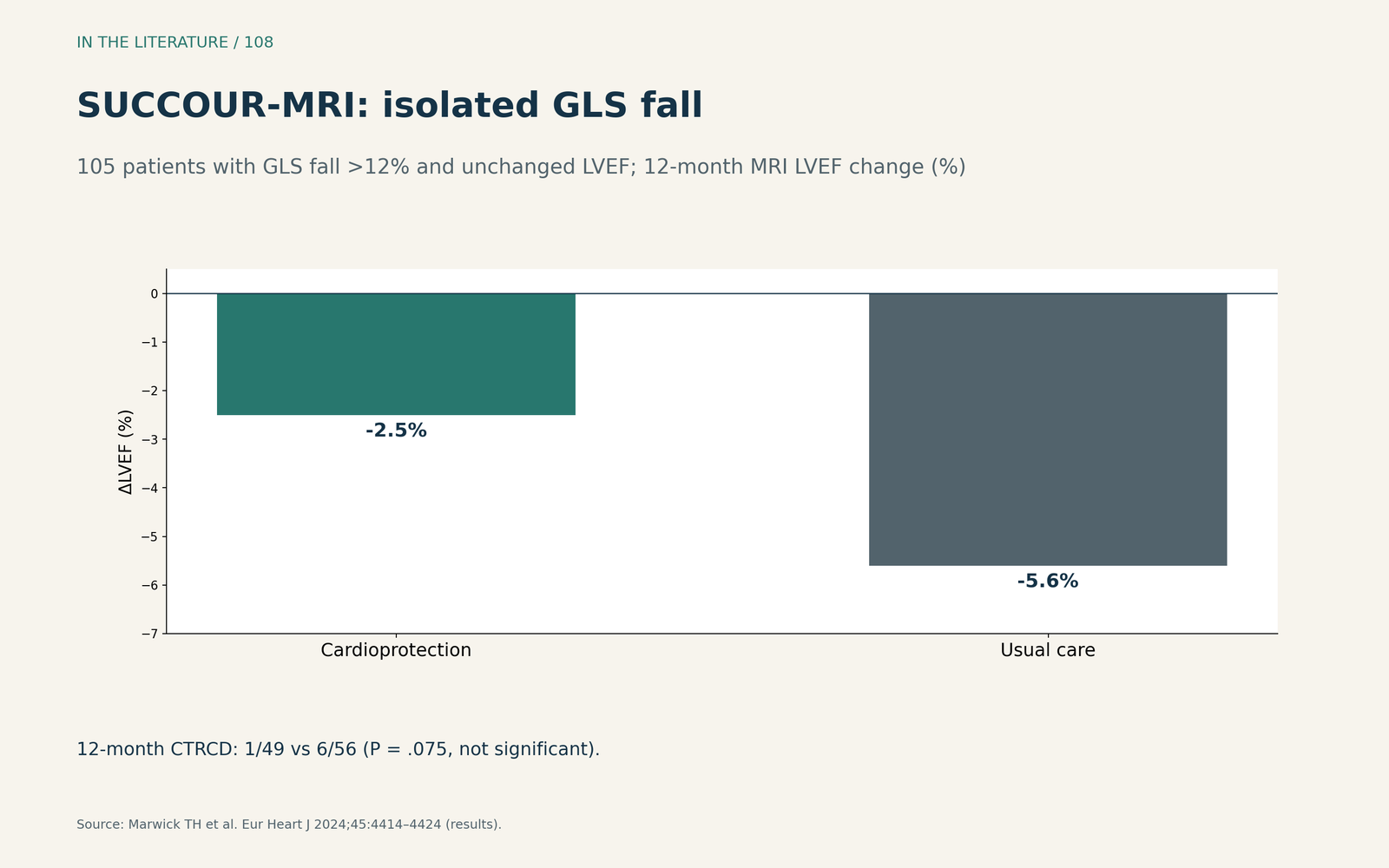
Figure 3. Cardioprotection after an isolated GLS fall. Redrawn from the results of SUCCOUR-MRI (Marwick et al.).[4] View larger.

When a GLS fall may not matter
Context changes the meaning of a fall. In PROTECT HER2, which followed mostly low-risk patients on anti-HER2 therapy, no randomized patient developed dysfunction by ejection fraction criteria, and the authors concluded that GLS changes in low-risk patients do not always reflect a precursor of dysfunction.[5]
Children differ. In a meta-analysis of 42 studies and 5,430 children, Li and colleagues found that GLS fell during therapy and remained lower in survivors, but the predictive value of an early reduction has not been studied.[7] The ASE recommends closer follow-up when GLS becomes borderline or abnormal with a preserved ejection fraction, and states that more data are needed before strain declines are used in therapeutic decisions.[1]
Three takeaways
- Measure GLS the same way every time. A relative fall of more than 15% is meaningful only on the same vendor and software.[1], [4]
- A GLS fall is an early warning, not a diagnosis. Acting on it slowed ejection fraction decline in SUCCOUR-MRI, but long-term benefit is unproven.[2], [3], [4]
- Interpret the fall in context. Consider baseline risk and age; in low-risk adults and in children, a fall does not necessarily predict dysfunction.[5], [6], [7]
References
- Mertens L, Singh G, Armenian S, et al. Multimodality imaging for cardiac surveillance of cancer treatment in children: recommendations from the American Society of Echocardiography. J Am Soc Echocardiogr. 2023;36(12):1227–1253. doi:10.1016/j.echo.2023.09.009
- Thavendiranathan P, Negishi T, Somerset E, et al. Strain-guided management of potentially cardiotoxic cancer therapy. J Am Coll Cardiol. 2021;77(4):392–401. Abstract only. doi:10.1016/j.jacc.2020.11.020
- Negishi T, Thavendiranathan P, Penicka M, et al. Cardioprotection using strain-guided management of potentially cardiotoxic cancer therapy: 3-year results of the SUCCOUR trial. JACC Cardiovasc Imaging. 2023;16(3):269–278. Abstract only. doi:10.1016/j.jcmg.2022.10.010
- Marwick TH, Dewar E, Nolan M, et al. Strain surveillance during chemotherapy to improve cardiovascular outcomes: the SUCCOUR-MRI trial. Eur Heart J. 2024;45(41):4414–4424. doi:10.1093/eurheartj/ehae574
- Gong FF, Grunblatt E, Voss WB, et al. A strain-guided trial of cardioprotection in early-stage breast cancer patients on anti-HER2 therapy (PROTECT HER2). Cardiooncology. 2024;10(1):85. doi:10.1186/s40959-024-00291-5
- Siddiqui MU, Yaacoub Y, Hanson HA, et al. Echocardiographic predictors of symptomatic cardiotoxicity among patients undergoing chemotherapy: a systematic review and meta-analysis. Medicine (Baltimore). 2022;101(28):e29562. doi:10.1097/MD.0000000000029562
- Li VW, So EK, Wong WH, Cheung YF. Myocardial deformation imaging by speckle-tracking echocardiography for assessment of cardiotoxicity in children during and after chemotherapy: a systematic review and meta-analysis. J Am Soc Echocardiogr. 2022;35(6):629–656. doi:10.1016/j.echo.2022.01.017