
ITL-640950b21e8f
Juan Li · In the Literature · Updated November 16, 2026
Watch the video

Key points
- Persistent severe hypotension during or after TAVR should raise concern for coronary obstruction, even without a new ECG change.
- Echocardiography helps identify ventricular dysfunction and competing explanations such as tamponade or severe regurgitation; coronary angiography addresses coronary patency.
- BASILICA or coronary protection does not eliminate the need to reassess flow after valve implantation.
- Prepare coronary intervention and hemodynamic support in parallel. If flow cannot be restored promptly, the team needs an actionable escalation plan.
- Successful PCI is an anatomic and flow result, not a guarantee of survival or recovery. Reassess the patient after reperfusion.
Do not wait for the full ischemic picture
A persistent blood-pressure collapse during transcatheter aortic valve replacement (TAVR) demands an explanation while resuscitation proceeds. Coronary obstruction belongs in that assessment even when the ECG has not developed an obvious new abnormality.
Ribeiro and colleagues’ multicenter registry reported 44 symptomatic coronary obstructions among 6,688 procedures performed in 2007–2013. Among those 44 cases, 30 had severe persistent hypotension and 25 had ECG changes. These are descriptive findings in an early-device-era registry, not the sensitivity of a diagnostic test or a current incidence estimate. They explain why waiting for a characteristic ECG pattern can be misleading. The authors recommend prompt echocardiographic and/or coronary angiographic assessment when hypotension persists. [1]
Echo and angiography answer complementary questions
The 2011 EAE/ASE echocardiography recommendations describe several explanations for acute severe hypotension during TAVR that can be assessed with TEE: tamponade, left-ventricular dysfunction, and severe aortic regurgitation. New regional wall-motion abnormalities may indicate coronary ostial obstruction. Acute mitral regurgitation can also arise from valve-related interference, pacing-related dyssynchrony, or injury to the mitral apparatus. These findings help direct treatment; they should not be collapsed into a single diagnosis of “poor LV function.” [2]
Communicate the finding in concrete terms: new regional dysfunction, severe regurgitation, a new pericardial collection, or an abnormal prosthesis position. At the same time, the interventional team assesses coronary filling and the feasibility of restoring flow. The practical aim is to identify the reversible cause without making one investigation wait unnecessarily for another. This is a clinical synthesis of the registry and imaging recommendations. [1, 2]
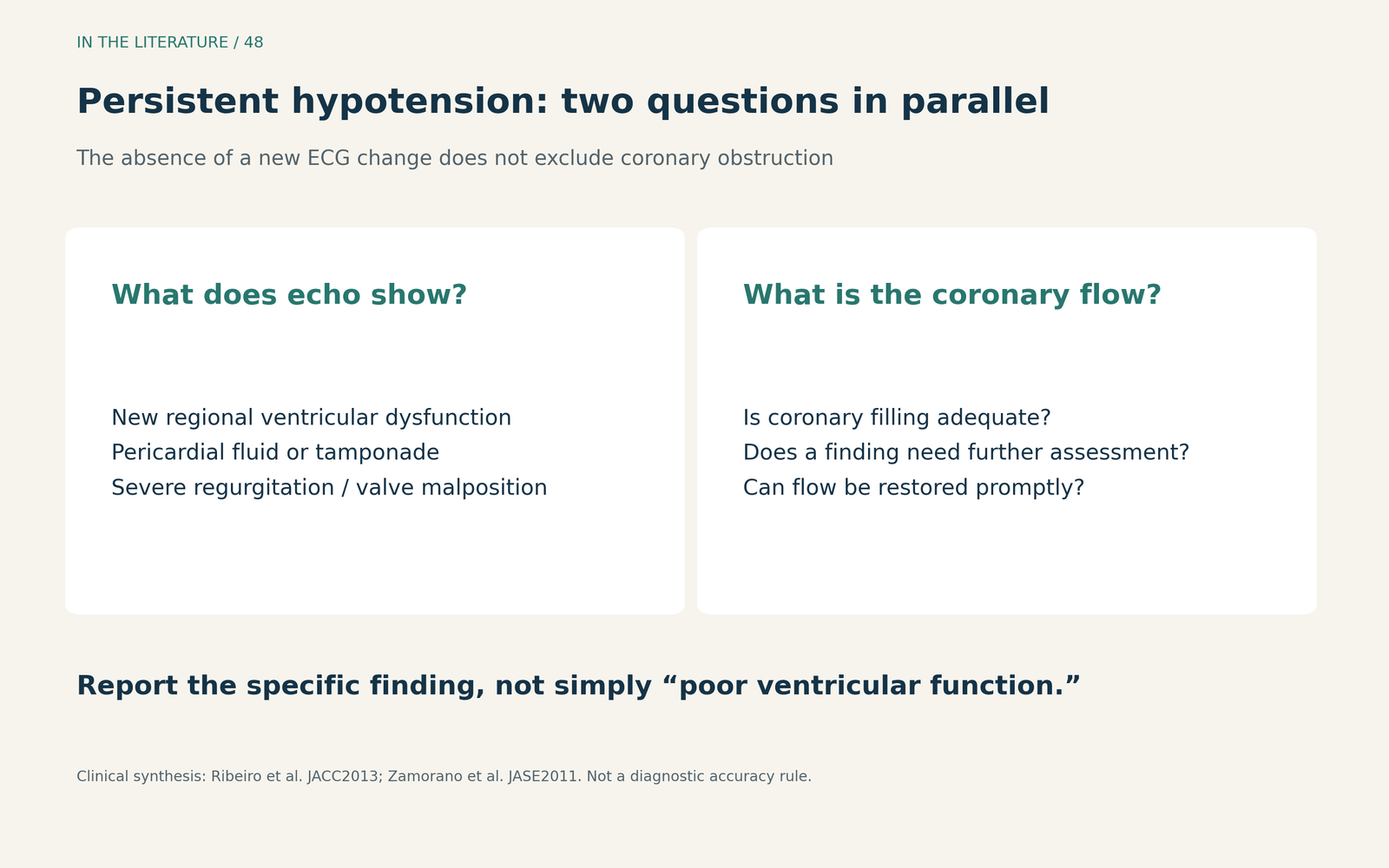
Figure 1. Complementary questions during sudden deterioration. Echo findings direct the differential diagnosis; they do not replace coronary assessment when obstruction remains suspected. [1, 2] View figure

Prevention does not close the diagnostic question
In the 2021 International BASILICA Registry, partial or complete culprit coronary obstruction occurred in 10/214 patients (4.7%) despite the preventive procedure. This was the culprit-obstruction endpoint assessed at catheterization-laboratory exit, not the broader 30-day coronary-obstruction endpoint. Six patients had partial leaflet obstruction presenting as angiographic haziness; all six were successfully treated with orthotopic stents. These aggregate observations show that residual obstruction can be subtle. They do not make haziness a specific diagnostic test or establish that stenting will always be feasible. [3]
The same report explains why a valve skirt or commissural post can still obstruct flow after leaflet modification. It also cautions that a protective coronary wire may temporarily hold a leaflet away from the ostium. Apparent patency with a wire in place therefore needs to be interpreted in the context of the final configuration. This is a mechanism to consider, not a blanket instruction to remove or retain a wire. [3]
For assessment after BASILICA-TAVR, the 2026 working-group position statement recommends selective coronary angiography when aortography inadequately opacifies the target vessel or persistent hypotension, ST elevation, or LV dysfunction raises concern. When obstruction remains uncertain despite abnormal angiography or these clinical findings, it recommends intravascular ultrasound. These recommendations address that procedural setting; they are not a validated universal algorithm for every episode of TAVR-related hypotension. [4]
Restore flow while arranging the support that may be needed
In Ribeiro’s registry, PCI was attempted in 33 of 44 patients and succeeded in 27 of those 33 attempts (81.8%). The success denominator is 33 attempted interventions, not all 44 obstruction cases. Success was defined by residual stenosis and restored TIMI grade 3 flow; it did not necessarily mean stent implantation. [1]
The authors regarded PCI as a reasonable initial revascularization strategy, while emphasizing readiness for circulatory support or urgent bypass surgery if flow could not be restored quickly. Treatment was not randomized, and the observed outcomes cannot identify a universally superior support device or rescue sequence. The team should establish who performs coronary rescue, who manages circulation, and how surgical or mechanical-support escalation will be activated. Support and reperfusion planning need to proceed together. [1]
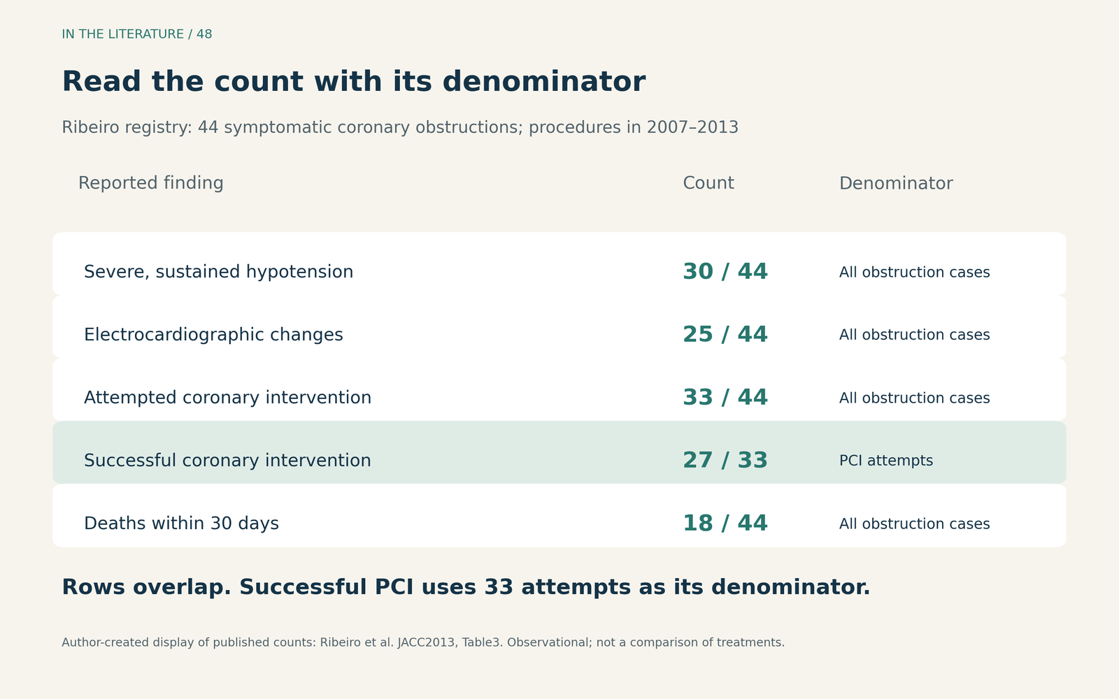
Figure 2. Author-created display of counts reported in Ribeiro et al., Table 3. The rows overlap and are not sequential outcome categories. PCI success uses the 33 attempted procedures; the other rows use the 44 obstruction cases. This historical registry does not establish comparative treatment effects. [1] View figure

A successful intervention does not end reassessment
Overall 30-day mortality in Ribeiro’s obstruction cohort was 18/44 (40.9%). This includes the entire obstruction cohort, not just patients with successful PCI. The result underscores the severity of the complication; it should not be used as a contemporary prognosis for an individual patient. [1]
The **2023 Spanish TAVI Registry—available here as an abstract only—**reported obstruction in 115 of 13,675 patients, with 83.5% occurring during the procedure. PCI was selected in 100 patients, with 78.0% technical success. In-hospital mortality was 37.4% with obstruction versus 4.1% without it. The registry also included events during hospitalization and follow-up, so these aggregate outcomes are not restricted to immediate intraprocedural obstruction. The observational comparison does not measure the causal benefit of a particular rescue intervention. [5]
After flow restoration, reassess blood pressure, ventricular function, regurgitation, prosthesis position, and the pericardial space. Persistent deterioration calls for continued investigation, including the possibility of another structural complication. A reassuring coronary result and a recovered patient are separate observations. [1, 2]
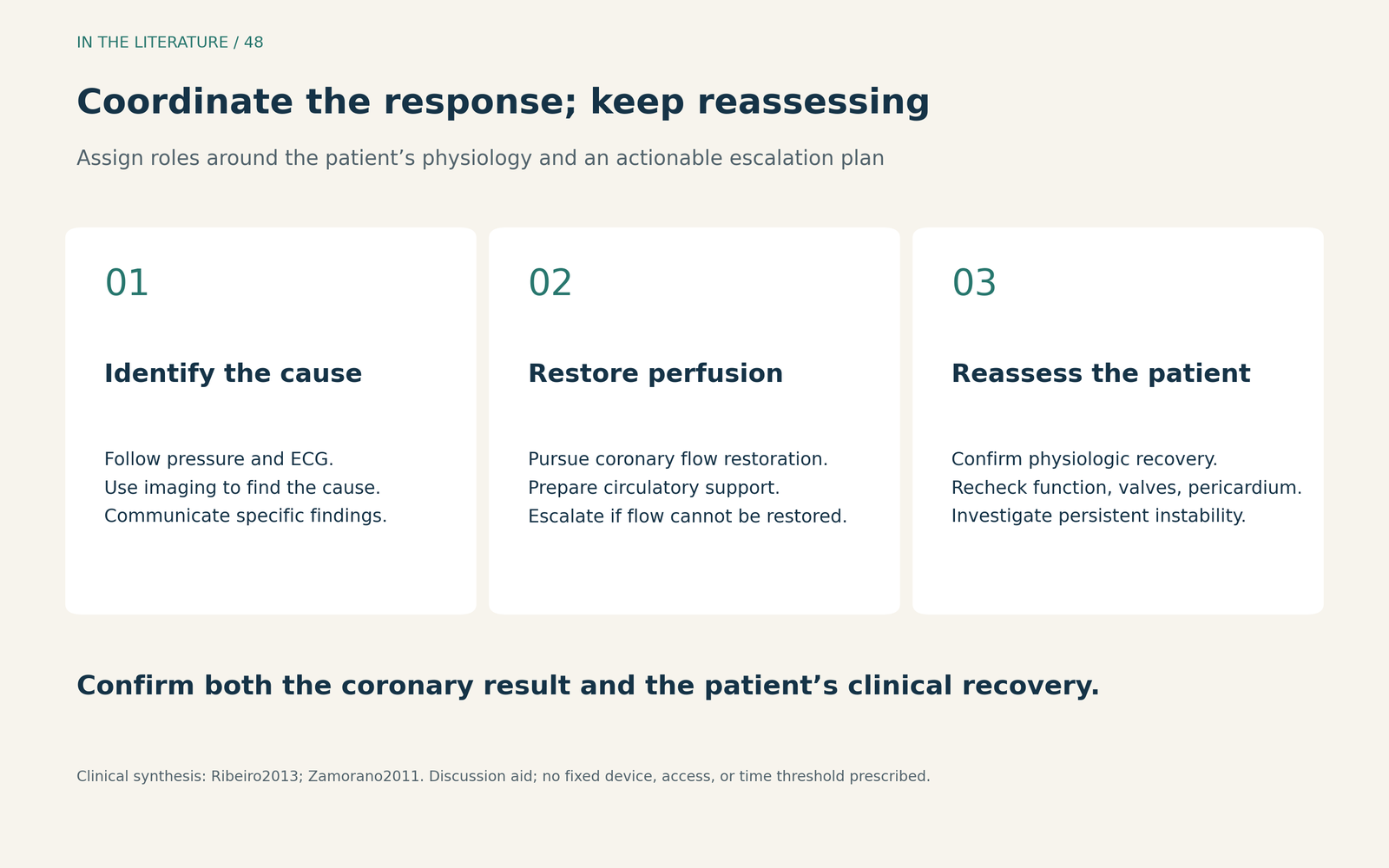
Figure 3. A team discussion aid for coordinated rescue and reassessment. It summarizes the clinical priorities without prescribing a fixed device, access route, or time threshold. [1, 2] View figure

Three takeaways
- Suspect obstruction before the ECG becomes convincing. Persistent severe hypotension warrants rapid assessment of coronary and structural causes.
- Pursue flow restoration with a ready escalation plan. PCI, circulatory support, and surgical rescue require coordinated roles; prevention does not guarantee patency.
- Reassess after reperfusion. Technical success does not establish clinical recovery, and continuing instability needs a continuing explanation.
References
- Ribeiro HB, Webb JG, Makkar RR, et al. Predictive Factors, Management, and Clinical Outcomes of Coronary Obstruction Following Transcatheter Aortic Valve Implantation: Insights From a Large Multicenter Registry. J Am Coll Cardiol. 2013;62(17):1552–1562. doi:10.1016/j.jacc.2013.07.040.
- Zamorano JL, Badano LP, Bruce C, et al. EAE/ASE Recommendations for the Use of Echocardiography in New Transcatheter Interventions for Valvular Heart Disease. J Am Soc Echocardiogr. 2011;24(9):937–965. doi:10.1016/j.echo.2011.07.003.
- Khan JM, Babaliaros VC, Greenbaum AB, et al. Preventing Coronary Obstruction During Transcatheter Aortic Valve Replacement: Results From the Multicenter International BASILICA Registry. JACC Cardiovasc Interv. 2021;14(9):941–948. doi:10.1016/j.jcin.2021.02.035.
- Rogers T, Halaby RN, Greenbaum AB, et al. Aortic Valve Leaflet Modification: A Working Group Position Statement on Best Practices and Step-by-Step Guide. JACC Cardiovasc Interv. 2026;19(11):1347–1377. doi:10.1016/j.jcin.2026.03.033.
- Ojeda S, González-Manzanares R, Jiménez-Quevedo P, et al. Coronary Obstruction After Transcatheter Aortic Valve Replacement: Insights From the Spanish TAVI Registry. JACC Cardiovasc Interv. 2023;16(10):1208–1217. doi:10.1016/j.jcin.2023.03.024. Abstract only.