ITL-0574a13f99d1
Juan Li, MD · Updated November 18, 2026
Key points
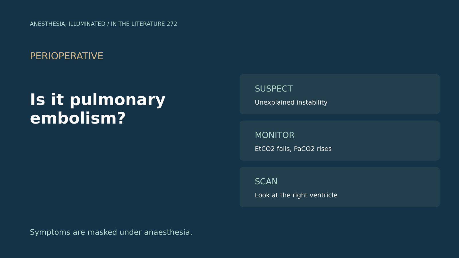
- Think of it early. Unexplained intraoperative haemodynamic instability must prompt consideration of PE, and up to a third of patients die within hours.[1]
- Watch the capnogram. A fall in end-tidal CO2 with a rise in arterial CO2 may be a useful clue.[2], [3]
- Echo signs rule in, not out. The McConnell sign was 100% specific but only 19% sensitive.[4]
- Know the definition. High-risk PE includes a systolic fall of more than 40 mm Hg not explained by other causes.[5]
- If CT cannot be done, scan. Guidelines advise echocardiography when CT is not immediately feasible.[6]
During a hip arthroplasty under general anaesthesia, saturation and blood pressure fall abruptly and end-tidal CO2 drops. When should the team suspect pulmonary embolism, and what should it do next?
Why PE is missed in the operating room
Up to a third of patients with PE die within hours, yet intraoperative PE is likely under-recognized: under anaesthesia the usual symptoms are masked, D-dimer will likely be elevated after surgery, and otherwise unexplained intraoperative haemodynamic instability must prompt consideration of PE. If no RV dysfunction is seen on echocardiography, an alternative diagnosis should be considered (Cormican et al., J Cardiothorac Vasc Anesth 2020).[1] In a case under general anaesthesia (Mao et al., BMC Anesthesiol 2017), saturation fell from 100% to 66% and CVP rose to 30 cmH2O, and the authors suggest that a fall in end-tidal CO2 with a rise in arterial CO2 may be a useful clue.[2] After non-cardiac surgery (Davis et al., Can J Anesth 2022), each 5 mm Hg increase in the arterial-to-end-tidal CO2 difference was associated with more complications (OR 1.20).[3]
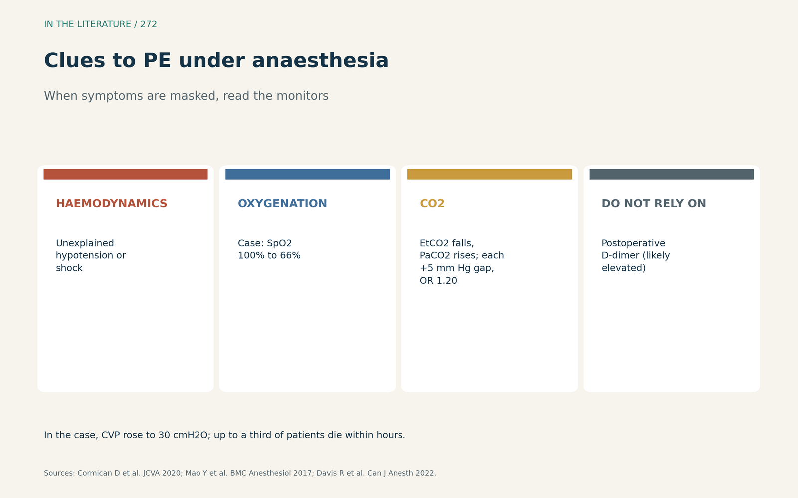
Figure 1. Symptoms are masked under anaesthesia, so monitors carry the signal. Synthesized from Cormican et al., Mao et al. and Davis et al.[1], [2], [3] View larger.

Bedside signs and definitions
The 60/60 sign had sensitivity 25% and specificity 94%, and the McConnell sign 19% and 100%; both are insensitive but reliable (Kurzyna et al., Am J Cardiol 2002).[4] Almost 20% of patients treated for PE die within 90 days; high-risk PE includes a fall in systolic pressure of more than 40 mm Hg not caused by sepsis, arrhythmia or hypovolaemia, and observational data support evaluating such patients for immediate reperfusion therapy (Kahn and de Wit, N Engl J Med 2022).[5] Across international guidelines (Falster et al., Lancet Haematol 2023), echo signs of RV strain have sensitivities below 70%, so their absence cannot rule out PE, but in unstable suspected PE, if CT pulmonary angiography is not immediately feasible, echocardiography should be performed.[6]
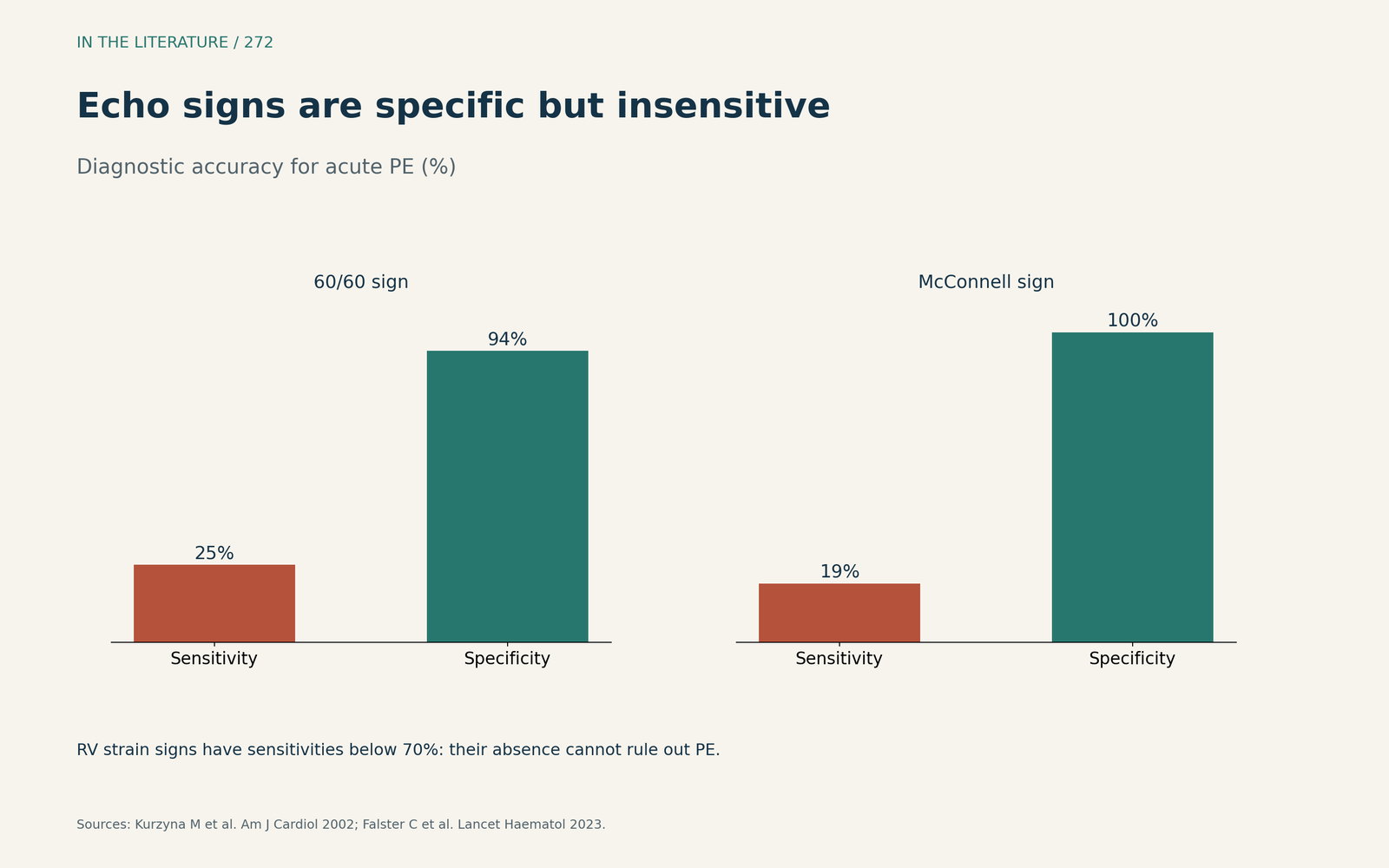
Figure 2. Sensitivity and specificity of two classic echo signs. Data from Kurzyna et al.[4] View larger.

Ultrasound to act in unstable patients
CT pulmonary angiography may be unsafe or impractical in evolving shock; multi-organ POCUS had a pooled sensitivity of 90% for PE in critically ill patients, can rapidly exclude other life-threatening causes, and a thrombus in transit provides diagnostic certainty; VA-ECMO may be considered as a bridge to reperfusion (Moreno-Loaiza et al., Crit Care 2026).[7] In an individual patient data meta-analysis of 9233 patients (Cimini et al., J Thromb Haemost 2026), short-term death was 7%, and only 1 abnormal RV parameter was not associated with increased mortality, whereas 2 or more were.[8]
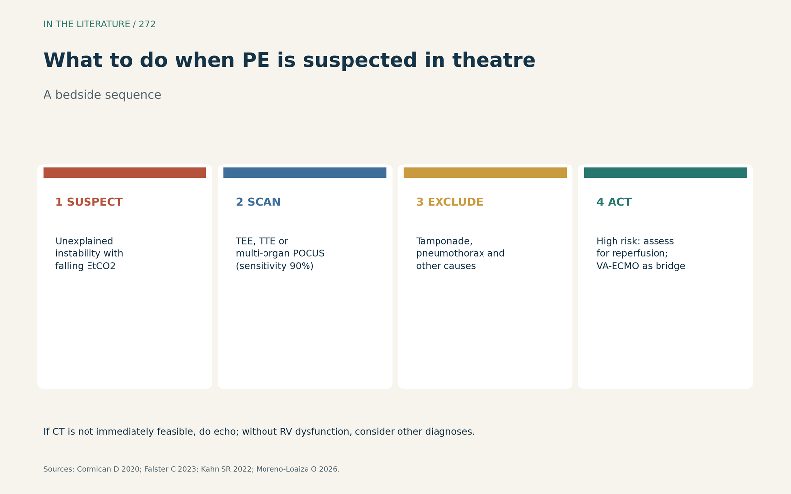
Figure 3. A bedside sequence for suspected perioperative PE. Synthesized from Cormican et al., Falster et al., Kahn and de Wit and Moreno-Loaiza et al.[1], [6], [5], [7] View larger.

Three takeaways
- In the operating room, unexplained hypoxaemia, hypotension and a falling end-tidal CO2 should prompt PE, because symptoms are masked and D-dimer is unhelpful.[1], [2]
- Look at the right ventricle: signs such as McConnell are specific but insensitive, so a normal scan does not exclude PE.[4], [6]
- In shock, use echo or POCUS to decide on reperfusion rather than waiting for CT.[5], [6], [7]
References
- Cormican D, Morkos MS, Winter D, et al. Acute perioperative pulmonary embolism—management strategies and outcomes. J Cardiothorac Vasc Anesth. 2020;34(7):1972–1984. doi:10.1053/j.jvca.2019.11.018
- Mao Y, Wen S, Chen G, et al. Management of intra-operative acute pulmonary embolism during general anesthesia: a case report. BMC Anesthesiol. 2017;17(1):67. doi:10.1186/s12871-017-0360-0
- Davis R, Jewell E, Engoren M, Maile M. Difference between arterial and end-tidal carbon dioxide and adverse events after non-cardiac surgery: a historical cohort study. Can J Anaesth. 2022;69(1):106–118. doi:10.1007/s12630-021-02118-8
- Kurzyna M, Torbicki A, Pruszczyk P, et al. Disturbed right ventricular ejection pattern as a new Doppler echocardiographic sign of acute pulmonary embolism. Am J Cardiol. 2002;90(5):507–511. doi:10.1016/S0002-9149(02)02523-7
- Kahn SR, de Wit K. Pulmonary embolism. N Engl J Med. 2022;387(1):45–57. doi:10.1056/NEJMcp2116489
- Falster C, Hellfritzsch M, Gaist TA, et al. Comparison of international guideline recommendations for the diagnosis of pulmonary embolism. Lancet Haematol. 2023;10(11):e922–e935. doi:10.1016/S2352-3026(23)00181-3
- Moreno-Loaiza O, de Melo CB, Moreno-Loaiza M, et al. Point-of-care ultrasound in hemodynamically unstable pulmonary embolism. Crit Care. 2026;30:271. doi:10.1186/s13054-026-06097-4
- Cimini LA, Pruszczyk P, Jiménez D, et al. Right ventricular dysfunction on echocardiography to predict mortality in acute pulmonary embolism: an individual patient data meta-analysis. J Thromb Haemost. 2026;24:1377–1387. doi:10.1016/j.jtha.2025.11.020