
ITL-2c88e8e68fa6
Watch the video

Figure 1. Conceptual illustration of route-dependent contrast filling; not a representation of cardiac anatomy.
Key points
- A dilated coronary sinus is a useful clue to persistent left superior vena cava (PLSVC), but it also occurs with other causes, including elevated right atrial pressure.[1]
- A right-arm injection may follow a normal right superior vena cava and miss an abnormal left-sided pathway; the injection site belongs in the interpretation.[1, 2]
- When the right superior vena cava is absent, injections from either arm can opacify the coronary sinus before the right atrium.[3]
- Early left-heart filling after a left-arm injection raises concern for systemic venous drainage into the left atrium; it should not automatically be labeled a patent foramen ovale.[2]
- Before an intervention, define the drainage site, right superior vena cava, bridging vein and coronary sinus outlet, using additional imaging when needed.[4]
An unexpectedly large coronary sinus appears on TEE, or a left-sided venous catheter takes an unfamiliar course. A previous bubble study was reported as negative. Does that exclude PLSVC?
No: a bubble study shows the route taken by the injected contrast. Its meaning depends on where the injection entered the circulation and which structures were observed first. A study aimed at detecting an atrial septal shunt may not answer a question about left-sided systemic venous drainage.[1, 2]
Follow the route, not just the presence of bubbles
In the common configuration, a PLSVC drains into the coronary sinus and then the right atrium. With a left-arm injection, the expected sequence is opacification of the PLSVC and coronary sinus before the right atrium. If a normal right superior vena cava is also present, a right-arm injection can enter the right atrium directly.[1]
Moore and colleagues illustrated this in a 2025 minimally invasive cardiac surgery case. After unsuccessful left-sided pulmonary artery catheter placement, TEE identified a markedly enlarged coronary sinus. Left-arm agitated saline first filled the PLSVC, then the coronary sinus and right heart; the right superior vena cava was separately visualized.[1]
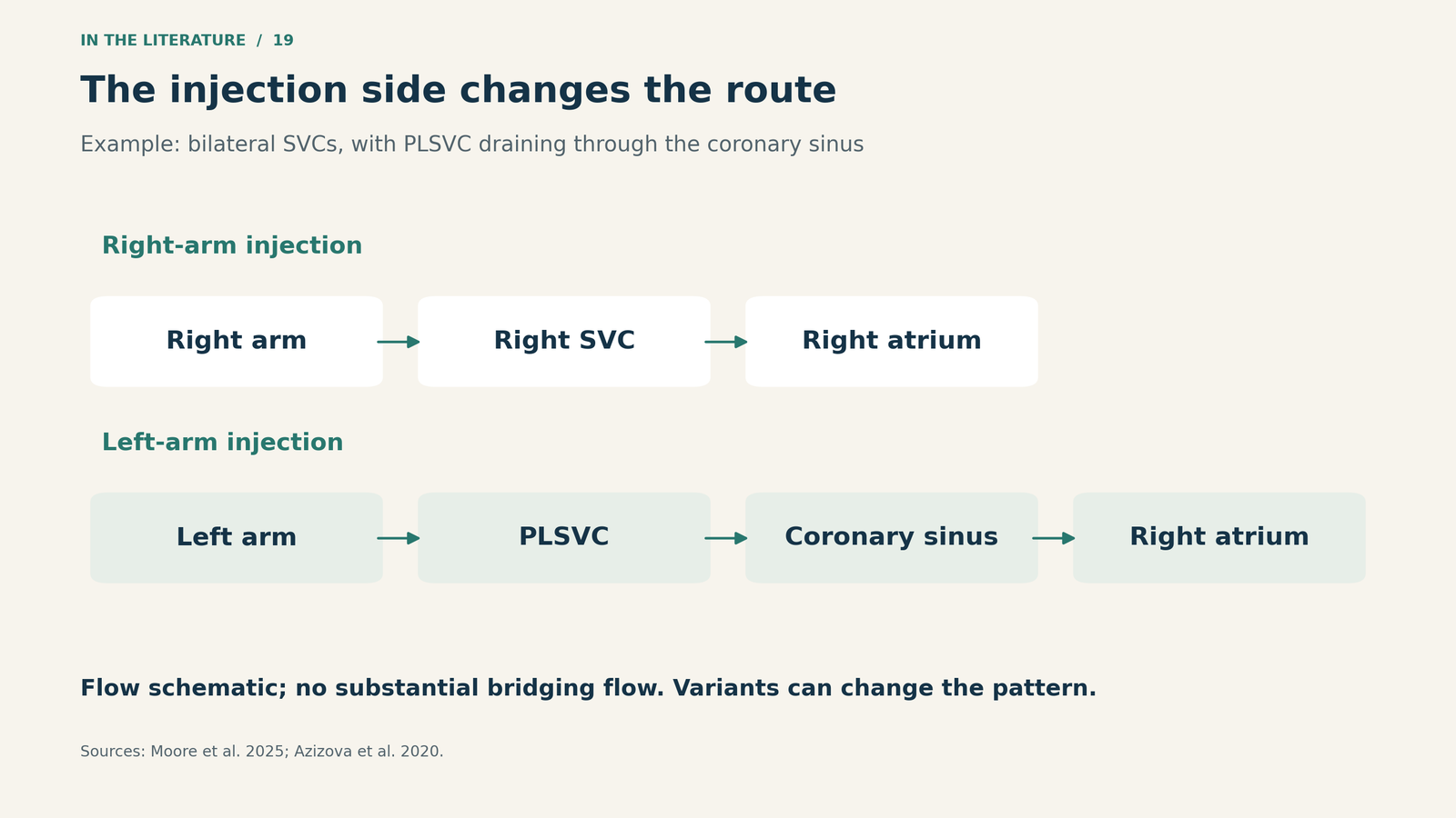
Figure 2. Expected routes in one common configuration: two superior venae cavae, PLSVC draining through the coronary sinus, and no substantial bridging flow. This is a flow schematic, not an anatomical drawing or a rule covering every variant.[1, 4]

Early left-heart contrast asks a different question
Park and colleagues reported a patient with recurrent stroke whose left-arm saline injection produced immediate left atrial opacification. Cardiac MRI subsequently demonstrated a PLSVC communicating with the left atrium and a completely unroofed coronary sinus.[2]
That sequence suggested a systemic venous connection to the left heart. It did not establish that bubbles crossed the interatrial septum through a patent foramen ovale. The entry route and subsequent anatomical imaging were central to identifying the lesion.[2]
The distinction matters: PLSVC draining through an intact coronary sinus into the right atrium and PLSVC communicating with the left atrium have different implications. The latter can provide a route for paradoxical embolism. These reports demonstrate a diagnostic mechanism; they do not provide a sensitivity estimate or quantify an individual patient’s stroke risk.[2, 4]
Both arms can also produce a coronary-sinus-first pattern
A right-arm injection is not invariably unhelpful. Joshi and Singh described a patient in whom both right- and left-arm injections opacified the coronary sinus before the right atrium. CT showed an absent right superior vena cava, with both brachiocephalic veins draining into a large PLSVC.[3]
This is why the study should be described as an observed sequence with a documented injection site. The same shorthand label—“positive bubble study”—can conceal very different anatomy.
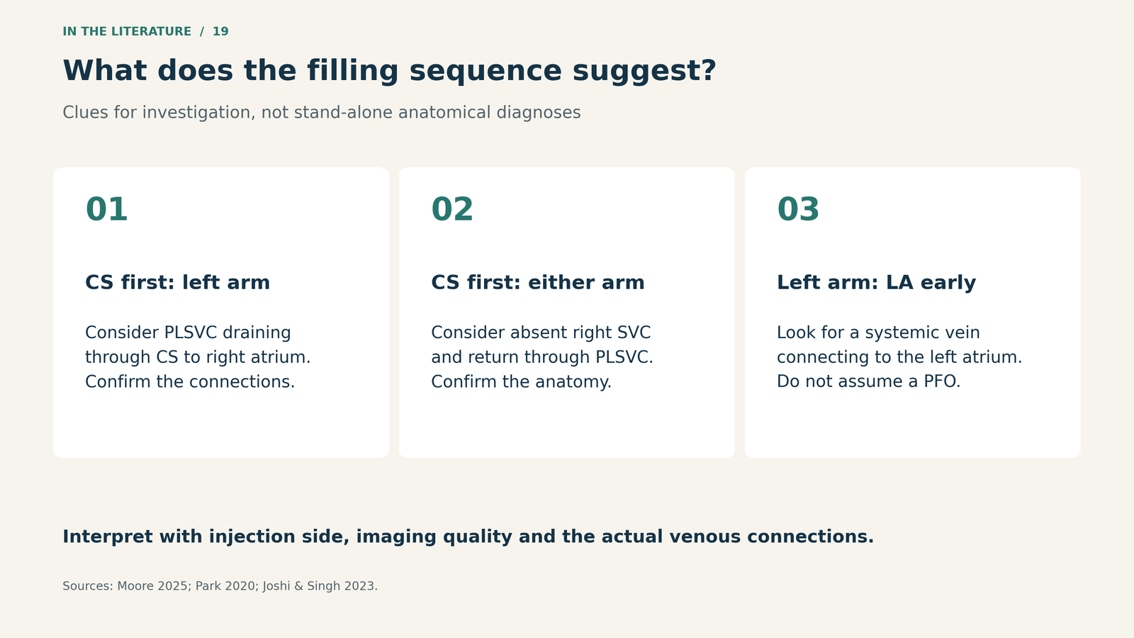
Figure 3. Patterns that guide further investigation. Each interpretation is conditional on adequate imaging and the actual venous connections; none is a complete anatomical diagnosis by itself.[1, 2, 3]

Finish with an anatomical answer that changes the plan
TEE can trace the vessel and its connections; CT or MRI can clarify anatomy that remains uncertain. The useful report answers where the PLSVC drains, whether a right superior vena cava exists, whether the two sides communicate through a bridging vein, and whether the coronary sinus outlet is patent.[4]
These details affect venous access, bypass drainage and myocardial protection. In the 2025 surgical case, recognition of PLSVC led to an additional venous cannula under imaging guidance. That was a response to the patient’s anatomy and operative plan, not evidence that every PLSVC requires the same cannulation strategy.[1]
An especially important exception is coronary sinus ostial atresia: the PLSVC may carry essential retrograde coronary venous drainage through a bridging vein toward the right superior vena cava. Ligation without understanding that pathway can interrupt cardiac venous drainage. Identifying the vessel is therefore only the beginning of the operative decision.[4]
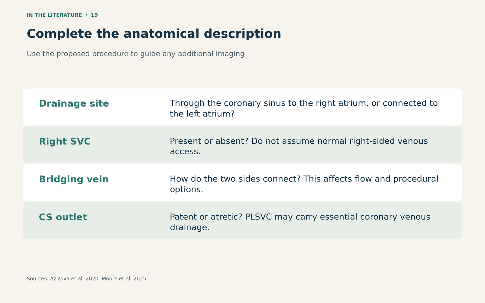
Figure 4. Turn an imaging suspicion into an actionable anatomical description. The investigation should resolve these questions to the extent needed for the proposed procedure.[1, 4]

Three takeaways
- Record the injection side and filling sequence. A “negative” result from one arm is not a universal exclusion of anomalous venous return.
- Treat early left-heart filling as a route to explain. Establish where contrast enters rather than assuming an atrial septal mechanism.
- Map the connections before changing the procedure. Drainage site, right SVC, bridging vein and coronary sinus outlet determine the practical significance.
References
- Moore C, Lamelas J, Jain P. Diagnosis of persistent left superior vena cava by transesophageal echocardiography during minimally invasive cardiac surgery. CASE (Phila). 2025;9(5):157–163. doi:10.1016/j.case.2024.11.007.
- Park CJ, Upadhya B, Pu M, Zhou L. Diagnosis of persistent left superior vena cava: lessons learned from a recurrent stroke case. CASE (Phila). 2020;4(5):320–323. doi:10.1016/j.case.2020.05.019.
- Joshi S, Singh A. Absent right superior vena cava and persistent left superior vena cava: an incidental finding. Ann Card Anaesth. 2023;26(4):433–434. doi:10.4103/aca.aca_91_23.
- Azizova A, Onder O, Arslan S, Ardali S, Hazirolan T. Persistent left superior vena cava: clinical importance and differential diagnoses. Insights Imaging. 2020;11:110. doi:10.1186/s13244-020-00906-2.