ITL-65ec55766f5c
Juan Li, MD · Updated November 17, 2026
Watch the video
Key points
- Echo is not a diagnostic test for pulmonary embolism. Guidelines do not recommend it as the primary means of diagnosis, because most right ventricular strain signs have sensitivities below 70%; McConnell's sign and the D-sign are specific but do not rule embolism out.[1], [2]
- In critically ill patients, point-of-care right ventricular dysfunction had a sensitivity of 63% and specificity of 85% against CT angiography.[3]
- In shock, echo changes decisions. Eight of 13 guidelines endorse bedside echo when CT is not feasible, and right ventricular strain can support reperfusion; a thrombus in transit gives diagnostic certainty.[2], [4]
- In stable patients, echo gives prognosis. Right ventricular dysfunction was associated with 3.3-fold higher odds of adverse events and fourfold higher odds of embolism-related death, but not with all-cause death, and it is present in over 37% of patients.[5]
- More signs mean more risk. In 9233 patients, abnormal right ventricular parameters identified higher risk of death, and the authors propose defining severe dysfunction as at least 2 abnormal parameters.[6]
A postoperative patient becomes breathless and hypotensive; a bedside scan shows a dilated right ventricle with a flattened septum. Does this prove pulmonary embolism, and how should the finding change what the team does next?[1], [2]
Echo signs and diagnosis
The ASE guideline on cardiac sources of embolism states that echocardiography is not recommended as a primary means of diagnosing pulmonary embolism, and notes that acute and chronic right ventricular abnormalities may exist in patients with chronic obstructive pulmonary disease.[1] Falster and colleagues compared 13 international guidelines: because right ventricular strain signs have sensitivities below 70%, their absence cannot rule out embolism, although the D-sign and McConnell's sign in particular are specific; only the ESC/ERS guideline treats a right heart thrombus as essentially confirming the diagnosis.[2]
Girardi and colleagues studied multiorgan point-of-care ultrasound in critically ill patients: right ventricular dysfunction on the bedside scan had 63% sensitivity and 85% specificity, and transthoracic echo was the most accurate single component.[3]
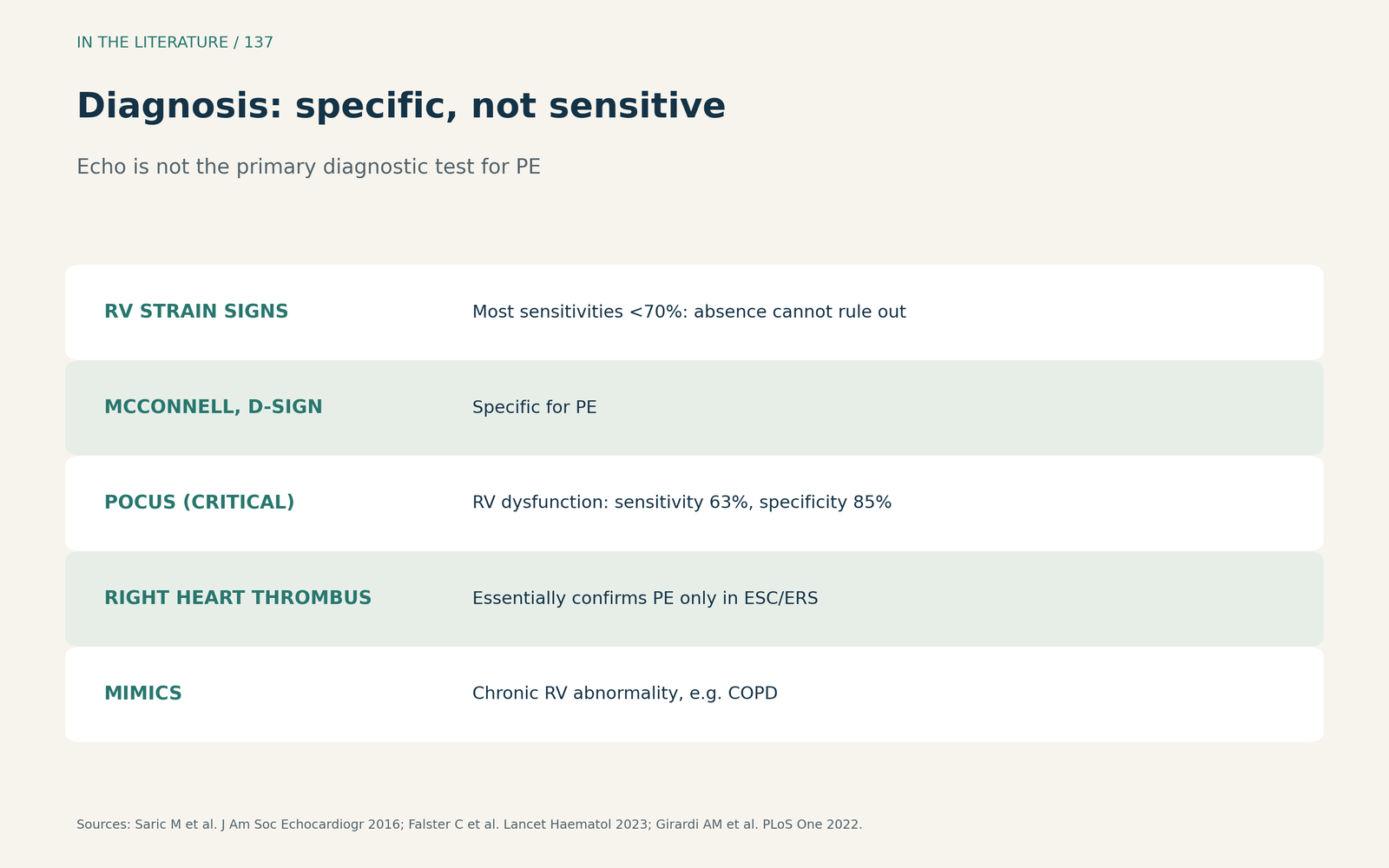
Figure 1. Echo signs are specific but insensitive. Synthesized from the ASE guideline, Falster et al. and Girardi et al.[1], [2], [3] View larger.

The unstable patient
When the patient is in shock and transport for CT may be unsafe, eight guidelines endorse bedside echocardiography, and right ventricular strain can support giving reperfusion therapy.[2] Moreno-Loaiza and colleagues argue that bedside ultrasound is not meant to establish certainty but may shift probability enough to cross a treatment threshold; a thrombus in transit provides diagnostic certainty, while pre-existing right ventricular dilation or dysfunction reduces the diagnostic value.[4]
Prognosis in stable patients
Prosperi-Porta and colleagues pooled 55 studies with 17,090 hemodynamically stable patients. Right ventricular dysfunction was associated with 3.3-fold higher odds of combined adverse events and fourfold higher odds of embolism-related death, but not with all-cause death; because its prevalence was over 37%, current definitions lack specificity.[5] In an individual-patient meta-analysis of 9233 patients, Cimini and colleagues found that abnormal right ventricular parameters identified patients at increased risk of death, with stronger associations when several were abnormal, and proposed defining severe dysfunction as at least 2 abnormal parameters.[6]
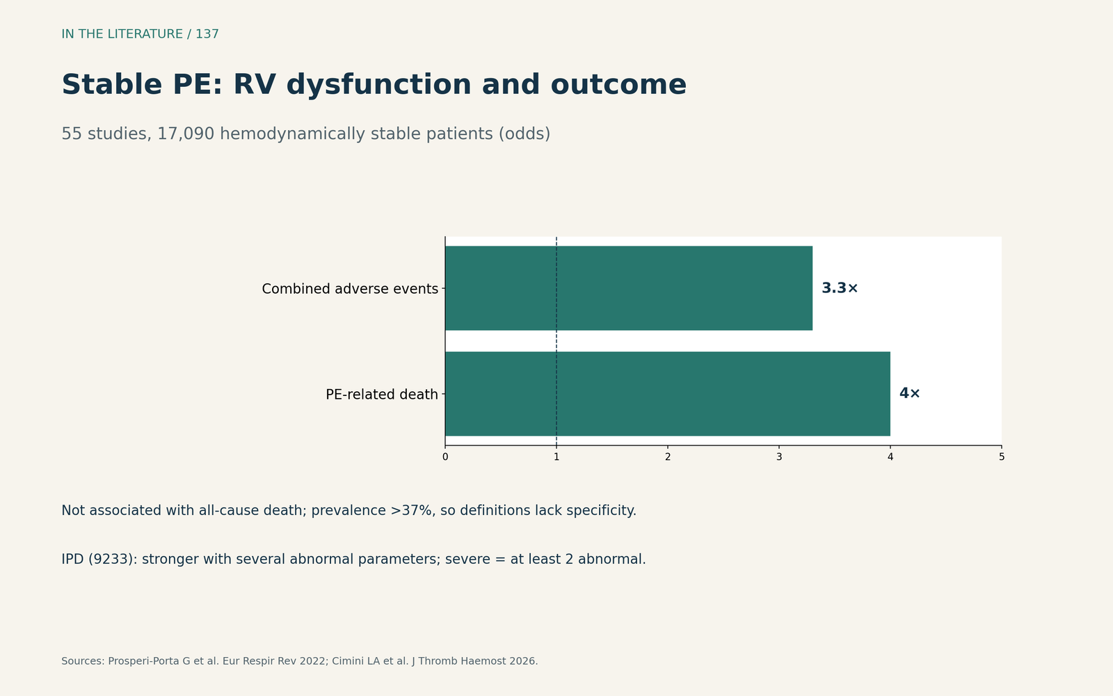
Figure 2. Right ventricular dysfunction and outcome in stable patients. Redrawn from Prosperi-Porta et al. and Cimini et al.[5], [6] View larger.

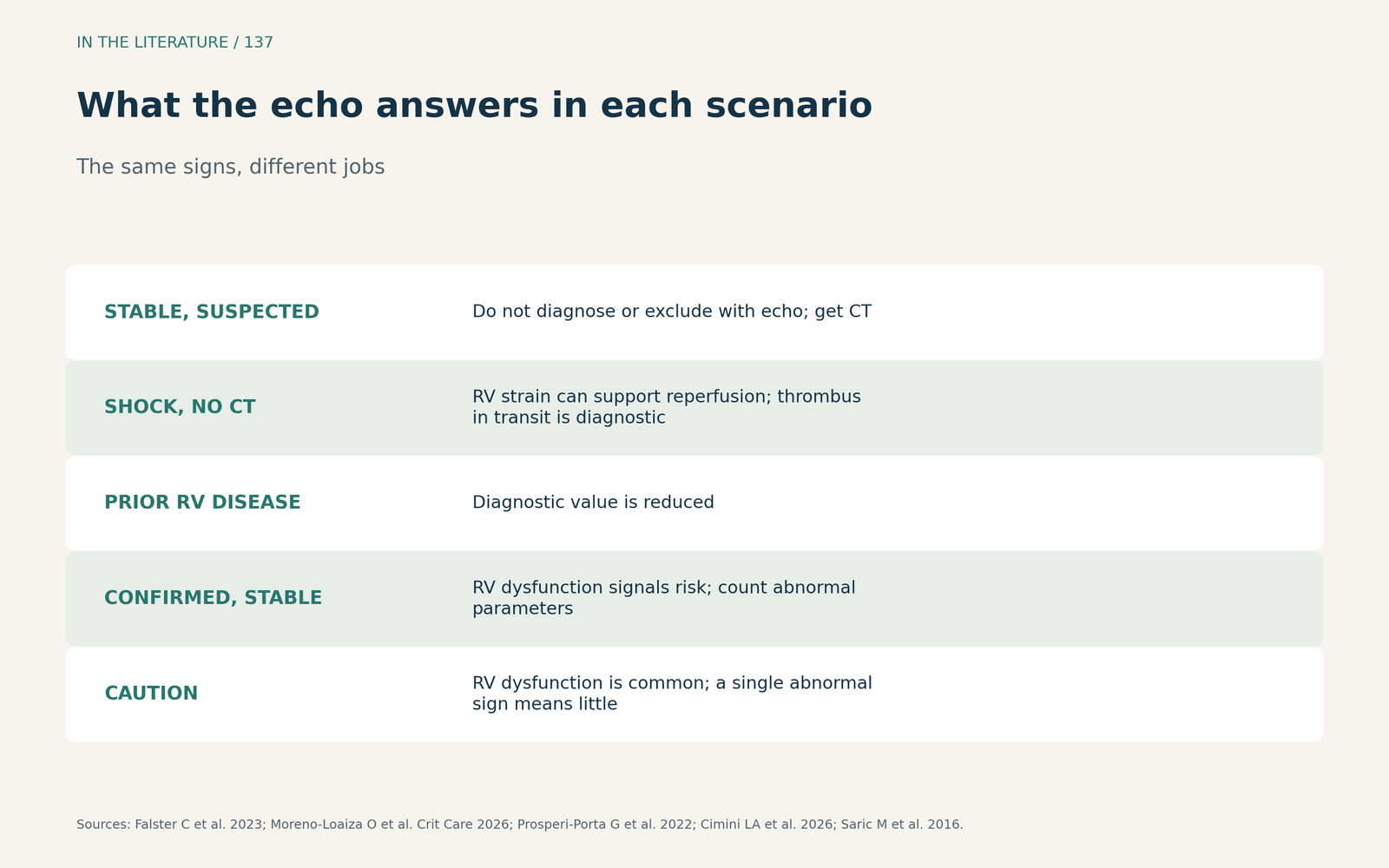
Figure 3. What the echo answers in each scenario. Synthesized from Falster et al., Moreno-Loaiza et al., Prosperi-Porta et al., Cimini et al. and the ASE guideline.[2], [4], [5], [6], [1] View larger.

Three takeaways
- A normal right ventricle does not exclude embolism. Echo signs are specific, not sensitive.[1], [2]
- In shock, echo can justify action. Right ventricular strain or a thrombus in transit can support reperfusion when CT is not feasible.[2], [4]
- In stable patients, echo is for risk. Several abnormal signs predict worse outcome; check for chronic disease that mimics strain.[5], [6], [1]
References
- Saric M, Armour AC, Arnaout MS, et al. Guidelines for the use of echocardiography in the evaluation of a cardiac source of embolism. J Am Soc Echocardiogr. 2016;29(1):1–42. doi:10.1016/j.echo.2015.09.011
- Falster C, Hellfritzsch M, Gaist TA, et al. Comparison of international guideline recommendations for the diagnosis of pulmonary embolism. Lancet Haematol. 2023;10(11):e922–e935. doi:10.1016/S2352-3026(23)00181-3
- Girardi AM, Turra EE, Loreto M, et al. Diagnostic accuracy of multiorgan point-of-care ultrasound compared with pulmonary computed tomographic angiogram in critically ill patients with suspected pulmonary embolism. PLoS One. 2022;17(10):e0276202. doi:10.1371/journal.pone.0276202
- Moreno-Loaiza O, de Melo CB, Moreno-Loaiza M, Bamberg S. Point-of-care ultrasound in hemodynamically unstable pulmonary embolism. Crit Care. 2026;30(1):271. doi:10.1186/s13054-026-06097-4
- Prosperi-Porta G, Ronksley P, Kiamanesh O, et al. Prognostic value of echocardiography-derived right ventricular dysfunction in haemodynamically stable pulmonary embolism: a systematic review and meta-analysis. Eur Respir Rev. 2022;31(166):220120. doi:10.1183/16000617.0120-2022
- Cimini LA, Pruszczyk P, Jiménez D, et al. Right ventricular dysfunction on echocardiography to predict mortality in acute pulmonary embolism: an individual patient data meta-analysis. J Thromb Haemost. 2026;24(4):1377–1387. doi:10.1016/j.jtha.2025.11.020