
ITL-d7cd10e7dcd8
Juan Li · In the Literature · Updated November 16, 2026
Watch the video

Key points
- A negative or inconclusive TEE does not exclude endocarditis when clinical suspicion remains high after TAVR.
- Obtain blood cultures promptly, preferably before antibiotics, and involve the Endocarditis Team early.
- Repeat TTE and/or TEE within 5–7 days under the ESC pathway; suspected new complications require earlier reassessment.
- Cardiac CTA and FDG-PET/CT answer complementary anatomical and metabolic questions.
- Interpret early prosthetic FDG uptake within a named diagnostic framework; neither a positive nor a negative scan replaces clinical judgment.
The clinical problem is unresolved suspicion
After TAVR, persistent bacteremia or an otherwise unexplained infectious syndrome may remain concerning even when TEE shows no convincing vegetation. “No vegetation seen” is an imaging finding, not a final exclusion of infection. Atypical presentations can delay recognition, and the initial assessment still needs clinical findings, cultures and both transthoracic and transesophageal echocardiography. [1]
The transcatheter valve frame can obscure the valve and surrounding structures. Infection may also involve the stent frame or another valve. The 2023 ESC guideline therefore supports a multimodality approach when echocardiography leaves the diagnosis uncertain. The next test should address what remains unanswered. [2]
Preserve the microbiological evidence and reassess the echo
The ESC guideline advises at least three blood-culture sets before antibiotic therapy. There is no reason to wait for a fever spike. Obtain samples promptly and communicate the suspected diagnosis to microbiology; this must not become a reason to delay urgently needed antimicrobial treatment. [2]
Review the original images with an experienced echocardiographer, including image quality, the valve frame, adjacent valves and any new regurgitation or perivalvular abnormality. Compare with previous studies where available. If the initial study is negative or inconclusive and suspicion remains high, repeat TTE and/or TEE within 5–7 days. [2]
That interval is not permission to wait through deterioration. New heart failure, persistent fever or bacteremia, embolism, a new murmur or atrioventricular block warrants prompt repeat assessment and Endocarditis Team review. [2]
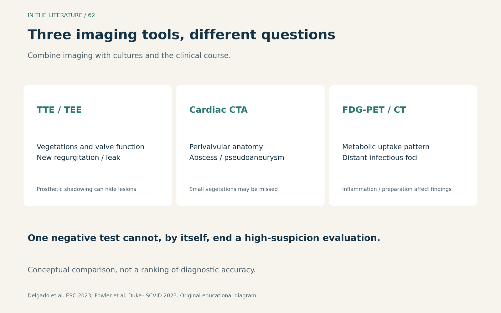
Figure 1. Original comparison of the questions each modality helps answer. These roles are complementary; the diagram does not rank overall test accuracy. [2] View figure

Choose CT for anatomy and PET/CT for additional metabolic information
Cardiac CTA is particularly useful when the concern is perivalvular or periprosthetic extension—such as an abscess or pseudoaneurysm—and TEE is inconclusive. It complements echocardiography rather than replacing it: small vegetations and some leaflet lesions remain better assessed with echo. [2]
FDG-PET/CT can support the diagnosis of prosthetic-valve infection when echocardiography is inconclusive, and can reveal distant infectious foci or an alternative explanation for the illness. Interpretation needs appropriate preparation, attention to the uptake pattern and experienced readers. If PET/CT is unavailable, the ESC guideline allows consideration of labeled-white-cell SPECT/CT in a high-suspicion case with negative or inconclusive echo. [2]
These investigations need not form a rigid sequence in which every patient waits for every preceding test. The team should select them according to the unresolved question, clinical urgency and local expertise. [2]
What does the direct TAVR evidence show?
In Wahadat and colleagues' multicenter retrospective study, 30 patients had suspected TAVR-related endocarditis. All underwent FDG-PET/CT; 14 also underwent cardiac CTA. Of the 22 initially classified as possible infection, additional imaging and team adjudication moved five to definite infection and five to rejected infection, while 12 remained possible. Overall, 10 of 30 patients—33%—changed classification. [3]
This is useful evidence that additional imaging can resolve some uncertainty. It is not an independent diagnostic-accuracy validation: the Endocarditis Team used the imaging findings when assigning the final diagnosis, and no patient underwent surgery to provide a surgical pathological reference. The study also documented negative PET reports among patients ultimately classified as definite infection. A negative PET result therefore needs the same clinical context as a negative TEE. [3]
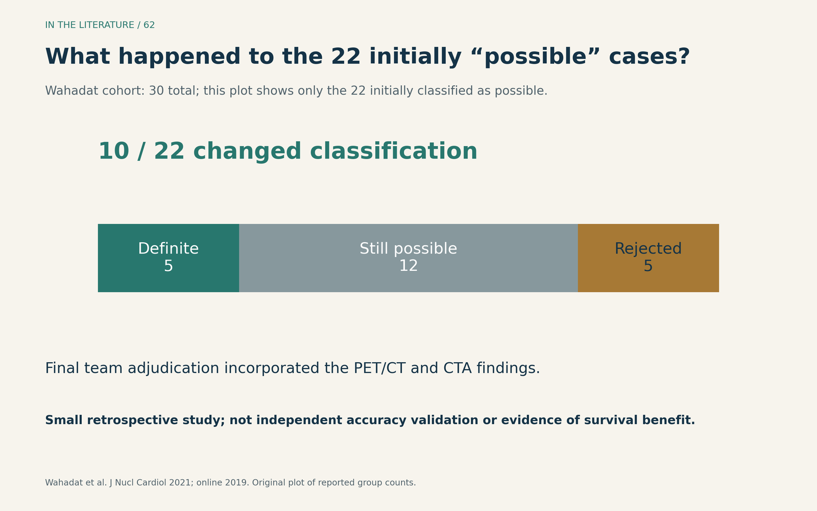
Figure 2. Original plot of reported group counts. The denominator here is the 22 initially possible cases, within a total cohort of 30. Reclassification is not the same as independently confirmed accuracy or improved survival. [3] View figure

A newer report by Boufoula and colleagues, published in 2025, compared diagnostic criteria in 92 patients from two French centers, with 82 definite and 10 rejected cases according to expert consensus. The following results are based on the formal published abstract only. Sensitivity increased from 65% with conventional Duke criteria to 77% with ESC 2023 criteria, while specificity decreased from 100% to 90%. Positive FDG-PET/CT was the most helpful added criterion, with 10 patients reported as correctly reclassified. [4]
The result supports further multimodality assessment, but the small rejected group and the abstract-only access limit how confidently one can judge accuracy and adjudication methods. These percentages compare diagnostic classifications; they do not establish that ordering PET/CT improves survival. [4]
Early after implantation, name the criteria you are using
Postprocedural inflammation complicates interpretation of FDG uptake. ESC 2023 accepts characteristic intense focal or heterogeneous prosthetic/periprosthetic uptake as a major criterion irrespective of the interval from surgery, provided it is interpreted appropriately. Duke-ISCVID 2023 places abnormal prosthetic-material uptake within the first three months in a minor imaging criterion; its major-criterion wording specifies characteristic uptake at least three months after surgical implantation. [2, 5]
These are different classification rules. They are not a validated TAVR-specific timing cutoff, and they do not instruct clinicians to postpone a needed investigation until three months have passed. Document the framework, discuss the pattern with imaging specialists, and integrate cultures, serial imaging and the clinical course. Duke-ISCVID explicitly treats criteria as a supplement to clinical judgment. [5]
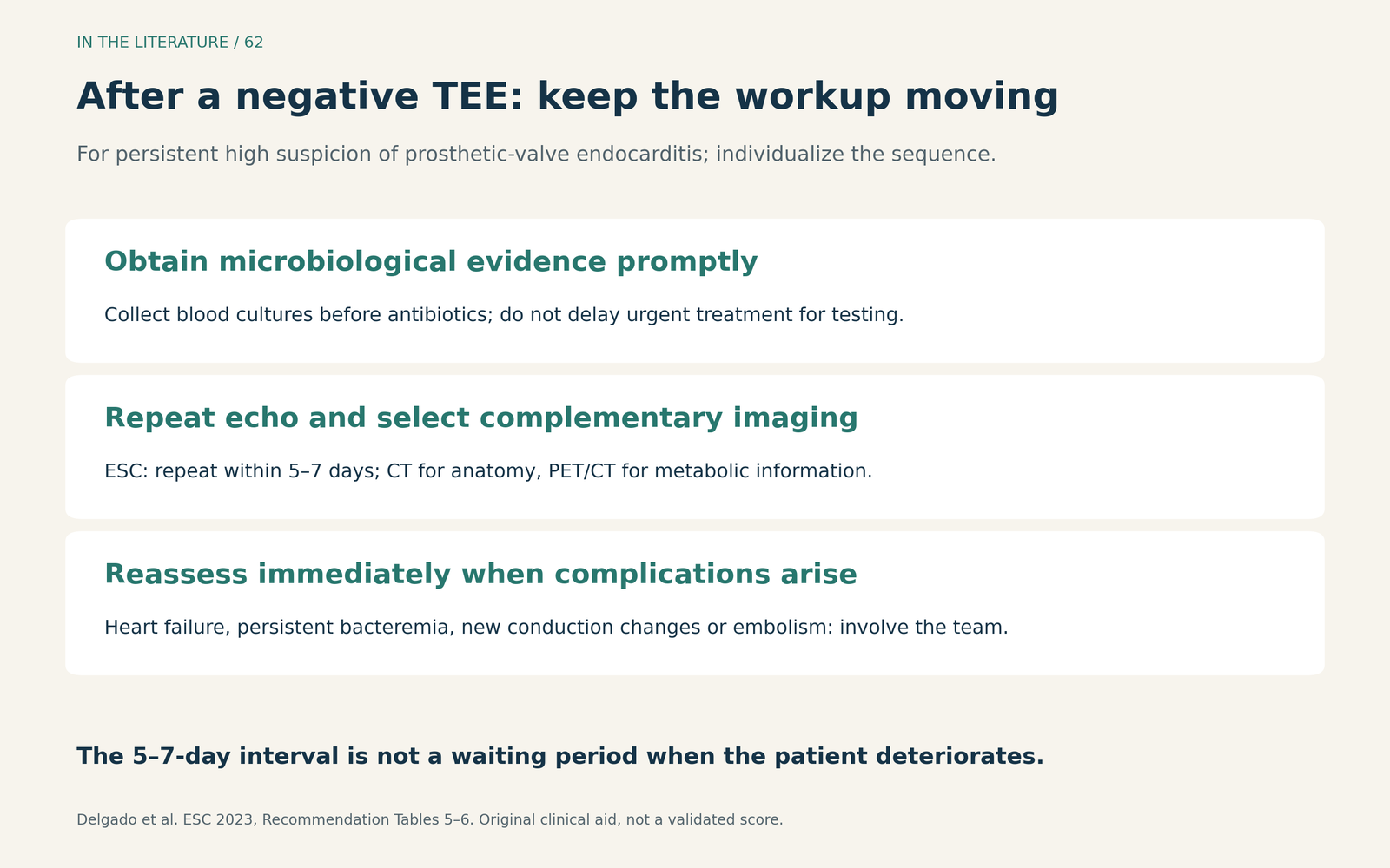
Figure 3. Original clinical aid based on the ESC diagnostic pathway. Clinical deterioration overrides a scheduled repeat-imaging interval. [2] View figure

Three takeaways
- Keep investigating when suspicion remains high. A negative TEE is insufficient to close a concerning post-TAVR case.
- Match the next image to the unresolved question. Repeat echo, cardiac CTA and FDG-PET/CT provide complementary evidence; escalate sooner if complications emerge.
- Use the Endocarditis Team to integrate the evidence. Early uptake, negative scans and diagnostic categories require interpretation alongside microbiology and the evolving clinical picture.
References
- Hussey PT, Meers JB. Infective Endocarditis After Transcatheter Aortic Valve Replacement: An Update for the Cardiothoracic Anesthesiologist. J Cardiothorac Vasc Anesth. 2023;37:1354–1357. doi:10.1053/j.jvca.2023.04.030.
- Delgado V, Ajmone Marsan N, de Waha S, et al. 2023 ESC Guidelines for the management of endocarditis. Eur Heart J. 2023;44(39):3948–4042. doi:10.1093/eurheartj/ehad193.
- Wahadat AR, Tanis W, Swart LE, et al. Added value of 18F-FDG-PET/CT and cardiac CTA in suspected transcatheter aortic valve endocarditis. J Nucl Cardiol. 2021;28(5):2072–2082. doi:10.1007/s12350-019-01963-x.
- Boufoula I, Philip M, Arregle F, et al. Comparison between Duke, European Society of Cardiology 2015, International Society for Cardiovascular Infectious Diseases 2023, and European Society of Cardiology 2023 criteria for the diagnosis of transcatheter aortic valve replacement-related infective endocarditis. Eur Heart J Cardiovasc Imaging. 2025;26(3):532–544. doi:10.1093/ehjci/jeae310. Formal abstract only.
- Fowler VG Jr, Durack DT, Selton-Suty C, et al. The 2023 Duke-International Society for Cardiovascular Infectious Diseases Criteria for Infective Endocarditis: Updating the Modified Duke Criteria. Clin Infect Dis. 2023;77(4):518–526. doi:10.1093/cid/ciad271.