ITL-01316102b81b
Juan Li, MD · Updated November 18, 2026
Key points
- TAPSE can fall while the RV keeps working. During aortic valve replacement, TAPSE fell from 20.8 to 9.1 mm while RVEF stayed at 27% vs 26%.[1]
- The fall is mostly longitudinal. Reductions were greater in long-axis measures across procedures, and FAC did not differ from baseline after CABG.[2]
- The RV changes how it contracts. Longitudinal impairment is compensated by circumferential contraction, and global parameters remained preserved after off-pump CABG.[3]
- Echo can also miss real problems. A fall in stroke volume with PEEP was not detected by any echo variable.[4]
- Use several measures. Postoperative RV assessment should be multiparametric and clinically contextual.[5]
After aortic valve replacement, the post-bypass TEE shows a TAPSE of 9 mm, down from 21 mm before sternotomy, while the RV looks normal in size and the patient is stable on low-dose support. Has right ventricular function really deteriorated?[1], [2]
What happens to TAPSE
Bootsma and colleagues found that during aortic valve replacement TAPSE fell from 20.8 to 9.1 mm while PAC-derived RVEF stayed at 27% vs 26%; the change in TAPSE did not correlate with the change in RVEF, may be a marker of geometric adaptation, and relying on a single RV parameter is inadequate.[1] Singh and colleagues found reduced RV function across procedures, approaches and reoperations, with greater reductions in long-axis measures such as TAPSE, while FAC did not differ from baseline after CABG.[2] Borde and colleagues found that after off-pump CABG global RV parameters remained preserved, that longitudinal impairment is compensated by circumferential contraction, and that measuring only longitudinal parameters may be inadequate.[3] Keller and colleagues found that global RVEF fell only slightly after mitral surgery (37 ± 13% vs 32 ± 9%, not significant), that after OPCAB only longitudinal RVEF fell significantly, and that longitudinal dysfunction might be compensated by other contraction.[6]
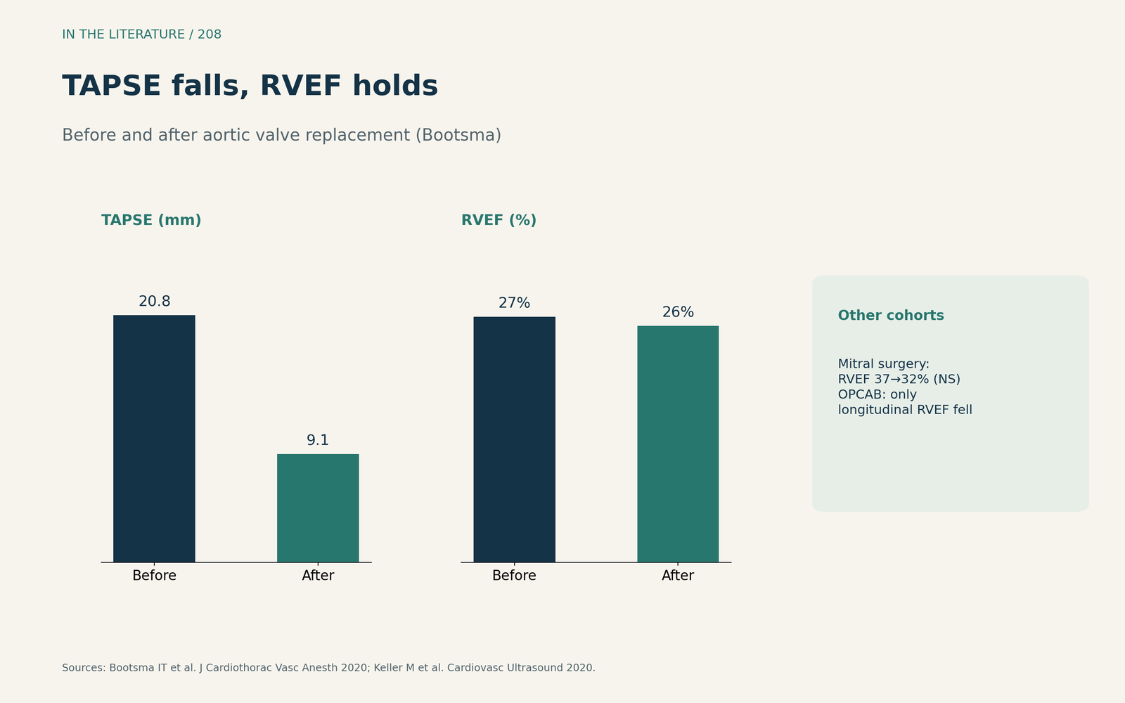
Figure 1. TAPSE falls, RVEF holds. Redrawn from Bootsma et al. and Keller et al.[1], [6] View larger.

Why the fall happens, and why echo can mislead
TAPSE, RV strain and FAC are all affected by loading conditions.[2] Grønlykke and colleagues consider longitudinal measures inadequate to evaluate RV function after cardiac surgery; VVI pacing decreased cardiac output by 24%, and a fall in stroke volume with PEEP was not detected by any echo variable.[4] Akkaya and colleagues studied 102 aortic surgery patients: TAPSE fell from 22.0 to 18.3 mm, and the relative fall in TAPSE exceeded that in FAC.[5]
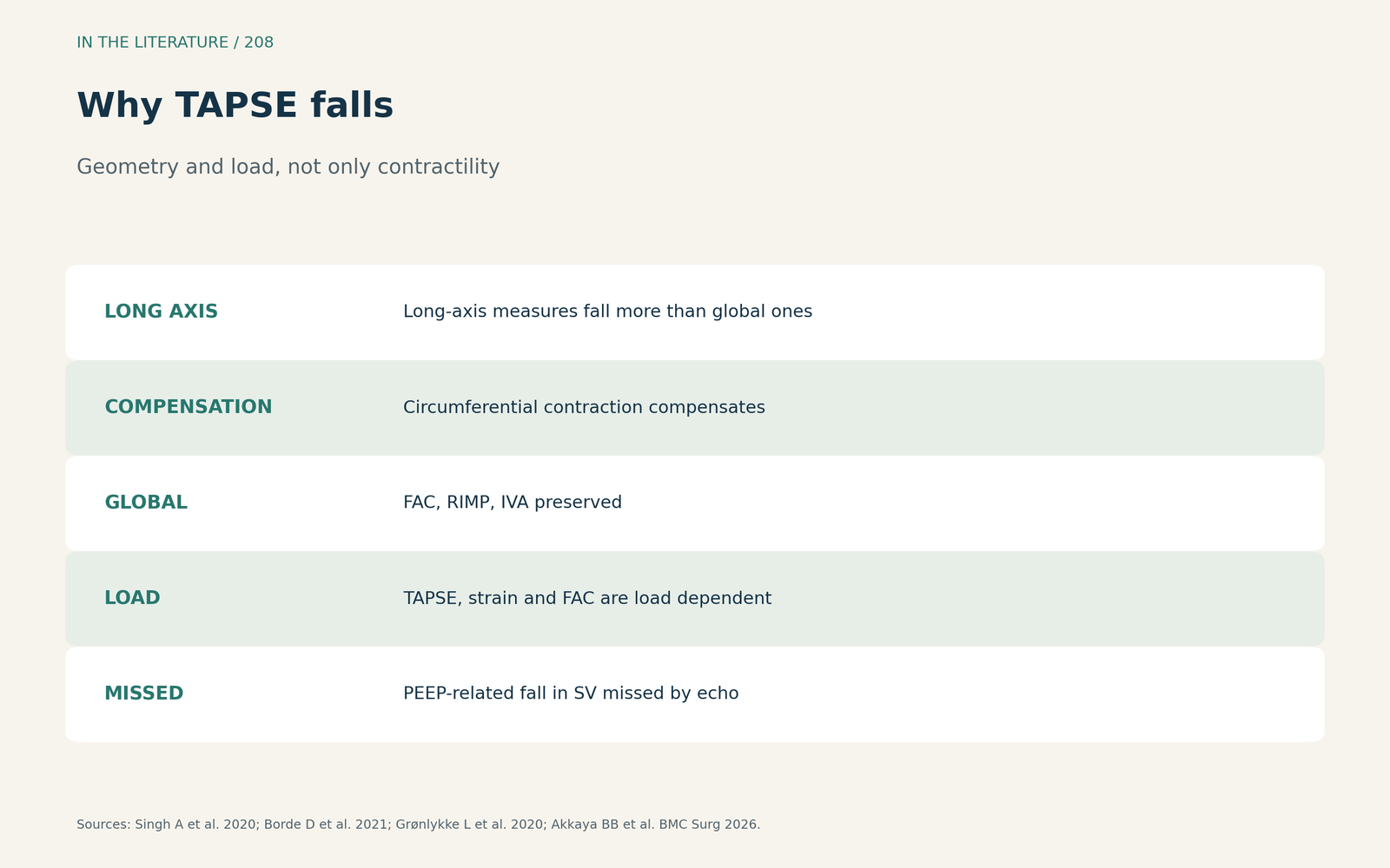
Figure 2. Geometry and load, not only contractility. Synthesized from Borde et al., Singh et al., Grønlykke et al. and Akkaya et al.[3], [2], [4], [5] View larger.

How to assess the RV after surgery
Akkaya and colleagues conclude that a TAPSE fall should not be read as proportional deterioration of all RV parameters and that postoperative RV assessment should be multiparametric and clinically contextual.[5] Bootsma and Borde and colleagues reach the same conclusion.[1], [3]
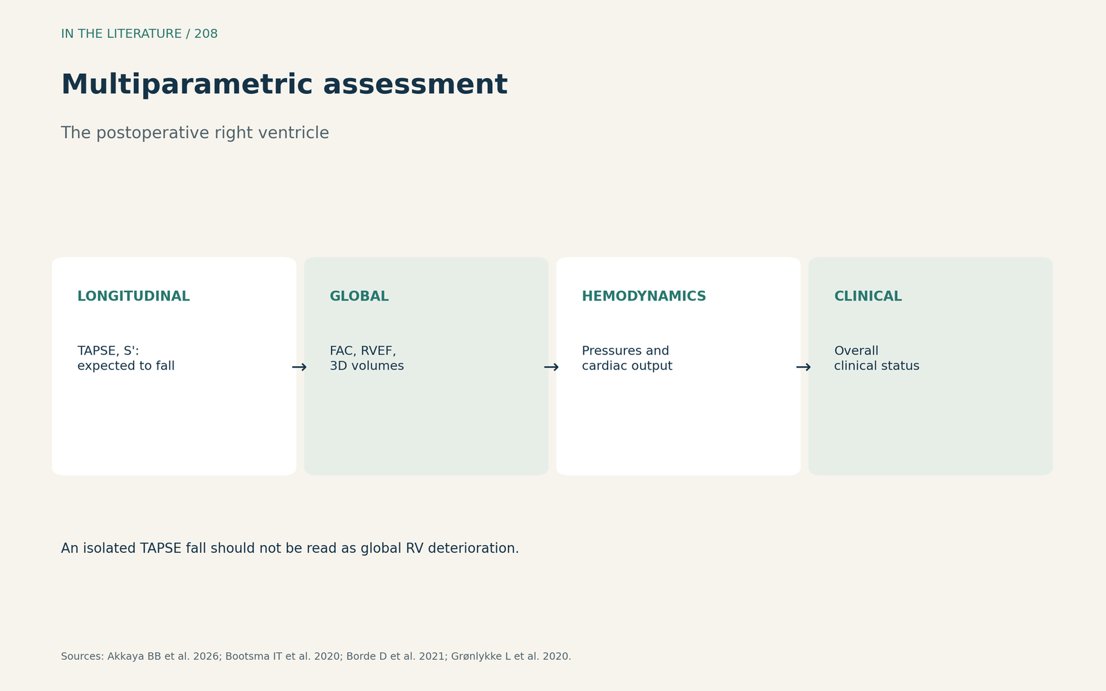
Figure 3. Global measures, hemodynamics, clinical picture. Synthesized from Akkaya et al., Bootsma et al., Borde et al. and Grønlykke et al.[5], [1], [3], [4] View larger.

Three takeaways
- A fall in TAPSE after cardiac surgery is expected and does not by itself mean RV failure.[1], [2]
- Judge the RV with global measures such as FAC or RVEF, plus hemodynamics.[3], [5]
- All echo indices are load dependent and can miss real falls in output; true RV failure still needs to be recognized.[2], [4]
References
- Bootsma IT, Scheeren TWL, de Lange F, et al. The reduction in right ventricular longitudinal contraction parameters is not accompanied by a reduction in general right ventricular performance during aortic valve replacement: an explorative study. J Cardiothorac Vasc Anesth. 2020;34(8):2140–2147. doi:10.1053/j.jvca.2020.01.012
- Singh A, Huang X, Dai L, et al. Right ventricular function is reduced during cardiac surgery independent of procedural characteristics, reoperative status, or pericardiotomy. J Thorac Cardiovasc Surg. 2020;159(4):1430–1438.e4. doi:10.1016/j.jtcvs.2019.04.035
- Borde D, Joshi P, Joshi S, et al. Changes in right ventricular function after off-pump coronary artery bypass grafting. J Cardiothorac Vasc Anesth. 2021;35(3):811–819. doi:10.1053/j.jvca.2020.06.086
- Grønlykke L, Korshin A, Gustafsson F, et al. The effect of common interventions in the intensive care unit on right ventricular function after cardiac surgery—an intervention study. J Cardiothorac Vasc Anesth. 2020;34(5):1211–1219. doi:10.1053/j.jvca.2019.11.043
- Akkaya BB, Sert DE, Yiğitbaşı MM, et al. Differential changes in longitudinal and area-based right ventricular function after aortic surgery: an exploratory echocardiographic study. BMC Surg. 2026;26(1):564. doi:10.1186/s12893-026-03941-w
- Keller M, Heller T, Lang T, et al. Acute changes of global and longitudinal right ventricular function: an exploratory analysis in patients undergoing open-chest mitral valve surgery, percutaneous mitral valve repair and off-pump coronary artery bypass grafting. Cardiovasc Ultrasound. 2020;18(1):32. doi:10.1186/s12947-020-00218-x