ITL-7bb732a0b254
Juan Li · In the Literature · Updated November 16, 2026
Watch the video
Key points
- Choose oxygen support, assess ventilation, and plan airway rescue as separate parts of the anesthetic strategy.
- Carbon dioxide can accumulate during deep TAVR sedation despite preserved oxygenation; a reassuring SpO₂ alone is insufficient.
- HIGH-OXY-TAVR found fewer desaturation events with high-flow nasal oxygen, without establishing prevention of hypercapnia or a reduction in manual ventilation.
- Supraglottic-airway studies evaluated general anesthesia strategies; they do not establish routine supraglottic-airway use during light sedation.
- Agree on the intended depth, monitoring, access to the airway, and escalation plan before the procedure starts.
First identify what needs support
During transfemoral TAVR under sedation, an acceptable oxygen saturation answers only part of the respiratory question. The clinician still needs to know whether the patient is moving air effectively and whether ventilation can be maintained throughout the procedure. Increasing oxygen delivery, relieving obstruction, and securing an airway are different interventions.
The 2026 ERAS Cardiac Working Group order set by Engelman and colleagues recommends an explicitly defined sedation target, titration with a validated sedation scale, continuous capnography, and the capability to rescue deeper-than-intended sedation. This expert framework makes monitoring and rescue part of the initial plan, rather than a response to a low saturation alone. [1]
Carbon dioxide can rise while oxygenation looks satisfactory
Chen and colleagues’ Open Heart 2024 retrospective study examined 201 patients with available intraoperative blood gases among 655 nonemergent transfemoral TAVR procedures. Sedation was predominantly deep: the mean bispectral index at valve deployment was 48, and almost all patients received both propofol and dexmedetomidine. [2]
PaCO₂ increased from baseline in 91% of patients with available baseline measurements; seven patients lacked a baseline sample. At valve deployment, 26% (52/201) had PaCO₂ above 60 mmHg. Arterial oxygen tension was not related to the change in carbon dioxide. These findings concern a selected, deeply sedated cohort receiving supplemental oxygen, rather than all TAVR patients receiving monitored anesthesia care. [2]
The practical inference is to assess ventilation independently of oxygenation. The study measured PaO₂ and PaCO₂; it did not test the diagnostic accuracy of SpO₂ or capnography. Its PaCO₂ categories were analysis thresholds, not validated triggers for intubation. Clinical assessment should integrate breathing, airway patency, sedation depth, carbon dioxide monitoring, and blood gases when indicated. [1, 2]
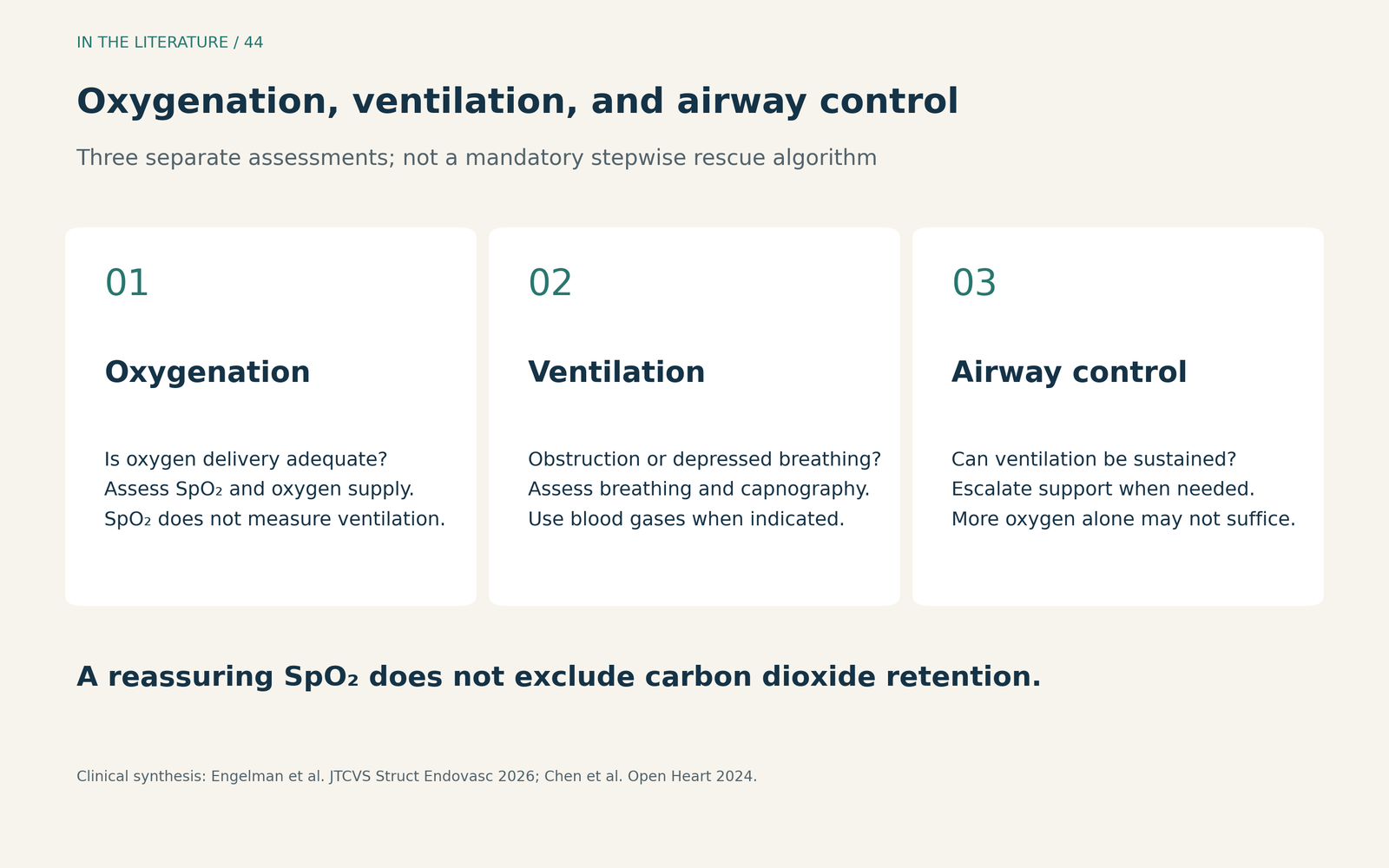
Figure 1. Assess three respiratory tasks separately. This clinical reasoning aid draws on the ERAS monitoring framework and the physiological observations of Chen et al.; it is not a validated escalation algorithm. [1, 2] View figure

What high-flow nasal oxygen actually improves
In Giménez-Milà and colleagues’ HIGH-OXY-TAVR trial, published in Journal of Clinical Medicine in 2025, 132 patients were randomized to high-flow nasal oxygen (HFNO) at 50 L/min with FiO₂ 0.6, or conventional nasal oxygen at 5 L/min. Patients received propofol–remifentanil sedation targeting a Richmond Agitation–Sedation Scale score of −4 to −2. These are the study settings, not a recommendation that every TAVR procedure requires this depth. [3]
The primary endpoint was at least one SpO₂ below 93% for more than 10 seconds. In the intention-to-treat analysis, this occurred in 14/66 (21%) with HFNO versus 35/66 (53%) with standard oxygen, with a risk ratio of 0.4 (95% CI, 0.24–0.67). The primary per-protocol analysis also favored HFNO: 13/64 (20%) versus 31/61 (51%). Seven randomized patients were excluded from that analysis because of vascular complications requiring general anesthesia or a procedure lasting less than 45 minutes. [3]
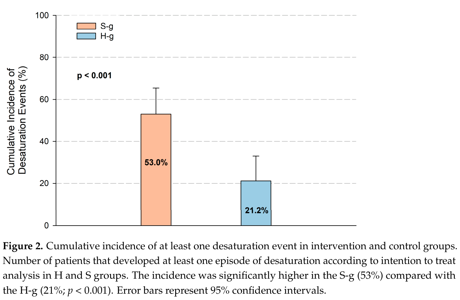
Figure 2. Original trial figure, cropped from Giménez-Milà et al., J Clin Med 2025, Figure 2; data and labels are unchanged. S-g is standard oxygen and H-g is HFNO. The bars retain the published 53.0% and 21.2%; the article text reports these as 53% and 21%. Error bars are 95% confidence intervals. Reproduced under CC BY 4.0. [3] View original figure

This is useful evidence for reducing desaturation in that setting. It does not establish that HFNO prevents hypoventilation. At 45 minutes, mean PaCO₂ was 6.42 versus 6.21 kPa with HFNO and standard oxygen, respectively (P = 0.390). Manual ventilation was required in 1 (2%) versus 3 (5%) per-protocol patients (P = 0.577). Neither comparison established a ventilation or rescue benefit. The oxygen strategies also differed in delivered oxygen concentration; the observed benefit cannot be attributed to flow alone. [3]
Why the earlier trial does not give the same answer
Scheuermann and colleagues’ Perioperative Medicine 2023 trial randomized 72 patients to HFNO at 50 L/min and FiO₂ 0.3, or conventional nasal oxygen at 2 L/min. It used remifentanil with regional analgesia and a Ramsay sedation target of 2–3, a different regimen from HIGH-OXY-TAVR. [4]
HFNO did not significantly improve the primary 30-minute percentage change in PaO₂ (P = 0.171). Carbon dioxide changes also did not differ significantly. Fewer patients desaturated and reported comfort was better, but the trial was not powered for these secondary endpoints. Differences in oxygen concentration, sedation depth, sampling, and the primary outcome help explain why the two trials should not be compressed into a single claim that every HFNO strategy has the same effect. [4]
For practice, HFNO is an oxygen-support option with direct TAVR trial evidence. It should accompany the same attention to airway patency and ventilation as other oxygen devices. Better oxygen saturation is not a reason to deepen sedation or postpone necessary ventilatory support. [1, 3, 4]
A supraglottic airway changes the anesthetic strategy
The supraglottic-airway (SGA) literature addresses another decision. Azad and colleagues’ JCVA 2020 study propensity-matched 31 SGA patients with 62 endotracheal-tube patients, all receiving general anesthesia. It found no significant difference in recovery-unit or hospital length of stay. This was a retrospective comparison of airway devices during general anesthesia. [5]
Musuku and colleagues’ JCVA 2021 study compared 148 matched pairs receiving general anesthesia with an SGA or monitored anesthesia care. Hospital stay did not differ significantly, while vasopressors were used in 97% of the SGA group versus 66% of the MAC group. Selection, anesthetic depth, and accompanying drugs differed; this finding cannot isolate a causal effect of the device. [6]
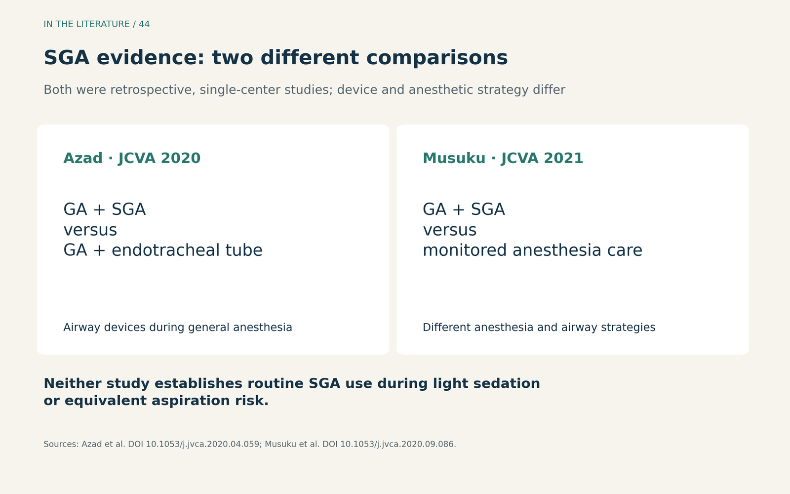
Figure 3. Read the comparator before choosing the device. Azad et al. compared SGA with endotracheal intubation during general anesthesia; Musuku et al. compared general anesthesia with an SGA against MAC. Both provide observational feasibility evidence in selected patients, without establishing equivalent aspiration risk. [5, 6] View figure

These studies support considering an SGA within an appropriate anesthesia plan and experienced team. They do not make it a routine substitute for nasal oxygen during light sedation. Nor does failure to detect a difference in uncommon complications establish equal protection from aspiration. [5, 6]
Make the plan before oxygen saturation falls
Before starting, agree on the required sedation depth, how ventilation will be assessed, who can reach the airway beneath the drapes, and how rescue or conversion to general anesthesia will occur. If ventilation becomes inadequate, reassess depth and airway patency, provide the required ventilatory support, and escalate according to the patient’s condition and the team’s airway plan. There is no requirement to exhaust a fixed sequence of oxygen devices before acting. This is a clinical synthesis of the monitoring guidance and study findings, not a trial-validated rescue protocol. [1, 2]
Three takeaways
- Treat oxygenation and ventilation as separate assessments. Preserved oxygenation during supplemental oxygen does not establish adequate carbon dioxide clearance.
- Use HFNO for the benefit demonstrated. It can reduce desaturation in selected TAVR sedation settings; it does not replace ventilation monitoring or rescue capability.
- Choose an airway within the complete anesthesia plan. SGA feasibility under general anesthesia is different from evidence for routine use during light sedation or equivalent aspiration protection.
References
- Engelman DT, Ailawadi G, Russo MJ, et al. Enhanced recovery after surgery turnkey order set for periprocedural care of the patient undergoing transcatheter aortic valve replacement. JTCVS Struct Endovasc. 2026;12:100146. doi:10.1016/j.xjse.2026.100146.
- Chen T, Asher S, Apruzzese P, et al. Hypercapnia during transcatheter aortic valve replacement under monitored anaesthesia care: a retrospective cohort study. Open Heart. 2024;11(2):e002801. doi:10.1136/openhrt-2024-002801.
- Giménez-Milà M, Manzano-Valls A, Abdul-Jawad O, et al. High-Flow Nasal Oxygenation During Sedation for Transcatheter Aortic Valve Replacement: The HIGH-OXY-TAVR Randomised–Controlled Trial. J Clin Med. 2025;14(23):8347. doi:10.3390/jcm14238347.
- Scheuermann S, Tan A, Govender P, et al. High-flow nasal oxygen vs. standard oxygen therapy for patients undergoing transcatheter aortic valve replacement with conscious sedation: a randomised controlled trial. Perioper Med (Lond). 2023;12(1):11. doi:10.1186/s13741-023-00300-8.
- Azad SS, Cobey FC, Price LL, et al. Supraglottic Airway Use for Transfemoral-Transcatheter Aortic Valve Replacement. J Cardiothorac Vasc Anesth. 2020;34(12):3243–3249. doi:10.1053/j.jvca.2020.04.059.
- Musuku SR, Capua CAD, Doshi I, et al. Outcomes of Transfemoral Transcatheter Aortic Valve Replacement Performed With General Anesthesia Using a Supraglottic Airway Versus Monitored Anesthesia Care. J Cardiothorac Vasc Anesth. 2021;35(6):1760–1768. doi:10.1053/j.jvca.2020.09.086.