
ITL-8d7f251e855b
Juan Li · In the Literature · Updated November 16, 2026
Watch the video

Key points
- A TTE-based TAVR pathway still requires a structured examination, experienced interpretation and access to additional imaging.
- Decide before the procedure whether the expected acoustic windows and procedural complexity favor planned TEE.
- Immediately after deployment, assess the prosthesis and look for new regurgitation, ventricular dysfunction and pericardial effusion.
- Unexplained instability or an unresolved imaging question should trigger reassessment and, when useful, escalation to TEE.
- Better image-quality scores do not establish better clinical outcomes; TEE under sedation also requires a specific airway and anesthesia plan.
Choose the examination that can answer the question
For an otherwise straightforward transfemoral TAVR, the team plans sedation and TTE. That can be a workable imaging pathway. The important question is whether the team can obtain the information needed before and after deployment—and promptly investigate anything unexpected.
The ASE competency recommendations describe an examination that can use either TTE or TEE. Baseline imaging establishes ventricular function, regional wall motion and associated valve disease. Immediately after implantation, reassess valve position, paravalvular regurgitation, annular integrity, new wall-motion abnormalities, and any pericardial effusion with its hemodynamic effect. [1]
A reassuring fluoroscopic valve position does not complete those assessments. In a TTE pathway, agree beforehand on the acquisition protocol, who interprets the images and how findings reach the operator. The modality should serve the clinical question, rather than determine how much of that question is answered. [1]
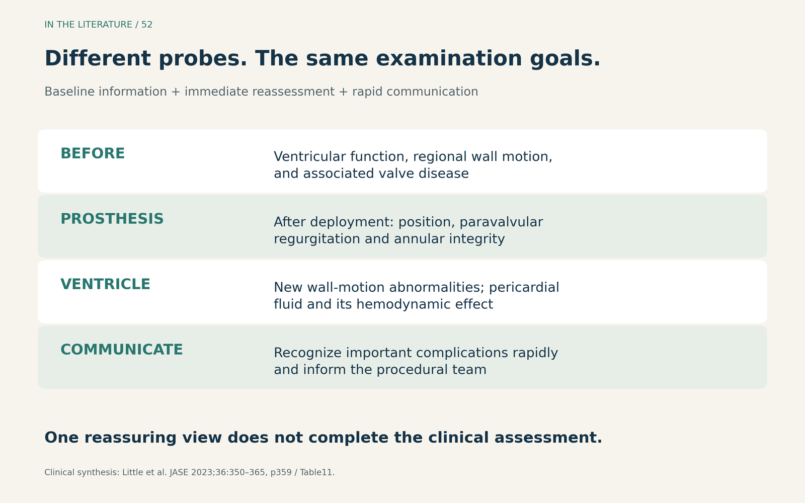
Figure 1. The shared examination goals, synthesized from the ASE competency recommendations. A normal result in one domain does not establish that all other domains are normal. [1] View figure

Identify the reasons to plan TEE before starting
TTE offers useful access without an esophageal probe, but acquisition may be intermittent and affected by positioning, the procedural equipment or poor windows. TEE permits continuous imaging, with its own invasiveness and acoustic limitations. Kinno and colleagues emphasize advance team discussion, experienced imagers and the ability to switch promptly when necessary. [2]
In their 2026 review, Chen and colleagues propose upfront TEE for selected high-risk anatomy, inadequate TTE windows or circumstances in which continuous imaging could guide the procedure. This is an expert selection framework, not a new graded recommendation that every patient must receive TEE. A contrast-sparing strategy may also increase the value of echo, but renal impairment alone does not decide the modality. Before choosing TEE, assess tolerance and whether esophageal disease makes probe placement unsafe; use another imaging strategy when TEE is contraindicated. [3]
Complex leaflet procedures illustrate a specific reason for planned TEE. Protsyk and colleagues describe multiplane and three-dimensional TEE during BASILICA to help align equipment, identify leaflet traversal and monitor laceration alongside fluoroscopy. Echo also checks the result and possible complications. That is a procedural imaging need to plan for; it is different from adding TEE routinely to an uncomplicated implantation. [4]
When the result is unclear, change the imaging plan
Suppose blood pressure remains low after deployment, or the apparent regurgitation does not fit the hemodynamics. Revisit the images and the clinical data together. New ventricular dysfunction, acute valve dysfunction or pericardial fluid may explain the deterioration and change the immediate response. The ASE recommendations emphasize rapid assessment and communication; Chen's review explains the value of continuous TEE in identifying the mechanism of instability. [1, 3]
If TTE cannot answer the question, obtain the additional imaging needed, including TEE when appropriate. A TTE-based workflow should already have the expertise and equipment for this transition. Use multiple planes and color-Doppler sweeps for regurgitation: one apparently reassuring view is insufficient. [2]
Resuscitation and investigation proceed together. This practical synthesis does not mean waiting for probe insertion before supporting an unstable patient. Nor does a reassuring echo alone exclude coronary obstruction; persistent suspicion may require direct coronary assessment. [1, 3, 4]
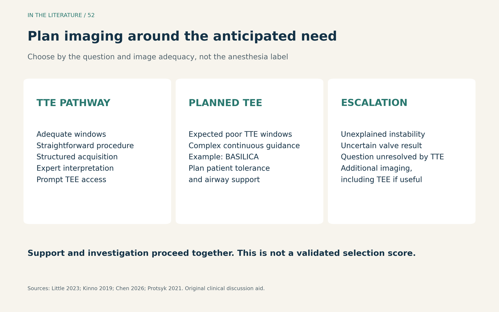
Figure 2. A clinical discussion aid based on the cited recommendations and reviews, not a validated selection score. Probe suitability, patient tolerance and the anesthesia plan remain part of the decision. [1–4] View figure

What the new comparison adds—and what it cannot settle
Khan and colleagues' 2026 retrospective study compared 30 TEE-guided with 30 TTE-guided transfemoral procedures. TEE was selected for inadequate preoperative TTE windows. Image quality was rated 5.0 versus 2.73 on a five-point scale, favoring TEE. This was a selected, nonrandomized comparison, without reported blinded image adjudication. [5]
The study supports feasibility in that center, not routine TEE superiority. Thirty patients per group and in-hospital follow-up cannot establish equivalent safety or reliably characterize uncommon harm. Higher image scores are not evidence that the imaging choice prevented complications. [5]
It also shows why TEE does not automatically mean general anesthesia, while avoiding the opposite assumption that ordinary light sedation will suffice. Despite the title's “conscious sedation” wording, the protocol used intravenous sedation with monitored depth and positive-pressure airway support in the TEE group. Applying this approach requires a deliberate patient-tolerance, airway and rescue plan; the paper is not a universal sedation prescription. [5]
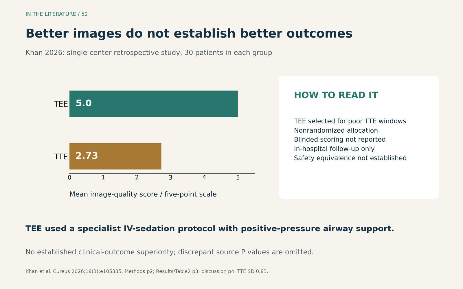
Figure 3. Original summary of Khan et al. Mean image-quality scores are shown without a significance value because the source's prose and table disagree on that value. The study reports TTE standard deviation 0.83; the chart does not imply an independently validated diagnostic-accuracy scale. [5] View figure

Three takeaways
- Make the imaging pathway complete. Adequate baseline and postdeployment assessment matters whichever probe is used.
- Anticipate difficult imaging and complex guidance. Plan TEE when its capabilities address a foreseeable need, and preserve prompt access when starting with TTE.
- Escalate for the unanswered question. Reassess instability or uncertain valve function promptly, while matching the probe choice to patient tolerance and the airway plan.
References
- Little SH, Rigolin VH, Garcia-Sayan E, et al. Recommendations for Special Competency in Echocardiographic Guidance of Structural Heart Disease Interventions: From the American Society of Echocardiography. J Am Soc Echocardiogr. 2023;36(4):350–365. doi:10.1016/j.echo.2023.01.014.
- Kinno M, Cantey EP, Rigolin VH. The transition from transesophageal to transthoracic echocardiography during transcatheter aortic valve replacement: an evolving field. J Echocardiogr. 2019;17(1):25–34. doi:10.1007/s12574-018-0409-1.
- Chen TM, Amanullah MR, Ewe SH. Intraprocedural echocardiographic guidance in transcatheter aortic valve implantation. J Cardiovasc Imaging. 2026;34:29. doi:10.1186/s44348-026-00094-5.
- Protsyk V, Meineri M, Kitamura M, et al. Echocardiographic Guidance of Intentional Leaflet Laceration prior to Transcatheter Aortic Valve Replacement: A Structured Approach to the Bioprosthetic or Native Aortic Scallop Intentional Laceration to Prevent Iatrogenic Coronary Artery Obstruction Procedure. J Am Soc Echocardiogr. 2021;34(6):676–689. doi:10.1016/j.echo.2021.01.015.
- Khan R, Ramadan E, Taha M, et al. Comparison of Transthoracic Echocardiography (TTE) and Transesophageal Echocardiography (TEE) Guidance for Transcatheter Aortic Valve Replacement (TAVR) Performed Under Conscious Sedation: A Retrospective Study. Cureus. 2026;18(3):e105335. doi:10.7759/cureus.105335.