
ITL-0c34c5783bf8
Juan Li, MD · Updated November 18, 2026
Key points
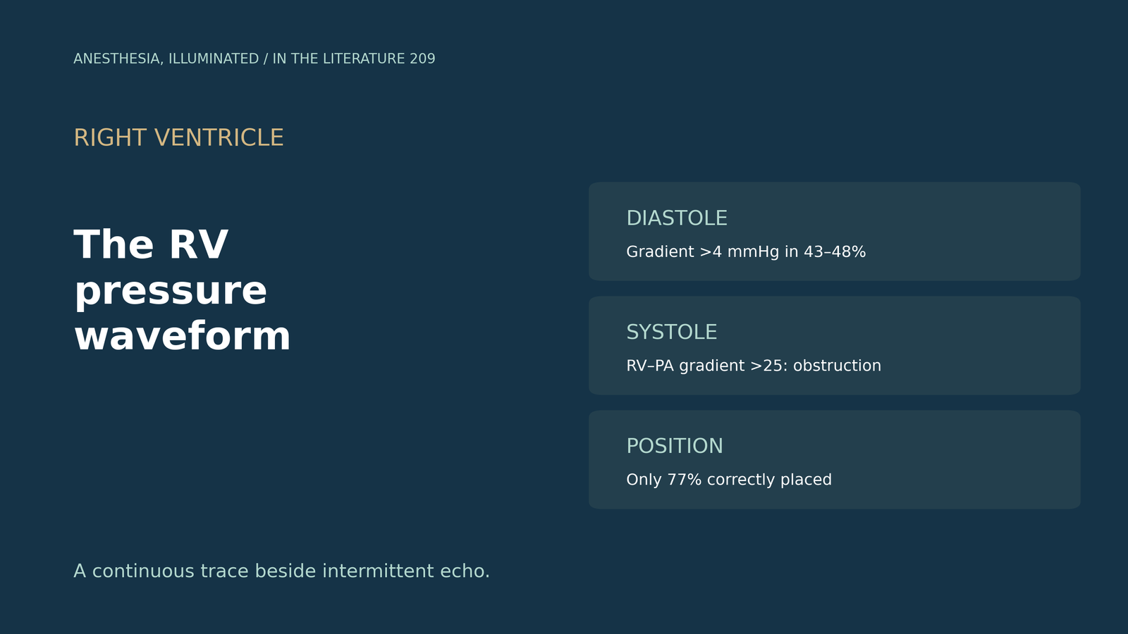
- The waveform is usually ignored. Most centers using PACs ignore the RV pressure waveform, yet an abnormal RV diastolic gradient (normal below 4 mmHg) occurs in 43 to 48% of cardiac surgical patients.[1]
- It warns against more volume. A square-root pattern reflects diastolic equalization, and in RV failure volume and transfusion can worsen right heart function.[1]
- It diagnoses outflow obstruction instantly. The normal RV-to-PA systolic gradient is below 6 mmHg, and obstruction is significant above 25 mmHg.[2]
- Check where the catheter is. Only 77% of paceport catheters were in adequate position, and normal-looking waveforms can occur despite malposition.[3]
- It is continuous where echo is intermittent. Echo needs an acoustic window, whereas catheter-based RV pressure monitoring is continuous.[4]
After mitral valve surgery, blood pressure falls in the ICU; a quick TTE has poor windows, but the pulmonary artery catheter has an RV port. What can the RV pressure waveform show that echo might miss?[1], [2]
What the waveform shows
Denault and colleagues note that most centers using PACs ignore the RV pressure waveform, although a mean PA pressure above 20 mmHg after induction occurs in 73% of patients and an abnormal RV diastolic gradient (normal below 4 mmHg) in 43 to 48%. Diastolic equalization produces a square-root sign, and in RV failure, volume and transfusion can worsen right heart function; they advise watching the RV visually, by echo and by pressure.[1] Zeng and colleagues add that the normal RV-to-PA systolic gradient is below 6 mmHg, that obstruction is significant above 25 mmHg, that RVOT obstruction can be diagnosed instantly from the waveform, and that it should be considered in any unstable patient after cardiac surgery.[2] Miles and colleagues detected dynamic RVOT obstruction with waveform monitoring during liver transplantation and note that normal RV dP/dt is above 400 mmHg/s.[5]
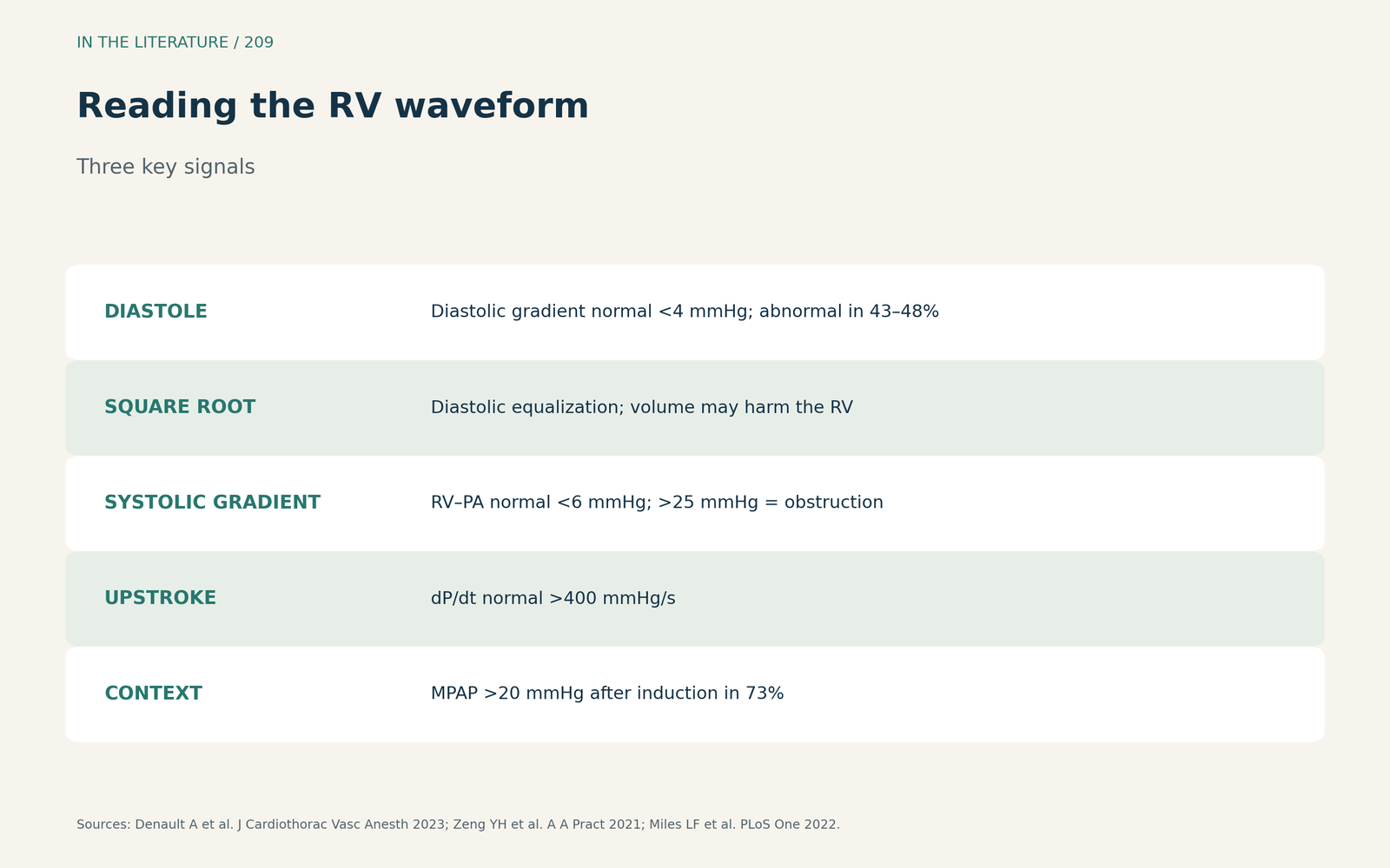
Figure 1. Diastolic slope, systolic gradient, upstroke. Synthesized from Denault et al., Zeng et al. and Miles et al.[1], [2], [5] View larger.

Catheter position pitfalls
Saade and colleagues found that only 91 of 118 paceport catheters (77%) were in adequate position, that normal-looking waveforms can occur despite malposition, and that if the catheter is advanced too far the RV port measures central venous pressure.[3] Miles and colleagues excluded eight recordings (16%) for catheter displacement.[5]
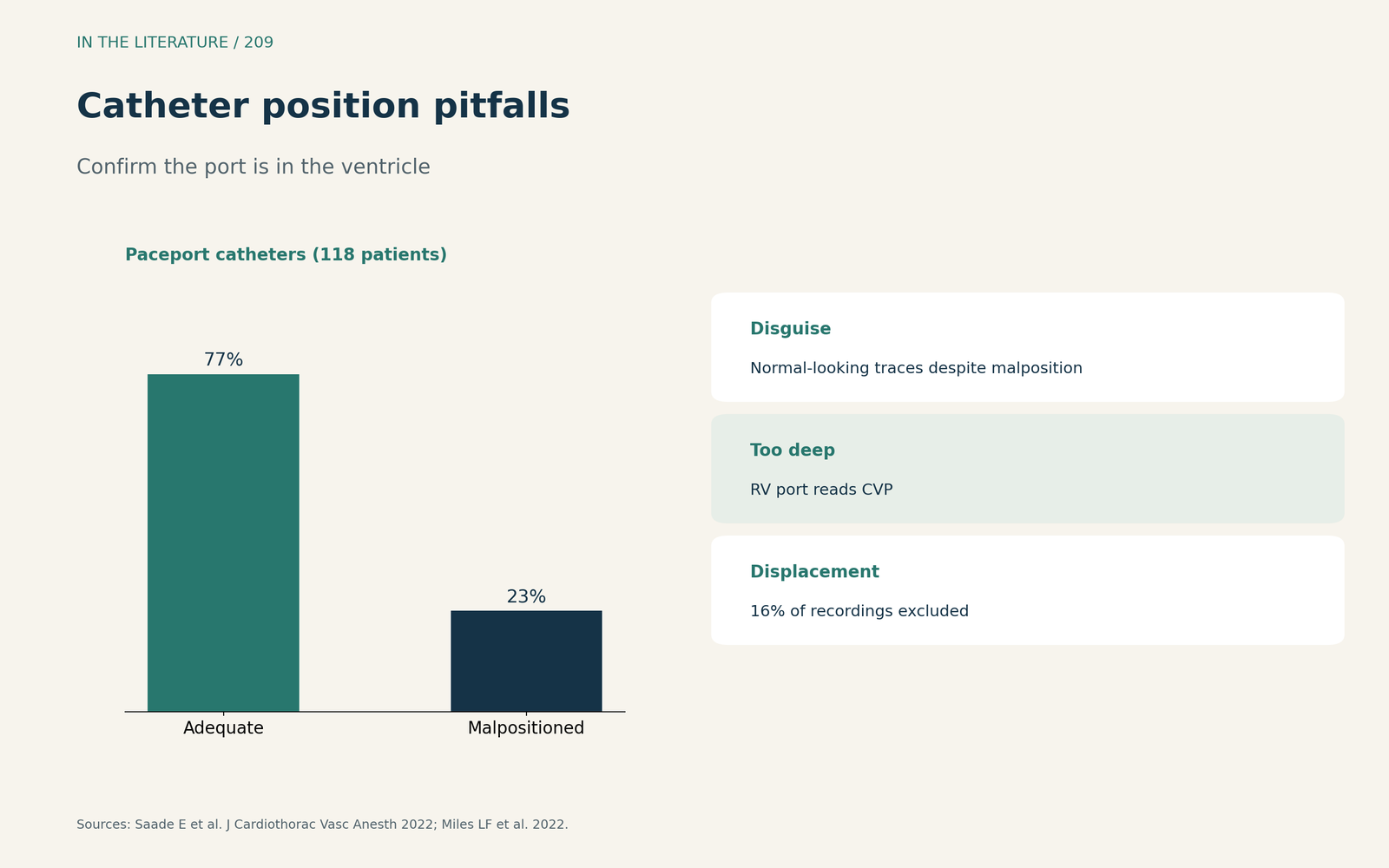
Figure 2. Confirm the port is in the ventricle. Redrawn from Saade et al. and Miles et al.[3], [5] View larger.

Continuous versus intermittent
In a pig model, Couture and colleagues found that RV dysfunction was tracked by dP/dtmax from the RV waveform while CVP changed little (6 to 9 mmHg). They note that echo needs an acoustic window and gives intermittent information, whereas catheter-based monitoring is continuous, but caution that animal findings need careful translation.[4]
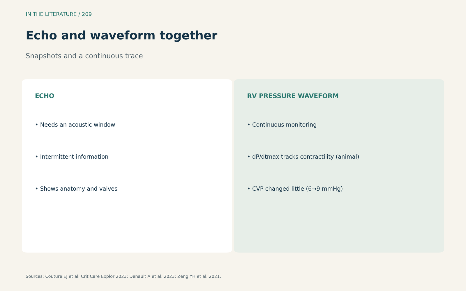
Figure 3. Snapshots and a continuous trace. Synthesized from Couture et al., Denault et al. and Zeng et al.[4], [1], [2] View larger.

Three takeaways
- The RV waveform adds continuous diastolic, systolic-gradient and contractility signals to intermittent echo.[1], [4]
- An RV-to-PA systolic gradient above 25 mmHg points to outflow obstruction.[2]
- Verify catheter position first; evidence is from small series and expert opinion.[3], [5]
References
- Denault A, Canevet M, Couture EJ. Pro: we should use a pulmonary artery catheter with right ventricular pressure waveforms in cardiac surgical patients. J Cardiothorac Vasc Anesth. 2023;37(4):659–662. doi:10.1053/j.jvca.2022.12.007
- Zeng YH, Calderone A, Beaubien-Souligny W, et al. Right ventricular outflow tract obstruction in the intensive care unit: a case report of 2 patients. A A Pract. 2021;15(12):e01532. doi:10.1213/XAA.0000000000001532
- Saade E, Couture EJ, Robillard J, Denault A. A word of caution on right ventricular pressure monitoring using the paceport pulmonary artery catheter. J Cardiothorac Vasc Anesth. 2022;36(11):4223–4226. doi:10.1053/j.jvca.2022.06.029
- Couture EJ, Moses K, Monge García MI, et al. Longitudinal validation of right ventricular pressure monitoring for the assessment of right ventricular systolic dysfunction in a large animal ischemic model. Crit Care Explor. 2023;5(1):e0847. doi:10.1097/CCE.0000000000000847
- Miles LF, Couture EJ, Potes C, et al. Preliminary experience with continuous right ventricular pressure and transesophageal echocardiography monitoring in orthotopic liver transplantation. PLoS One. 2022;17(2):e0263386. doi:10.1371/journal.pone.0263386