ITL-c05d8e12246f
Juan Li, MD · Updated November 17, 2026
Watch the video
Key points
- Increased left ventricular (LV) wall thickness is characteristic of cardiac amyloidosis but not specific. Hypertension, aortic stenosis and hypertrophic cardiomyopathy (HCM) produce the same finding.[1], [2]
- Suspicion rests on a pattern of red flags: QRS voltage low relative to wall thickness, right ventricular and valve thickening, pericardial effusion, advanced diastolic dysfunction, reduced longitudinal strain, and extracardiac clues such as carpal tunnel syndrome.[1], [2]
- No single echocardiographic measurement settles the question. Strain ratios were developed in selected cohorts, and apical sparing identified only a minority of amyloidosis cases against HCM in one comparison.[2], [3]
- Confirmation follows a defined algorithm: bone scintigraphy together with serum and urine immunofixation and serum free light chains, with cardiac magnetic resonance or biopsy when results are discordant.[1]
- Amyloidosis is found in a meaningful minority of older patients with aortic stenosis, and can occur without increased wall thickness.[4], [5]
The preoperative echocardiogram shows a thick, non-dilated left ventricle with preserved ejection fraction in an older patient scheduled for valve or noncardiac surgery. Hypertensive heart disease is the usual explanation, but cardiac amyloidosis changes prognosis and treatment. What distinguishes amyloid infiltration from other hypertrophy, and what testing actually confirms it?[1], [6]
Thickness is the starting point, not the diagnosis
The European Society of Cardiology (ESC) Working Group position statement defines cardiac amyloidosis by amyloid fibrils in cardiac tissue. It recommends looking for amyloidosis in patients with increased wall thickness who have heart failure, aortic stenosis or red-flag signs, particularly if older than 65 years. In heart failure with preserved ejection fraction (HFpEF), HCM phenotypes and severe aortic stenosis, the statement notes that transthyretin amyloidosis (ATTR) has been found in up to 7–19% of patients.[1]
Thickness alone can also mislead in the other direction. Achten and colleagues prospectively screened 202 unselected patients with HFpEF using scintigraphy. Six (3%) had wild-type ATTR cardiomyopathy, and 2 of the 6 did not have increased wall thickness (≥12 mm); both had early-stage disease. Red flags were very common in the whole HFpEF cohort (81%), so their presence alone does not identify the few patients with amyloidosis.[5]
Red flags that point toward amyloid
The ESC statement lists cardiac and extracardiac red flags. Cardiac features include QRS voltage that is low relative to LV thickness, atrioventricular conduction disease, disproportionately high natriuretic peptide and persistent troponin elevation. Echocardiographic features include granular sparkling myocardium, increased right ventricular wall and valve thickness, pericardial effusion and reduced longitudinal strain with apical sparing. Extracardiac clues include bilateral carpal tunnel syndrome, lumbar spinal stenosis, polyneuropathy, macroglossia, skin bruising and proteinuria.[1]
The American Society of Echocardiography (ASE) practical points add that the discordance between wall thickness and QRS voltage is particularly suggestive, but normal voltage does not exclude amyloidosis. Other echocardiographic clues include grade II or worse diastolic dysfunction, increased interatrial septal thickness, diffuse valve thickening and paradoxical low-flow, low-gradient aortic stenosis. The ASE also notes that the "5-5-5" sign, in which all tissue Doppler velocities are below 5 cm/s, may be insensitive in early disease.[2]
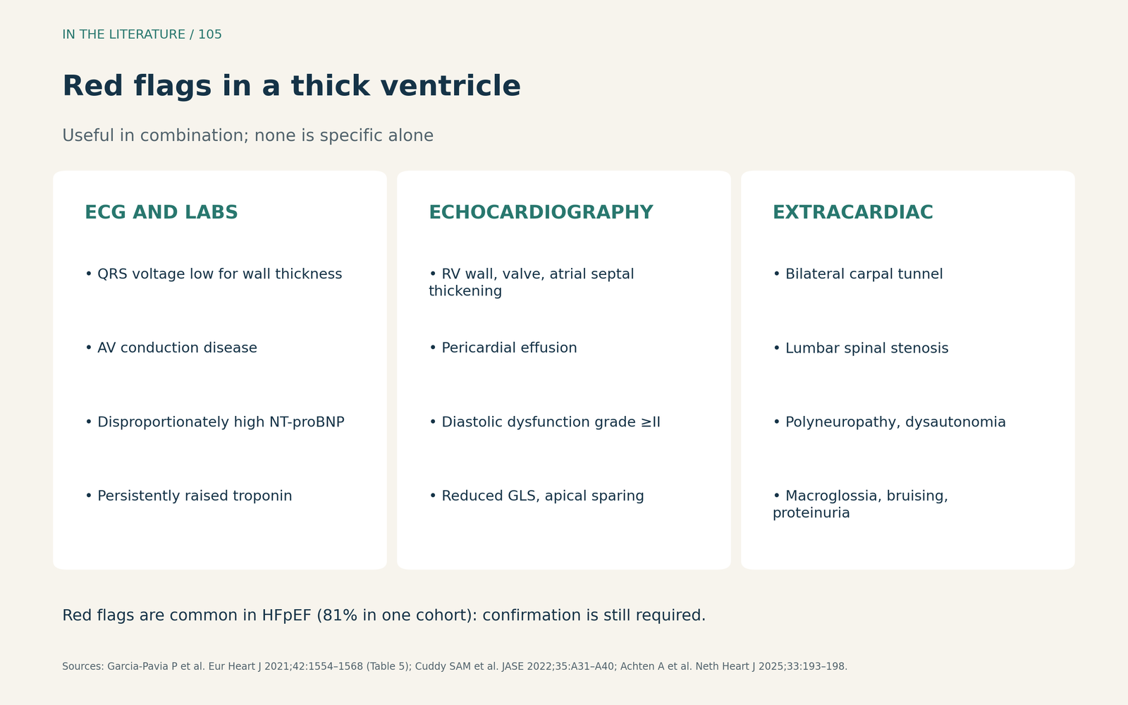
Figure 1. Red flags for cardiac amyloidosis. Summarized from the ESC position statement (Table 5) and the ASE practical points; their value lies in combination, not in any single feature.[1], [2] View larger.

Echo criteria and scores: useful, with limits
The ESC statement gives echocardiographic criteria for use alongside an extracardiac biopsy showing amyloid. These require unexplained LV thickness of at least 12 mm plus either two characteristic findings or a multiparametric score of 8 or more. The characteristic findings are grade 2 or worse diastolic dysfunction, tissue Doppler s′, e′ and a′ velocities below 5 cm/s, and an absolute global longitudinal strain below 15%. The score includes relative wall thickness, E/e′, tricuspid annular excursion, global strain and the apex-to-base strain ratio. The statement notes that the score had not yet been externally validated.[1]
Single strain markers are less discriminating than their popularity suggests. The ASE notes that strain ratio cut-offs were derived with one vendor's software in selected populations with increased wall thickness and have not been validated as screening tools. Stassen and colleagues compared patients with amyloidosis and HCM. Relative apical sparing of 1 or more identified only 31 of 83 (37%) amyloidosis cases. Global constructive myocardial work discriminated better (area under the curve 0.820), but this was a single comparison that needs confirmation.[2], [3]
How the diagnosis is confirmed
The ESC algorithm begins with bone scintigraphy, using technetium pyrophosphate, DPD or HMDP with SPECT, together with tests for a monoclonal protein. Those tests are serum and urine protein electrophoresis with immunofixation and the serum free light chain assay; combined, they have a reported sensitivity of 99% for the abnormal precursor in light-chain (AL) amyloidosis. The algorithm leads to four scenarios:[1]
- Grade 2–3 cardiac uptake with no monoclonal protein: ATTR cardiomyopathy can be diagnosed without biopsy, followed by genetic testing.
- Grade 1 uptake: non-invasive diagnosis is not possible; histology is required.
- Any abnormal monoclonal protein test: AL amyloidosis must be excluded promptly with hematology input, cardiac magnetic resonance and usually tissue diagnosis.
- No uptake and normal protein tests: amyloidosis is unlikely, although some hereditary variants and rare types can give negative scans; magnetic resonance or biopsy remains an option if suspicion persists.
Meers and Townsley review these modalities for cardiothoracic anesthesiologists. They note that scintigraphic uptake can be falsely positive in AL amyloidosis, which is why monoclonal protein testing is essential before scintigraphy is used to establish an ATTR diagnosis.[1], [6]
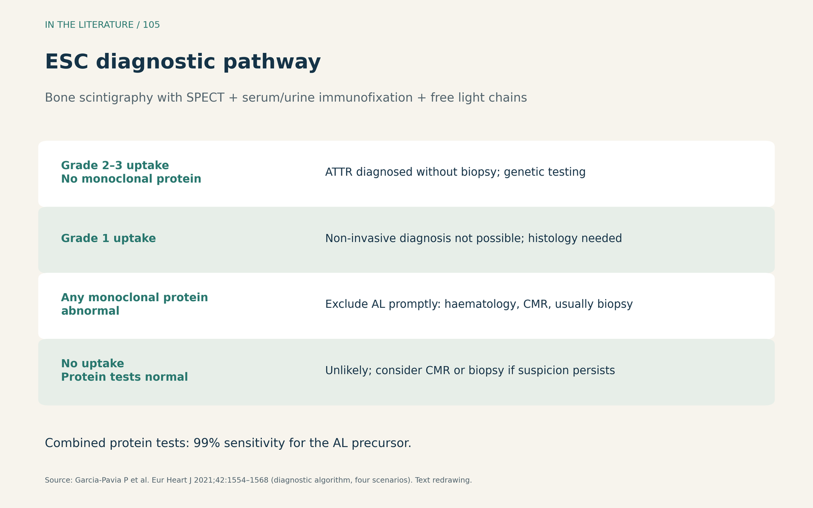
Figure 2. The ESC diagnostic pathway. Redrawn as text from the four scenarios described in the position statement; not a substitute for hematology or specialist review.[1] View larger.

Aortic stenosis and the cardiac surgical patient
Aortic stenosis is the overlap most relevant to cardiac anesthesiologists. Nies and colleagues prospectively performed bone scintigraphy after aortic valve replacement, almost all transcatheter, in 131 patients aged 60 years or older with septal thickness of at least 12 mm. Pathological uptake was found in 21 (16%), and 11 (8.4%) were diagnosed with ATTR cardiomyopathy. The RAISE screening score of 2 or more had a sensitivity of 76% and a specificity of 56% for pathological uptake. This was lower than in the cohort where the score was derived. All patients diagnosed with ATTR cardiomyopathy were 83 years or older.[4]
The authors favored valve replacement first and evaluation for amyloidosis afterward, and argued that transcatheter replacement should not be withheld because of ATTR cardiomyopathy. Their study was single-center and observational, and some patients with grade 1 uptake did not undergo biopsy.[4]
For the anesthesiologist, the practical step is recognition and referral. A thick ventricle with several red flags, especially in an older patient with aortic stenosis or HFpEF, warrants a note to the treating team that amyloidosis has not been excluded. The diagnosis should come from the confirmatory pathway, not from intraoperative imaging alone.[1], [6]
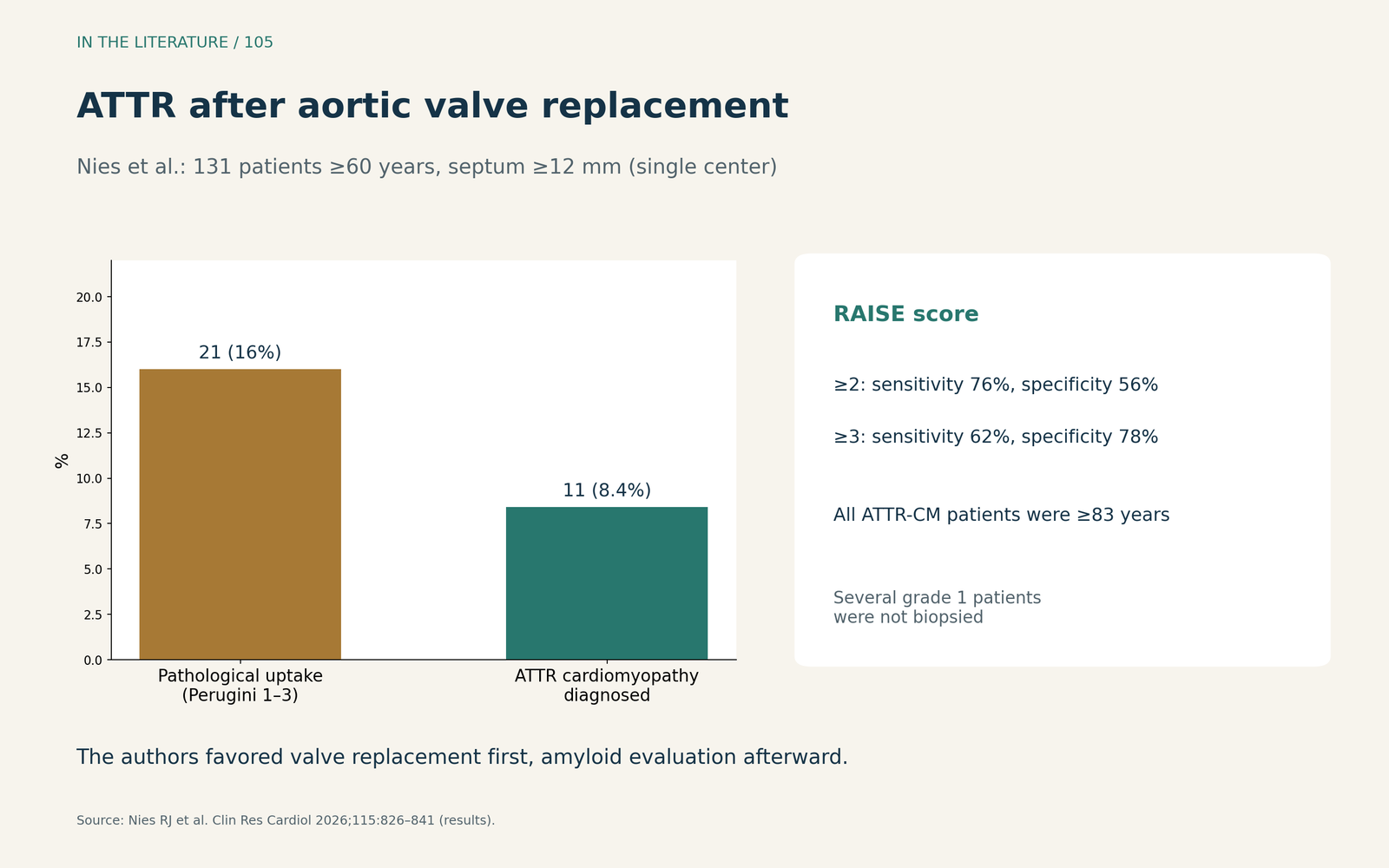
Figure 3. Amyloidosis in aortic stenosis. Redrawn from the results of Nies et al. (n = 131 patients ≥60 years with septum ≥12 mm after aortic valve replacement; single center).[4] View larger.

Three takeaways
- A thick ventricle is a reason to look, not a diagnosis. Combine cardiac, echocardiographic and extracardiac red flags; amyloidosis can also occur without increased thickness.[1], [2], [5]
- Do not lean on one strain index. Scores and ratios help, but cut-offs come from selected cohorts and single-vendor software.[2], [3]
- Confirm with the scintigraphy-plus-light-chain pathway. Monoclonal protein testing is mandatory before scintigraphy can establish ATTR; older patients with aortic stenosis deserve particular attention.[1], [4], [6]
References
- Garcia-Pavia P, Rapezzi C, Adler Y, et al. Diagnosis and treatment of cardiac amyloidosis: a position statement of the ESC Working Group on Myocardial and Pericardial Diseases. Eur Heart J. 2021;42(16):1554–1568. doi:10.1093/eurheartj/ehab072
- Cuddy SAM, Chetrit M, Jankowski M, et al. Practical points for echocardiography in cardiac amyloidosis. J Am Soc Echocardiogr. 2022;35(9):A31–A40. doi:10.1016/j.echo.2022.06.006
- Stassen J, Tjahjadi C, Adam R, et al. Left ventricular myocardial work to differentiate cardiac amyloidosis from hypertrophic cardiomyopathy. J Am Soc Echocardiogr. 2023;36(2):252–254. doi:10.1016/j.echo.2022.08.015
- Nies RJ, Ney S, Nies JF, et al. Cardiac transthyretin amyloidosis in aortic valve replacement: RAISE score performance in the postoperative setting. Clin Res Cardiol. 2026;115(5):826–841. doi:10.1007/s00392-025-02766-6
- Achten A, van Empel VPM, Weerts J, et al. Prevalence of transthyretin amyloid cardiomyopathy in an unselected cohort with heart failure with preserved ejection fraction. Neth Heart J. 2025;33(6):193–198. doi:10.1007/s12471-025-01954-3
- Meers JB, Townsley MM. Imaging cardiac amyloidosis: an update for the cardiothoracic anesthesiologist. J Cardiothorac Vasc Anesth. 2021;35(7):1911–1916. doi:10.1053/j.jvca.2021.02.040