ITL-a89a0f22ca92
Watch the video
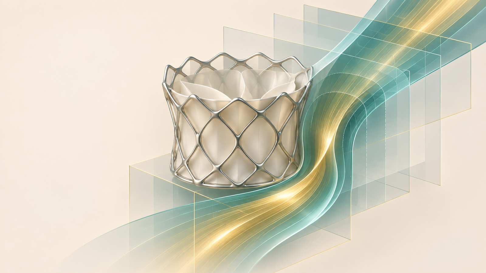
Figure 1. Conceptual planning illustration, not a patient scan or a specific device model.
Key points
- The predicted neo-LVOT is the residual outflow space after virtual valve implantation; it depends on anatomy, device, position, and cardiac phase.[1]
- The frequently quoted 170 mm² threshold came from a particular TMVR cohort and is not a universal boundary between safe and unsafe implantation.[2, 3]
- When leaflet modification is contemplated, assess the skirt neo-LVOT as well: splitting the leaflet does not remove the prosthetic skirt.[1, 3, 4]
- A long, thick, or calcified anterior leaflet can create problems that a single area measurement does not fully describe.[3, 4]
- CT planning and procedural TEE answer complementary questions; the actual implant and hemodynamic result still require assessment.[1, 3]
A planning report gives a predicted neo-LVOT of 180 mm². Is that enough space for TMVR? The answer depends on how the area was generated, what valve is being implanted, and what will obstruct the outflow tract after deployment.
A useful report therefore needs more than a favorable number. It should allow the Heart Team to understand the assumptions behind that number and the consequences if the final implant differs from the simulation.[3, 4]
What the classic threshold tells us
In Yoon and colleagues' study of 194 TMVR patients, the reported incidence of LVOT obstruction varied markedly: 1.9% after valve-in-valve, 8.0% after valve-in-ring, and 54.1% after valve-in-MAC. A predicted neo-LVOT ≤1.7 cm²—equivalent to 170 mm²—identified obstruction with 96.2% sensitivity and 92.3% specificity.[2]
Those results are available here from the published abstract. The study defined obstruction as an increase in mean LVOT gradient of at least 10 mmHg from baseline. This definition and the mix of procedures matter when interpreting the threshold. The finding supports careful risk assessment; it does not validate applying 170 mm² to every modern device, simulation method, or patient.[2, 3]
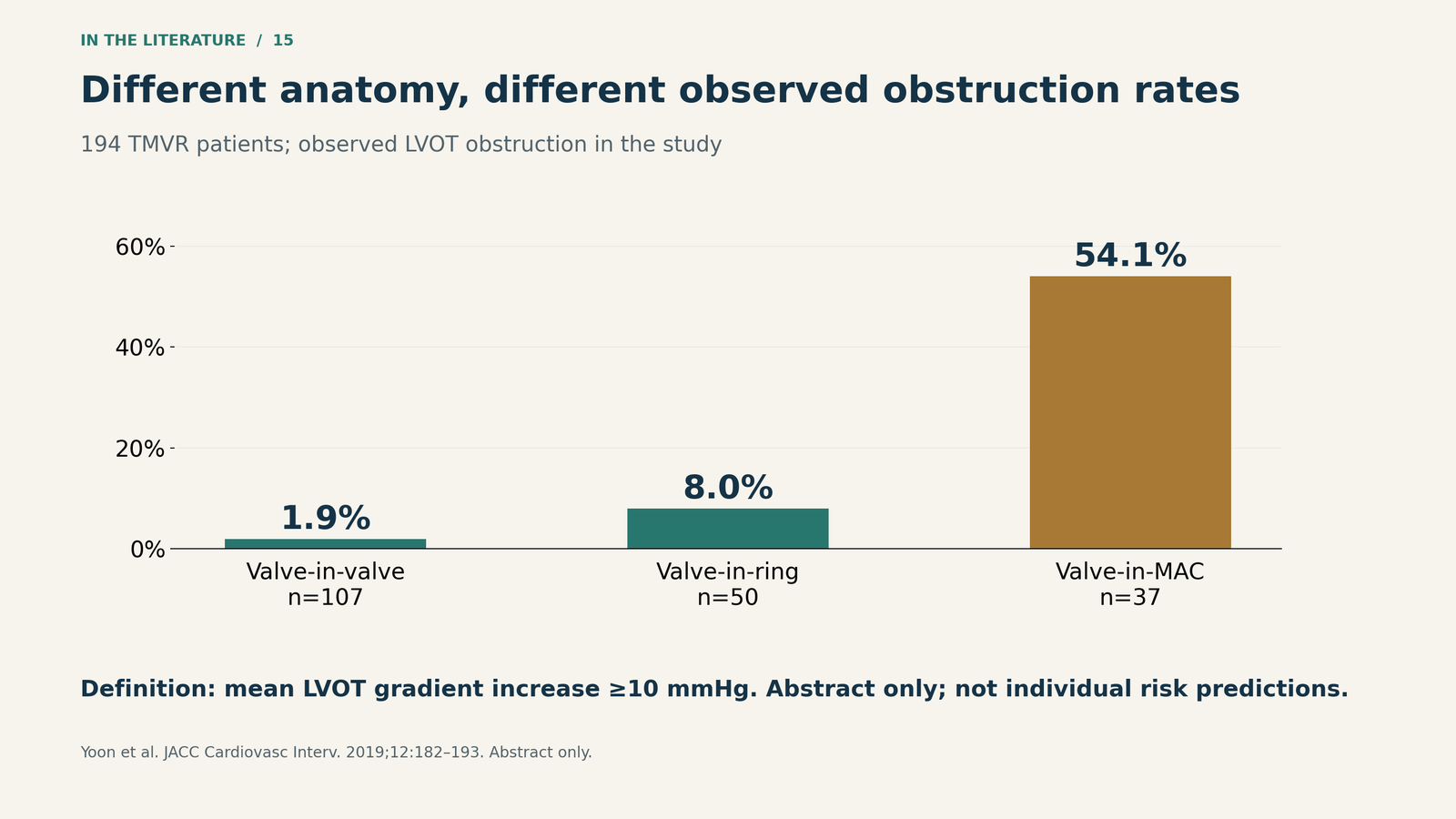
Figure 2. Different anatomical settings within one study. Group sizes were 107, 50, and 37, respectively. These are observed cohort rates, not current individual predictions. Source: Yoon et al., published abstract.[2]

Ask how the simulation was constructed
The November 2026 ESCR/NASCI recommendations emphasize systolic neo-LVOT measurement, documentation of the cardiac phase, and device-specific assessment. The outflow tract changes during systole; a value from one phase cannot be interpreted as though it were measured in another.[3]
Implant depth and tilt also matter. A device extending farther into the ventricle can reduce the remaining outflow space. The virtual valve should represent the intended device and size, and the team should examine plausible deployment variation rather than select the most favorable configuration. The 2026 leaflet-modification review explicitly argues for planning around an unfavorable plausible position.[3, 4]
A 2022 JCVA review also highlights the difficulty of transferring prediction methods between balloon-expandable valves and dedicated TMVR systems. This is why a report should identify the platform and the measurement method, not simply label an area “adequate.”[1]
Distinguish leaflet obstruction from skirt obstruction
After TMVR, the displaced anterior mitral leaflet can restrict the outflow channel. LAMPOON intentionally splits that leaflet so blood can pass through open frame cells. However, flow remains constrained by the covered portion of the prosthesis. The skirt neo-LVOT estimates that residual space after effective leaflet modification.[1, 3, 4]
This distinction changes planning. A small conventional neo-LVOT with adequate skirt clearance may lead to consideration of leaflet modification. If the skirt clearance is also small, leaflet splitting alone may leave important obstruction. Septal reduction, another device or approach, or a different treatment strategy may need discussion.[3, 4]
The 2026 expert algorithm uses skirt neo-LVOT <200 mm² as a reason to consider preemptive septal reduction in its high-risk planning pathway. This is an expert decision framework, not a universally validated cutoff. Likewise, dense confluent leaflet calcium or inadequate leaflet splay may undermine an otherwise attractive LAMPOON plan.[4]
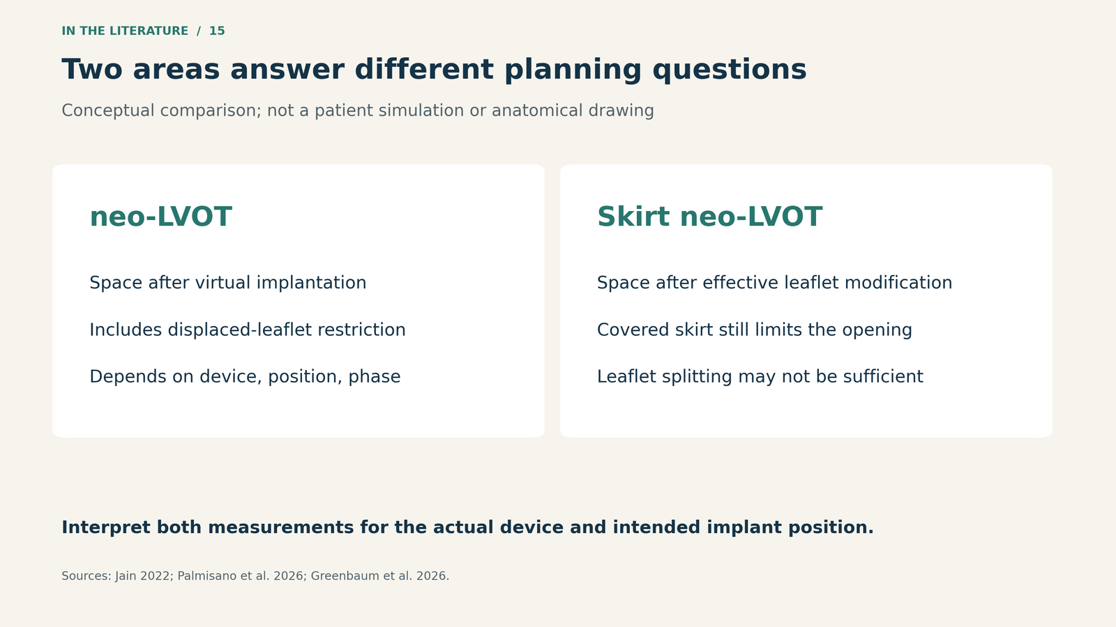
Figure 3. Two different questions about remaining outflow space. This conceptual comparison does not predict an individual patient's result.[1, 3, 4]

Turn the CT result into a procedural plan
Before proceeding, the team should be able to describe the expected obstruction mechanism, the range of plausible implant positions, and the intended preventive strategy. A small predicted area is most useful when it changes that discussion.[3, 4]
TEE then evaluates what actually happened: device position, leaflet motion, residual regurgitation, transvalvular hemodynamics, and evidence of LVOT narrowing or obstruction. The anatomical appearance and Doppler findings must be interpreted together with the circulation. A technically successful valve deployment does not, by itself, demonstrate an acceptable outflow result.[1]
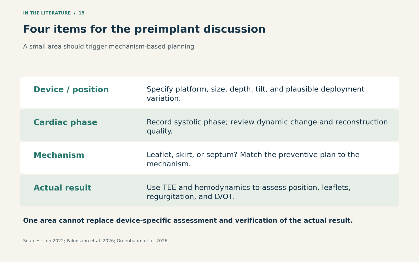
Figure 4. A practical planning discussion derived from the imaging recommendations and procedural reviews; not a substitute for device-specific assessment.[1, 3, 4]

Three takeaways
- Interrogate the number. Record the device, intended position, measurement phase, and anatomical context before interpreting neo-LVOT area.
- Identify what will obstruct flow. Leaflet modification may address leaflet obstruction while leaving skirt-related obstruction unresolved.
- Connect prediction with verification. Agree on prevention before implantation and use procedural imaging and hemodynamics to assess the actual result.
References
- Jain P, Mohammed AN, Fischer C, Aljure O. Current and investigational transcatheter mitral valve replacement systems: a narrative review for the cardiac anesthesiologist. J Cardiothorac Vasc Anesth. 2022;36:4108–4128. doi:10.1053/j.jvca.2022.05.019.
- Yoon SH, Bleiziffer S, Latib A, Eschenbach L, Ancona M, Vincent F, et al. Predictors of left ventricular outflow tract obstruction after transcatheter mitral valve replacement. JACC Cardiovasc Interv. 2019;12(2):182–193. doi:10.1016/j.jcin.2018.12.001. Abstract only.
- Palmisano A, Francone M, Rajiah P, Leipsic J, Feuchtner G, Eberhard M, et al. CT and MRI in periprocedural transcatheter mitral valve replacement: expert recommendations by the European Society of Cardiovascular Radiology (ESCR) and the North American Society for Cardiovascular Imaging (NASCI). Eur Radiol. Published online November 6, 2026. doi:10.1007/s00330-026-12955-0.
- Greenbaum AB, D’Angelo JT, Lederman RJ, Hebbo E, Eric Chan KY, Chiu HOAC, et al. Current evidence, emerging evidence, and decision-making: an algorithm for leaflet modification procedures. JACC Cardiovasc Interv. 2026;19(11):1378–1392. doi:10.1016/j.jcin.2026.04.023.