ITL-c45a3b25ef66
Juan Li, MD · Updated November 17, 2026
Watch the video
Key points
- Transesophageal echocardiography (TEE) locates the puncture in three directions. The bicaval view shows superior–inferior, the aortic valve short-axis view shows anterior–posterior, and the four-chamber view shows height above the mitral annulus.[1], [2]
- The right site depends on the procedure. Mitral edge-to-edge repair needs a posterior puncture with height matched to the jet, appendage occlusion an inferior–posterior one, and large-sheath ablation a central, more inferior one. Consensus statements give no validated millimetre targets.[2], [3]
- Look for traps before the needle moves: an aneurysmal septum, a Chiari network, leads and an anterior fossa near the aortic root. During the puncture, keep the needle tip in view and see it in the left atrium before the sheath follows.[1], [3], [4], [5]
- TEE improved needle position compared with fluoroscopy alone, and in a before-and-after cohort tamponade was less frequent with TEE guidance. Neither study was randomized.[6], [7]
- The septal hole matters afterwards. After mitral edge-to-edge repair, more than 20% of iatrogenic atrial septal defects persisted at 12 months and were associated with heart failure rehospitalization, but routine closure has not shown benefit.[2], [8]
A patient is under general anesthesia for transcatheter mitral edge-to-edge repair. The interventionalist withdraws the needle from the superior vena cava and asks you where it is. Which views tell you where the tip sits, where should it be for this procedure, and what must you watch for to keep the puncture safe?[1], [2]
Three views, three directions
The PITLOC protocol, described by Katsiampoura and colleagues, builds the puncture on three mid-esophageal views: bicaval, aortic valve short-axis and four-chamber.[1] The 2026 European consensus statement places the bicaval view at about 90° to 110° for the superior–inferior axis and the short-axis view at about 30° to 45° for the anterior–posterior axis. On the four-chamber view, it advises measuring the puncture height as the distance between the tenting and the mitral annulus.[2] The Italian imaging society consensus gives slightly wider angle ranges.[3]
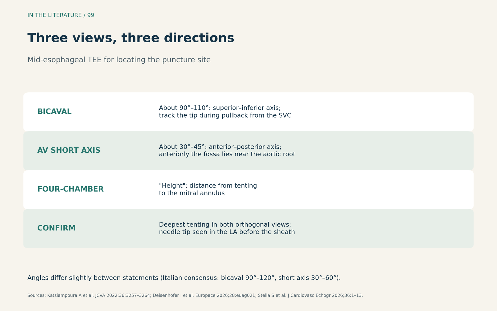
Figure 1. Three views, three directions. Synthesized from the PITLOC protocol and the European and Italian consensus statements.[1], [2], [3] View larger.

Choose the site for the procedure
The puncture is the first step of the procedure that follows. The European statement advises a posterior puncture for mitral edge-to-edge repair, a posterior and inferior one to achieve coaxial alignment for appendage occlusion in conventional anatomy, and a central, more inferior one for large-sheath ablation so that the right inferior pulmonary vein can be reached.[2]
The Italian consensus adds detail for edge-to-edge repair. A central jet calls for a posterior, slightly superior site; medial jets need more height and lateral jets less. It recommends an inferior–posterior site for most appendage occlusions and a mid-posterior site for transseptal mitral valve-in-valve.[3] Neither document gives a validated height in millimetres.
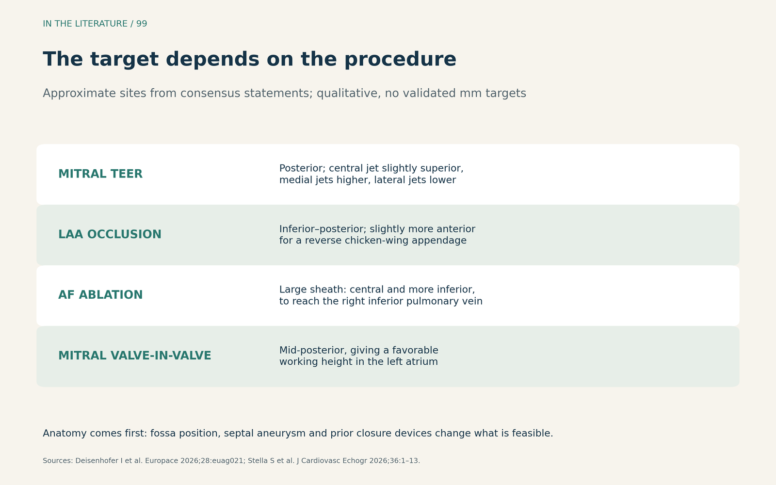
Figure 2. The target depends on the procedure. Qualitative sites summarized from the European and Italian consensus statements; individual anatomy can change them.[2], [3] View larger.

Before the needle moves: look for traps
A full TEE before the case matters. Mayl and colleagues described a Chiari network pulled through the puncture site that looked like catheter thrombus in the left atrium, and they stressed comprehensive TEE evaluation before beginning the case.[4] Katsiampoura and colleagues advise identifying existing intracardiac leads before the needle goes in.[1]
An aneurysmal septum, defined in the European statement as protrusion more than 10 mm beyond the septal plane, raises the risk of puncturing the lateral atrial wall.[2] In Chen and colleagues' case, pushing the dilator against such a septum placed its tip very near the lateral left atrial wall, and the authors describe radiofrequency needles as a way to reduce tenting.[5] An anteriorly placed fossa ovalis may lie immediately behind the aortic root.[3]
During the puncture
Keep the needle tip in view continuously and avoid the thick part of the septum, which can cause a septal hematoma.[1] Before the sheath is advanced, confirm the needle tip in the left atrium and the release of tenting; the Italian consensus also advises confirming left atrial pressure.[1], [3]
The main complications are anatomical. The European statement reports tamponade in 0.5% to 2%, largely from whole procedures, and describes how a posterior puncture can exit the right atrium into the pericardium before entering the left atrium. Such perforations may only appear after sheath removal. Aortic puncture occurs in less than 0.1% of cases.[2] The Italian consensus reports aortic puncture in 0.05% and advises continuing TEE monitoring for some minutes after sheath removal.[3]
Does TEE guidance help?
The evidence is observational. In 150 patients undergoing left atrial electrophysiology procedures, Katov and colleagues first positioned the needle under fluoroscopy and then checked it with TEE. Fluoroscopy alone achieved optimal positions in 32.7% and poor or dangerous ones in 24.0%; after TEE adjustment, 59.3% were optimal and none were poor or dangerous. There was no control group.[6]
In a before-and-after comparison of 932 patients from the same center, Teumer and colleagues found fewer tamponades requiring intervention with TEE guidance than with fluoroscopy alone (0.5% vs 1.8%; adjusted odds ratio 0.103, 95% CI 0.013–0.819). TEE itself caused complications in 0.9%.[7] Because the groups were treated in different periods, this is not randomized evidence. The European statement advises TEE or intracardiac echocardiography when a specific location is targeted, and notes that advantages of intracardiac echocardiography over TEE remain to be proven.[2]
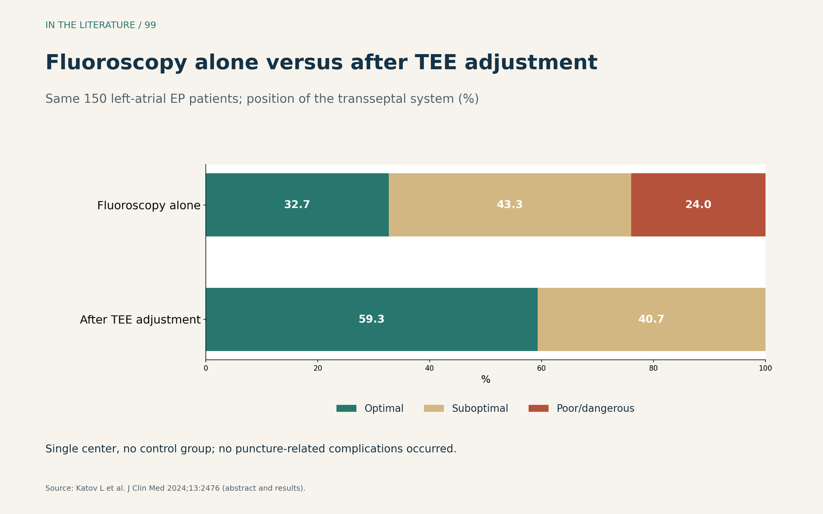
Figure 3. TEE refined the puncture position. Redrawn from the results of Katov et al. (same patients assessed first by fluoroscopy, then with TEE; no control group).[6] View larger.

After crossing: the iatrogenic septal defect
Large sheaths leave a hole. The European statement cites a 42% incidence of iatrogenic atrial septal defect at 19 months after mitral edge-to-edge repair, all with left-to-right shunts.[2] In a meta-analysis by Lei and colleagues, persistence occurred in more than 20% at 12 months and was associated with heart failure rehospitalization (odds ratio 2.71; 95% CI 1.42–5.18), with no significant difference in mortality (odds ratio 1.48; 95% CI 0.86–2.56). Closure was not superior to conservative therapy in the MITHRAS trial.[8]
Three takeaways
- Locate the needle in three directions. Use the bicaval, short-axis and four-chamber views, and keep the tip in view.[1], [2]
- Match the site to the procedure and the anatomy. Posterior for mitral repair, inferior–posterior for appendage occlusion; check for an aneurysmal septum, a Chiari network and the aortic root first.[2], [3], [4]
- Watch beyond the crossing. Confirm the tip in the left atrium before the sheath, keep imaging after sheath removal, and assess the residual septal defect.[3], [7], [8]
References
- Katsiampoura A, Mufarrih SH, Sharkey A, et al. A sequential approach for echocardiographic guidance of transseptal puncture: the PITLOC protocol. J Cardiothorac Vasc Anesth. 2022;36(8):3257–3264. doi:10.1053/j.jvca.2021.12.025
- Deisenhofer I, Abdelhamid M, Adamo M, et al. Transseptal puncture in cardiovascular interventions: a clinical consensus statement of the European Heart Rhythm Association, the Heart Failure Association, the European Association of Percutaneous Cardiovascular Interventions, the European Association of Cardiovascular Imaging of the ESC, and the Association for European Paediatric and Congenital Cardiology. Europace. 2026;28(5):euag021. doi:10.1093/europace/euag021
- Stella S, Cameli M, Cusma'-Piccione M, et al. How to monitor transseptal puncture: a consensus statement of the Italian Society of Echocardiography and Cardiovascular Imaging. J Cardiovasc Echogr. 2026;36(1):1–13. doi:10.4103/jcecho.jcecho_17_26
- Mayl J, Peters L, Zhao D, Richardson K. Chiari network or catheter-associated thrombus? A rare complication of transseptal access. CASE (Phila). 2020;4(1):21–24. doi:10.1016/j.case.2019.10.005
- Chen CB, Sandhu HS, Keane MG, et al. Dramatic aneurysmal atrial septum identified with intracardiac echo complicating transseptal puncture. CASE (Phila). 2020;4(3):115–118. doi:10.1016/j.case.2019.11.005
- Katov L, Teumer Y, Lederbogen K, et al. Transesophageal echocardiography improves precision in transseptal puncture compared to fluoroscopy in left atrial electrophysiological procedures. J Clin Med. 2024;13(9):2476. doi:10.3390/jcm13092476
- Teumer Y, Eckart D, Katov L, et al. Transesophageal echocardiography-guided transseptal puncture reduces pericardial tamponade in electrophysiological procedures. Diagnostics (Basel). 2024;14(22):2495. doi:10.3390/diagnostics14222495
- Lei D, Zhou C, Zhou J, et al. Iatrogenic atrial septal defect following transcatheter edge-to-edge mitral valve repair: a systematic review and meta-analysis. Medicine (Baltimore). 2025;104(52):e46486. doi:10.1097/MD.0000000000046486