
ITL-2e9cfe33f9f6
Watch the video

Figure 1. Conceptual illustration of flow, motion and pressure; not an anatomical model or diagnostic tracing.
Key points
- Tricuspid E/e′ is one component of an integrated right-heart examination; an elevated value alone does not establish the mechanism or severity of RV diastolic dysfunction.[1]
- A filling pattern, intrinsic chamber stiffness and measured filling pressure describe different aspects of diastole.[2]
- A 2025 pressure–volume validation study found important limitations of conventional RV diastolic indices, but its human cohort contained only nine patients.[2]
- Rhythm, respiration, heart rate, loading conditions and significant tricuspid valve disease can change the interpretation.[1]
- Under positive-pressure ventilation, IVC collapse is unreliable for estimating right atrial pressure; clinically consequential uncertainty may require invasive measurement when feasible.[1]
An echocardiogram reports a tricuspid E/e′ of 8. Is the right ventricle stiff? Is right atrial pressure elevated? These are reasonable questions, but the ratio cannot answer both by itself.
The practical task is to identify what the measurement suggests, what may have altered it, and whether the rest of the examination supports the same interpretation.
First separate the three questions
The E wave measures early transtricuspid inflow velocity; e′ measures early diastolic motion of the lateral tricuspid annulus. Their ratio combines flow and tissue motion. It does not directly measure pressure or the pressure–volume relationship.[1, 2]
Relaxation describes the active fall in ventricular pressure. Stiffness describes the passive pressure response to filling. Filling pressure describes the pressure present under the current physiological conditions. Elastic recoil also contributes to early RV filling. These processes interact, but a high filling pressure cannot simply be renamed “increased stiffness.”[2]
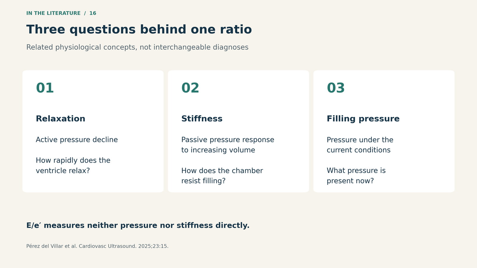
Figure 2. Separate the physiological question before interpreting an ultrasound surrogate. This is a conceptual framework, not a diagnostic grading algorithm.[2]

What the guideline asks us to integrate
The 2025 ASE guideline includes tricuspid E/e′ >6 among abnormal diastolic findings. It places this alongside tricuspid inflow, deceleration time, hepatic venous flow and right atrial, right ventricular and IVC morphology. It explicitly cautions against interpreting any single diastolic parameter in isolation.[1]
For transtricuspid inflow, the guideline recommends end-expiratory measurements and averaging at least five consecutive beats. Respiratory phase and heart rate matter; atrial arrhythmias and hemodynamically significant tricuspid valve disease can confound the pattern. Atrial fibrillation, for example, prevents the usual interpretation of an E/A ratio because coordinated atrial contraction is absent.[1]
Thus, an E/e′ of 8 is a reason to look more closely at the whole right-heart examination. It is not a calibrated reading of right atrial pressure, nor a stand-alone measurement of myocardial stiffness.
Why the direct validation study matters
Pérez del Villar and colleagues compared echocardiographic indices with invasive pressure–volume measurements in 13 pigs exposed to acute hemodynamic interventions and nine patients: five with repaired tetralogy of Fallot and pulmonary regurgitation, and four with pulmonary hypertension.[2]
An instructive example comes from the animal experiment. With volume loading, the reported mean right atrial pressure increased from 2.6 to 10.3 mmHg, while the modeled mean E/e′ remained 4.3. The intervention samples differed—13 at baseline and nine during volume loading—so these group estimates should not be mistaken for identical paired responses in every animal.[2]
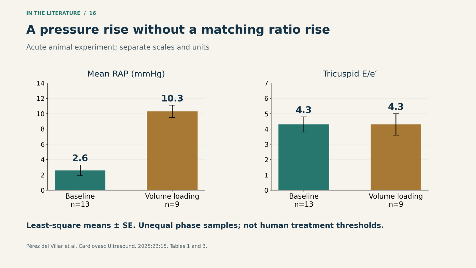
Figure 3. Two measurements need not move together. Least-square means ± standard errors from the animal experiment; baseline n=13 and volume loading n=9. Separate vertical scales preserve the different units. These are not human treatment thresholds. Data: Tables 1 and 3.[2]

In the clinical cohort, the tested echocardiographic indices did not show statistically significant correlations with intrinsic RV diastolic properties. E/e′ was not significantly associated with mean right atrial pressure or RV end-diastolic pressure. Some other indices correlated with right atrial pressure, illustrating that an association with pressure is not the same as validation against relaxation or stiffness.[2]
The study challenges an overly literal interpretation of Doppler ratios. It does not prove that every integrated right-heart assessment is ineffective: the human sample was small, and right atrial size, hepatic venous flow and combined multiparameter approaches were not tested.[2]
Make the report useful for the next decision
A useful interpretation describes the measured pattern and its context, then states whether other findings support elevated right-sided filling pressure. The conclusion should follow the combined evidence, rather than attach a definitive physiological diagnosis to one ratio.[1, 2]
In a ventilated perioperative patient, substituting IVC collapse for E/e′ does not solve the problem. The ASE guideline specifically warns that IVC collapse cannot reliably estimate right atrial pressure during positive-pressure ventilation. When that pressure is clinically relevant and remains uncertain, invasive measurement may be appropriate if feasible.[1]
This distinction also matters for serial examinations: a change in a Doppler ratio may reflect a change in loading or acquisition conditions. Comparing the surrounding physiology is essential before describing it as recovery or worsening of intrinsic RV diastolic function.[1, 2]
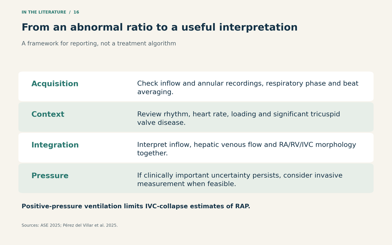
Figure 4. A reporting framework based on the guideline and validation study. It organizes interpretation rather than prescribing treatment.[1, 2]

Three takeaways
- Name what was measured. An abnormal E/e′ is an ultrasound finding, not a direct measurement of stiffness or pressure.
- Look for agreement across the examination. Rhythm, respiration, valve disease and loading conditions determine how much confidence to place in the pattern.
- Resolve the pressure question when it changes care. Use appropriate corroborating findings and, when necessary and feasible, invasive hemodynamics rather than forcing certainty from a single ratio.
References
- Mukherjee M, Rudski LG, Addetia K, Afilalo J, D’Alto M, Freed BH, et al. Guidelines for the echocardiographic assessment of the right heart in adults and special considerations in pulmonary hypertension: recommendations from the American Society of Echocardiography. J Am Soc Echocardiogr. 2025;38(3):141–186. doi:10.1016/j.echo.2025.01.006.
- Pérez del Villar C, Prieto-Arévalo R, García-Carreño J, Martínez-Legazpi P, Rodríguez-Pérez D, Benito Y, et al. Validation of noninvasive indices of right ventricular diastolic function. Simultaneous echocardiography and pressure-volume catheterization studies. Cardiovasc Ultrasound. 2025;23:15. doi:10.1186/s12947-025-00351-5.