
ITL-f0f7cc7a95a5
Juan Li, MD · Updated November 17, 2026
Watch the video

Key points
- Assess the mechanism and severity of tricuspid regurgitation (TR) across several views, adding three-dimensional imaging when feasible.1
- A low TR jet velocity does not exclude severe regurgitation; rapid right ventricular–atrial pressure equalization can reduce the measured gradient.1
- A single vena contracta width can miss an irregular orifice's larger dimension; biplane imaging helps, but does not make two-dimensional and three-dimensional measurements interchangeable.2
- Three-dimensional vena contracta area (VCA) depends on acquisition and analysis settings. A smaller value on follow-up may reflect a technical change.3
- Discordance between echocardiography and cardiac magnetic resonance (CMR) warrants reconciliation of methods and conditions; it does not, by itself, identify which test is correct.4
The right heart looks enlarged, the regurgitant jet is conspicuous, yet one measured vena contracta width suggests less important TR. Should that single number settle the report? The useful next step is to ask what the measurement captured, under which conditions, and whether the other findings support it. Avoiding underestimation means resolving the discrepancy, rather than automatically choosing the largest number.
Start with the valve and the imaging window
The 2025 American Society of Echocardiography right-heart guideline recommends several two-dimensional planes and three-dimensional echocardiography when feasible to identify TR mechanism and severity. Transthoracic echocardiography (TTE) remains the initial approach. With a good acoustic window, it may image the anteriorly positioned tricuspid valve better than transesophageal echocardiography (TEE); TEE can still be necessary to clarify leaflet or annular pathology.1
This distinction matters when comparing an awake TTE with an intraoperative TEE. The modality, loading conditions, ventilation and timing may differ. A lower intraoperative measurement should prompt review of those differences before concluding that the valve disease has improved. Conversely, the intraoperative examination may reveal geometry missed by the earlier study.2
Low Doppler velocity is another potential source of false reassurance. In severe TR, early pressure equalization between the right ventricle and right atrium can produce a truncated envelope and an underestimated pressure gradient. Regurgitant severity and the pressure driving the jet are different quantities. Severe TR can occur without severe pulmonary hypertension, and pulmonary pressure alone cannot grade the regurgitation.1
Check whether the plane sampled the relevant geometry
Vena contracta width describes a linear dimension. It cannot fully characterize an elongated or irregular flow orifice from one plane. Biplane imaging adds an orthogonal dimension; three-dimensional multiplanar reconstruction permits assessment of the cross-section and its orientation.2
Singh and colleagues compared these approaches in 30 selected patients undergoing elective tricuspid surgery, with TEE images acquired after induction of general anesthesia. Biplane widths from all three standard midesophageal views were smaller, on average, than the three-dimensional average width. The right ventricular inflow–outflow view agreed best with that reference among the tested biplane views.2
The practical lesson is to examine additional planes when a single width does not fit the rest of the study. It is not a mandate to treat one view as universally accurate. The study selected good-quality datasets, used three-dimensional measurements as its comparator, and lacked an independent gold standard. Its findings also do not validate applying a width threshold directly to an area measurement.2
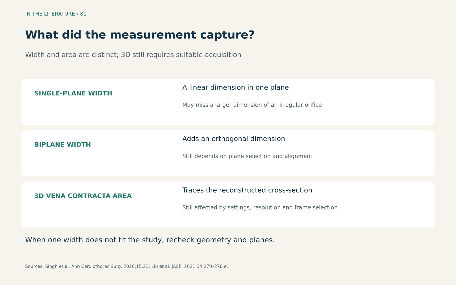
Figure 1. The measurement should match the question. Additional dimensions help reveal geometry, but three-dimensional imaging still requires suitable acquisition and correct reconstruction.2,3 View larger

Before interpreting a change, check the settings
Three-dimensional VCA avoids assuming a circular orifice, but it is not independent of the machine. In Liu and colleagues' prospective study of 69 patients scheduled for surgery, datasets were acquired on at least two of three platforms. Color gain, baseline shifts and vendor differences affected the measured VCA.3
The within-platform comparison is particularly instructive. In the same 39 patients examined on the GE E95, mean VCA was 1.11 ± 0.81 cm² at default settings, 1.20 ± 0.79 cm² with a baseline shift, and 0.65 ± 0.57 cm² at minimal gain. These were measurements of the same acquired frames under different settings—not responses to treatment.3
This does not establish that the smallest or largest setting-derived area is the true value, nor prescribe a universal gain setting. It shows why serial reports need comparable acquisition and analysis. Liu and colleagues recommend using the same vendor and settings for longitudinal VCA assessment in the same patient; timing within systole and respiratory or rhythm variation also deserve attention.3
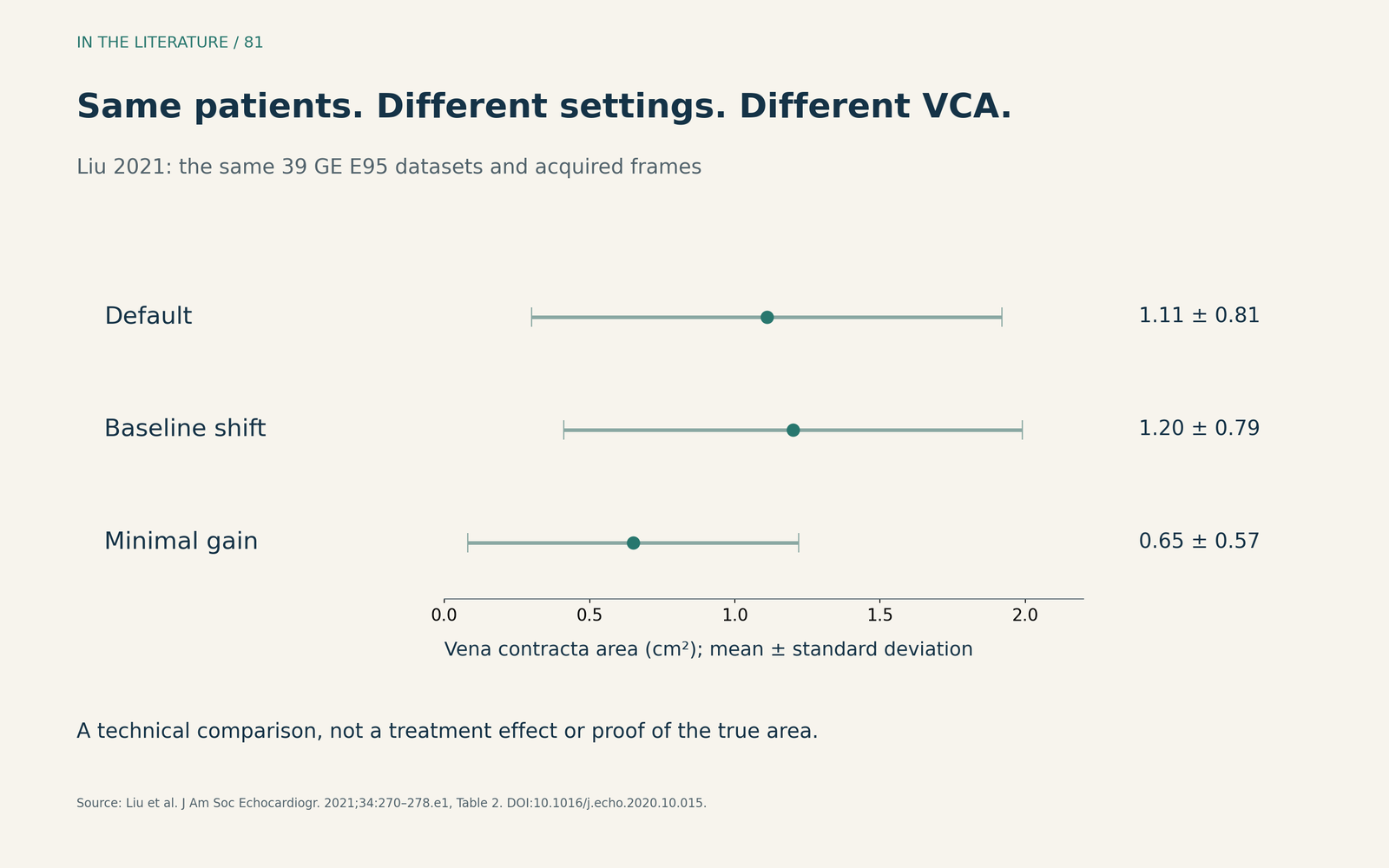
Figure 2. Original display of Liu et al.'s reported GE E95 measurements. Points show means and bars show standard deviations, not confidence intervals. The three rows represent the same patients and acquired frames. This is a technical comparison, not a treatment effect.3 View larger

If the discrepancy persists, reconcile the modalities
CMR can provide complementary regurgitant-volume, regurgitant-fraction and right ventricular information. However, switching modality does not automatically resolve every disagreement. Sularz and colleagues prospectively enrolled 52 patients with at least moderate TR on their screening TTE and compared echocardiographic grading with CMR incorporating four-dimensional flow. Classification was frequently discordant, with CMR generally assigning lower grades under the study's chosen thresholds.4
The examinations were not simultaneous: the median interval was 2 days, with an interquartile range of 1–37 days. Patients with permanent pacemakers, end-stage renal disease or other important valve disease were excluded. The study therefore does not establish that CMR should replace echocardiography in every patient, or that all discordant echocardiograms overestimate TR. The clinical implications of the disagreement remained unresolved.4
For an individual patient, the useful synthesis is explicit: describe the mechanism, the reliable measurements and their acquisition conditions, the right-heart response, and the specific information that remains uncertain. Recheck image quality and geometry before using another modality to answer that remaining question. This is a clinical synthesis of the guideline and comparison studies, rather than a validated scoring algorithm.1–4
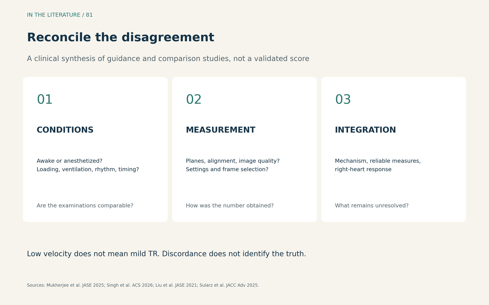
Figure 3. A practical way to organize reassessment. Chamber and venous findings add context; no single finding or modality automatically overrides the integrated assessment.1–4 View larger

Three takeaways
- Resolve the mismatch: a low jet velocity or a narrow width in one plane should not dismiss otherwise concerning TR findings.
- Make serial measurements comparable: record the view, modality, relevant settings and physiological conditions before interpreting a numerical change.
- Use complementary imaging to answer a specific question: reconcile what each method measures and state remaining uncertainty, instead of automatically accepting the highest grade or the newest modality.
References
- Mukherjee M, Rudski LG, Addetia K, et al. Guidelines for the Echocardiographic Assessment of the Right Heart in Adults and Special Considerations in Pulmonary Hypertension: Recommendations from the American Society of Echocardiography. J Am Soc Echocardiogr. 2025;38(3):141–186. doi:10.1016/j.echo.2025.01.006
- Singh R, Meineri M, Zakhary W, et al. Comparison of biplanar- with 3D-vena contracta and vena contracta area for the assessment of tricuspid valve regurgitation by intraoperative transesophageal echocardiography. Ann Cardiothorac Surg. 2026;15(2):23. doi:10.21037/acs-2025-1-72-tvd
- Liu Y, Chen B, Zhang Y, et al. Sources of Variability in Vena Contracta Area Measurement for Tricuspid Regurgitation Severity Grading: Comparison of Technical Settings and Vendors. J Am Soc Echocardiogr. 2021;34(3):270-278.e1. doi:10.1016/j.echo.2020.10.015
- Sularz A, Negm AS, Chavez Ponce A, et al. Prospective Quantification of Tricuspid Regurgitation With Echocardiography vs 4D Flow Cardiac Magnetic Resonance. JACC Adv. 2025;4(6 Pt 1):101759. doi:10.1016/j.jacadv.2025.101759